
Recommended
More Related Content
What's hot
What's hot (20)
Viewers also liked
Viewers also liked (18)
Similar to Development of the Inner Ear and Common Malformations
Similar to Development of the Inner Ear and Common Malformations (20)
Recently uploaded
Recently uploaded (20)
Development of the Inner Ear and Common Malformations
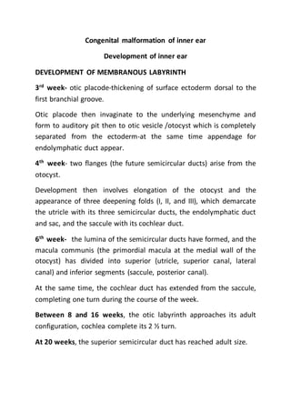
- 1. Congenital malformation of inner ear Development of inner ear DEVELOPMENT OF MEMBRANOUS LABYRINTH 3rd week- otic placode-thickening of surface ectoderm dorsal to the first branchial groove. Otic placode then invaginate to the underlying mesenchyme and form to auditory pit then to otic vesicle /otocyst which is completely separated from the ectoderm-at the same time appendage for endolymphatic duct appear. 4th week- two flanges (the future semicircular ducts) arise from the otocyst. Development then involves elongation of the otocyst and the appearance of three deepening folds (I, II, and III), which demarcate the utricle with its three semicircular ducts, the endolymphatic duct and sac, and the saccule with its cochlear duct. 6th week- the lumina of the semicircular ducts have formed, and the macula communis (the primordial macula at the medial wall of the otocyst) has divided into superior (utricle, superior canal, lateral canal) and inferior segments (saccule, posterior canal). At the same time, the cochlear duct has extended from the saccule, completing one turn during the course of the week. Between 8 and 16 weeks, the otic labyrinth approaches its adult configuration, cochlea complete its 2 ½ turn. At 20 weeks, the superior semicircular duct has reached adult size.
- 2. The organ of Corti is differentiated to such a degree by 20 weeks that the fetus can “hear” and respond to fluid-borne sounds. The organ of Corti approximates the adult structure by 25 weeks. DEVELOPMENT OF OTIC CAPSULE develops from the precartilage (compacted mesenchyme that is differentiating into embryonic cartilage) surrounding it. Started by the end of fourth week as cell density of the mesenchyme enveloping otic capsule increases. By 8th week cartilaginous model of otic capsule formed. A total of 14 ossification centers eventually appear and fuse to complete the ossification of the otic capsule. CONGENITAL MALFORMATION OF INNER EAR Due to Interruption during first trimester of pregnancy- inborn genetic error or teratogenic. Genetic errors- may be either autosomal dominant or recessive and may manifest as sensorineural hearing loss (SNHL) alone or be associated with any of a number of syndromes Teratogenic - in utero viral infection (e.g,rubella), chemical teratogens (e.g, thalidomide), and radiation exposure Derangement of the otic capsule ossification process alone does not appear to be a major mechanism in congenital hearing loss. Ossification of the labyrinthine lumen, however, is a common finding in early acquired deafness, typically arising as a consequence of meningitis. Incidence
- 3. malformations limited to the membranous labyrinth predominate-cochlea>SCC>vestibular aqueduct. Note that otic capsule deformity is radiographically detected but not membranous deformity. Classification Key terms are 1) aplasia, complete lack of development; 2) hypoplasia, incomplete development; and 3) dysplasia, aberration in development. Congenital anomalies of the inner ear may be considered in two broad categories: malformations with pathologic changes limited to the membranous labyrinth and malformations that involve both the osseous and membranous labyrinth I. Malformations limited to the membranous labyrinth A. Complete membranous labyrinthine dysplasia (Siebenmann-Bing) B. Limited membranous labyrinthine dysplasia 1. Cochleosaccular dysplasia (Scheibe) 2. Cochlear basal turn dysplasia (Alexander) II. Malformations of the osseous and membranous labyrinth A. Complete labyrinthine aplasia (Michel) B. Cochlear anomalies 1. Cochlear aplasia
- 4. 2. Cochlear hypoplasia 3. Incomplete partition (Mondini) 4. Common cavity C. Labyrinthine anomalies 1. Semicircular canal dysplasia 2. Semicircular canal aplasia D. Aqueductal anomalies 1. Enlargement of the vestibular aqueduct 2. Enlargement of the cochlear aqueduct E. Internal auditory canal abnormalities 1. Narrow internal auditory canal 2. Wide internal auditory canal. MALFORMATION LIMITED TO MEMBRANOUS LABYRINTH account for over 90% of congenital deafness, the bony labyrinth is normal A. COMPLETE MEMBRANOUS LABYRINTHINE DYSPLASIA (BING- SIEBENMANN) extremely rare, has been reported in association with the cardioauditory and Usher's syndromes B. LIMITED MEMBRANOUS LABYRINTHINE DYSPLASIA COCHLEOSACCULAR DYSPLASIA (SCHEIBE) The cochlear duct is usually collapsed, with Reissner's membrane adherent to the limbus.
- 5. The stria vascularis is typically degenerated and may contain colloidal inclusions. Cochlear changes may be severe in the base turn and gradually lessen in intensity towards the apex, or they may be severe throughout. The saccule is usually collapsed and has degenerated sensory epithelium. COCHLEAR BASAL TURN DYSPLASIA May be related to familial high frequency SNHL Malformation of the membranous and osseous labyrinth C. COMPLETE LABYRINTHINE APLASIA Extremely rare, developmental arrest before appearance of otic vesicle. Associated with anencephaly and thalidomide exposure. Radiographucally may be confused with labyrinthine ossification. D. COCHLEAR ANOMALIES Incidence Incomplete partition (Mondini) -55% Common cavity -26% Cochlear hypoplasia- 15% Cochlear aplasia- 3% Complete labyrinthine aplasia (Michel)- 1%. COCHLEAR APLASIA
- 6. Cochlea completely absent, presumably arrest at 5th week gestation. Radiographically only vestibule and SSC present. COCHLEAR HYPOPLASIA Arrest during 6th week of gestation Hypoplastic cochlea with single turn or less Radiographically a small bud (1-3mm) protrude from the vestibule which is frequently enlarged Hearing is variable, may be remarkably good. INCOMPLETE PARTITION (MONDINI DEFORMITY) Arrest at 7th week gestation Cochlea only 1.5 turns, measuring 5-6mm (normal=8-10mm vertically) Radiographically cochlea smaller than normal and partially or completely lack interscaler septum. Three variants of incomplete partition IP1- lack the entire modiolus and interscalar septa and demonstrate a cystic appearance IP2- normal basal turn but cystic apex IP3- deficient modiolus and partial interscalar septation at the cochlea’s periphery. Hearing is variable, normal to profound SNHL E. COMMON CAVITY Cochlea and vestibule are confluent forming an ovoid cystic space without an internal structure. F. VESTIBULAR AQUEDUCT ENLARGEMENT Diameter > 2mm (N=0.4-1mm)-determined radiographically
- 7. VA is short and broad and lack vascularity and rugosity Large VA syndrome usually b/l, often associated with other radiographic anomalies of the inner ear. Hearing is mild to profound SNHL, gradual deterioration of auditory function from childhood to adulthood Management- steroid administration, shunt procedure, obliteration of endolymphatic sac. G. DEVELOPMENTASL ANOMALIES OF IAC WIDE IAC: >10mm diameter Important of wide IAC is its association with spontaneous CSF leak and the occurrence of csf gusher during stapes surgery. NARROW IAC: <3mm diameter, may indicate failure of 8th nerve development.