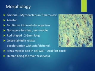
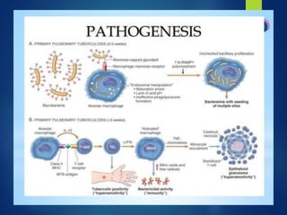
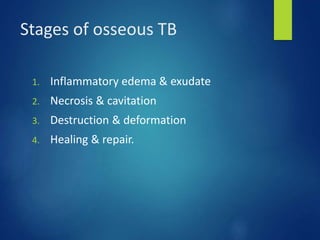
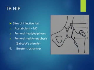
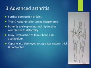
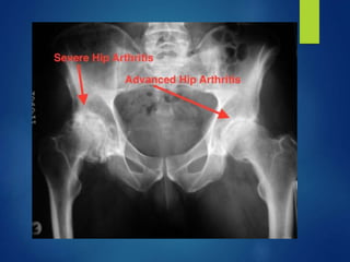
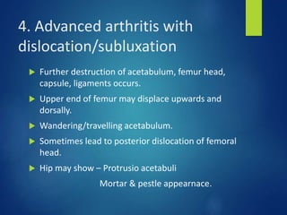
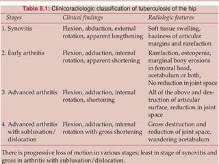
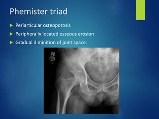
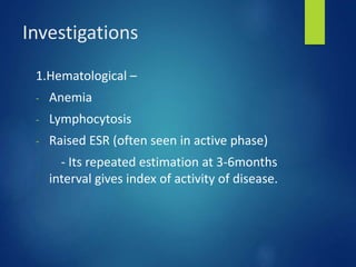
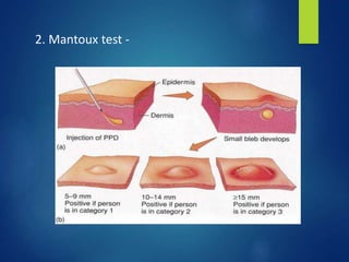
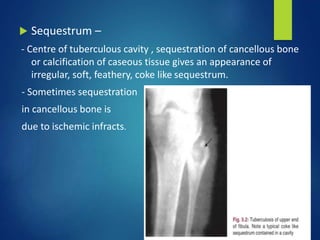
TUBERCULOSIS OF
HIP
BY – DR. SANTOSH M S
ASSISTANT PROFESSOR
DEPT. OF ORTHOPAEDICS
History
Morphology
 Bacteria – Mycobacterium Tuberculosis
 Aerobic
 facultative intra-cellular organism
 Non-spore forming , non-motile
 Rod shaped : 2-5mm long
 Once stained it resists
decolorization with acid/alchohol.
 It has mycolic acid in cell wall – Acid fast bacilli
 Human being the main resorviour
Pathology of TB
 Entry of infection into body
 Accumulation of macrophages
 TB bacilli phagocytosed – lipid from cell wall dispersed int
cytoplasm
 Epitheloid cells – large cells with abundant cytoplassm
 Group of epitheloid cells – form Langhans gaint cell.
 Tubercle – Lymphocytes surround gaint cells in periphery
which appear as nodule in reticulo-endothelial cells.
 Tubercle – 2 types : Hard , Soft
 Soft type associated with caseous necrosis. Its presence is
diagnostic of Tb pathology. `
At risk groups
 Immunocompromised
 Diabetics
 Anticancer chemotherapy
 Immunosuppressive medication
 Chronic alchoholics
 Long term steroid therapy
 Malnourished children
Areas of infective foci
 In descending order –
1. Spine
2. Hip Least common areas :
3. Knee 1. Mandible
4. Foot 2. TM joint
5. Elbow
6. Hand
7. Shoulder
8. Bursal sheaths
Cold abcess
 Collection of – serum, WBC , Caseous material, bone debri,
TB bacilli
 It feels warm on palpation, though not as significant as septic
infection.
 If it bursts – form ulcer/sinus.
 It can form in the joint – usually weaker aspect of capsule
area
 Around hip – Femoral triangle, Medial / lateral aspect of
thigh.
Stages of osseous TB
1. Inflammatory edema & exudate
2. Necrosis & cavitation
3. Destruction & deformation
4. Healing & repair.
TB HIP
 Sites of infective foci
1. Acetabulum – MC
2. Femoral head/epiphyses
3. Femoral neck/metaphysis
(Babcock’s triangle)
4. Greater trochantrer
 TB of GT may involve overlying trochantric bursa
without involving hip joint for long time.
 Foci in upper end of femur – intracapsular – joint
involved rapidly.
 Foci in acetabular roof – joint involvement is late and
mild symptoms.
 Cold abcess forms in joint perforates the inferior
weaker part of capsule and presents any where
around joint.
Features specific to hip
 MC age group – first 3 decades
 Pain
 Limping – Earliest & MC
 Fullness over joint & painful/decreased
ROM.
 Deformity.
 Night cries
 Discharging sinus
Stages of TB Hip
 5 stages –
1. Tubercular synovitis
2. Early arthritis
3. Advanced arthritis
4. Advanced arthritis with subluxation/dislocation.
5. Terminal or Aftermath of arthritis.
1. Tubercular synovitis
 Irritable hip
 Joint is held in position of maximum capacity –
F-AB-ER
 Apparent lengthening present with no true shortening.
 Terminal movements at hip painful, restricted.
 X ray- soft tissue swelling, haziness of articular margin
 USG- Soft tissue sweling.
 MRI- Synovial effusion.
 Biopsy – can be done to confirm by HPE &
Microbiological methods.
Differential diagnosis
1. Transient synovitis
2. Septic arthritis
3. Perthe’s disease
4. SCFE
5. Ilio-psoas abcess
6. Osteomylitis of femur head.
2.Early arthritis
 Destruction of articular cartilages sets in
 Spasm of adductors & flexors occurs which gives
deformity picture – F-AD-IR.
 Apparent shortening & true shortening usually not
>1cm
 Global restriction ROM.
 X ray- Localised osteoporosis, decrease in joint
space, erosion of articular cartilage.
 MRI- Synovial effusion, minimal areas of bone
desrtuction with bone edema.
3.Advanced arthritis
 Further destruction of joint
 True & Apparent shortening exaggerated
 Pt tends to sleep on normal hip further
contributes to deformity.
 X ray- Destruction of femur head and
acetabulum.
 Capsule also destroyed to a greater extent- thick
& contracted.
4. Advanced arthritis with
dislocation/subluxation
 Further destruction of acetabulum, femur head,
capsule, ligaments occurs.
 Upper end of femur may displace upwards and
dorsally.
 Wandering/travelling acetabulum.
 Sometimes lead to posterior dislocation of femoral
head.
 Hip may show – Protrusio acetabuli
Mortar & pestle appearnace.
5. Terminal or aftermath of
arthritis
 It is ankylosis of joint.
 Articular margins are adapted to the deformed
position
 Degenerative arthritic changes will be present.
 Grossly , joint may appear irregular , cobbled ,
deformed , pock-marked and devoid of articular
cartilage.
Phemister triad
 Periarticular osteoporosis
 Peripherally located osseous erosion
 Gradual diminition of joint space.
Investigations
1.Hematological –
- Anemia
- Lymphocytosis
- Raised ESR (often seen in active phase)
- Its repeated estimation at 3-6months
interval gives index of activity of disease.
2. Mantoux test -
3. Biopsy –
- More reliable procedure specially to prove
disease in early stages.
- Can be synovium, lymph node, bone
tissue.
- HPE – granulomatous inflammation with
epitheloid cells and gaint cells.
4. Synovial fluid analysis –
- Lymphocytosis
- Glucose levels reduced
- Protein levels increased
- Excellent source for PCR
technique.
5.Bacteriological investigations –
- Specimen sample stained for AFB, C/s
- Media used for growth – Lowenstein-
Jensen
- BACTEC –
 Detects microbe in 7-14 days.
6)Molecular -
 PCR –
- highly specific for TB bacilli , amplifies
even if single organsim is present.
- Diagnosis can be made in 3 days.
Radiological
1. X ray –
- AP & Lateral view of hip with CXR to be done.
- 1st sign : localised osteoporosis (in active disease).
- Articular margins & bony cortex become hazy and
gradually destructive changes occur.
- Soft tissue swelling – due to synovial fluid, thickened
synovium, capsule & peri-articular disease
involvement.
- Joint space reduced – articular cartilage involved.
X ray findings in Left Hip
 Sequestrum –
- Centre of tuberculous cavity , sequestration of cancellous bone
or calcification of caseous tissue gives an appearance of
irregular, soft, feathery, coke like sequestrum.
- Sometimes sequestration
in cancellous bone is
due to ischemic infracts.
2. CT scans –
- Helpful in demonstrating small destroyed
areas (lytic cavities) in bone and marginal
erosion.
- Soft tissue edema, granulations, exudation,
abcess formation can be diagnosed earlier
stage.
3. MRI scan –
- It shows pre destructive leisons like edema,
inflammation of bone in active disease
Management
 Two pathways –
1. Medical management
2. Surgical management.
Medical management
1. General
2. Anti-tb chemotherapy.
3. Rest
4. Traction ,if deformity present.
5. Mobilisation.
Anti TB drugs
Scheduled Anti TB drug treatment
3.Rest
 All pt. are adviced to sleep on
hard bed.
 In active stages ,joint is
given rest in “position of ease”.
 Pt. in early stages are
adviced intermittent active & passive exercises to improve
funtional arc of joint involved.
4.Traction
 It is one of the best modality to-
1. Correct deformity.
2. Maintain limb in functional position throughout treatment
3. Maintains joint space.
4. Relieves muscle spasm.
5. B/l traction to be put to stabilise the pelvis.
6. Prevent complications – dislocation/subluxation, widening of
acetabulum.
 Maintainace of traction and intermittent active and passive
motion of joint within range of tolerable pain during healing
process will –
1. Encourage development of healthy synovial membrane and
well lubricated fibrocartilage for joint function.
2. Induction of proliferating mesenchymal cells will metaplaise
the synovial cells.
 These process may permit good functional recovery of joint
even in joint damaged by tb lasting in healed status of
disease.
5.Mobilisation
 After starting the treatment in initial stages –
 1st 12 weeks : Non-weight bearing
 Next 12 weeks : Partial weight bearing
Ambulation with orthosis/crutches.
 18-24 months : Unprotected weight bearing can
be started.
If advanced arthritis ?
 The usual outcome is gross fibrous ankylosis.
 The limb should be immobilised with help of hip spica for
about 4-6 months.
 Ideal position of immobilsation –
1. Neutral between adduction & abduction.
2. External rotation of 5-10 degree
3. Flexion – In children – upto 10 degree
In adults – upto 30 degree.
 After 6 months – partial weight bearing with hip spica
 After 2 yrs – with orthrosis & crutches.
Surgical management
 It is used as adjunct to chemotherapy but not substitute.
 Indications :
1. Clinically non-responsive disease
2. Failure to obtain acceptable outcome / deformity after
chemotherapy.
3. To obtain tissue for diagnosis.
- Before taking up for surgery , make sure pt. recieves
chemotherapy for 4weeks and physically fit for surgery.
Types of surgery
1.Synovectomy
2.Joint debridement
3.Osteotomy
4.Arthrodesis : Intra-articular
Extra-articular
Combined
5.Excision arthroplasty
6.Tectoplasty
7.THR
Management in children
 In children with arthritis , deformity , subluxation / dislocation is
corrected or minimised by Traction.
 Rarely , children go for operative procedure.
 Failure of correction / minimise the deforming changes will
require –
1. Open arthrotomy
2. Joint debridement & synovectomy.
3. Improvement of displacement.
 After completion of growth plate potential, can be taken up for -
1. Arthrodesis
2. Excisional arthroplasty
 Children presenting with healed gross deformity requires
extra-articular corrective osteotomy to enable them to
walk better until skeletal maturity.
 If there is no gross deformity of hip joint –
1. Sub-total excision of contracted fibrous capsule is done.
2. Traction and repititive exercises.
 This may be helpful in restoring a useful range of
movements for few years.
 Incidence of reactivation will be least –
1. Healed status achieved with remineralisation and
restoration of destroyed bone.
2. Healing of articular surface with near complete funtion
or bony ankylosis of destroyed joint or painless
fibrous ankylosis.
 Prognosis of disease -
1. With evolution of chemotherapy , tretament protocols
of TB has been changed.
2. If diagnosed early and treated with strict adherence to
chemotherapy, healing can be expected with good
mobility of joint.
THANK YOU

tb hip.pptx

  • 1.
    TUBERCULOSIS OF HIP BY –DR. SANTOSH M S ASSISTANT PROFESSOR DEPT. OF ORTHOPAEDICS
  • 2.
  • 3.
    Morphology  Bacteria –Mycobacterium Tuberculosis  Aerobic  facultative intra-cellular organism  Non-spore forming , non-motile  Rod shaped : 2-5mm long  Once stained it resists decolorization with acid/alchohol.  It has mycolic acid in cell wall – Acid fast bacilli  Human being the main resorviour
  • 4.
    Pathology of TB Entry of infection into body  Accumulation of macrophages  TB bacilli phagocytosed – lipid from cell wall dispersed int cytoplasm  Epitheloid cells – large cells with abundant cytoplassm  Group of epitheloid cells – form Langhans gaint cell.  Tubercle – Lymphocytes surround gaint cells in periphery which appear as nodule in reticulo-endothelial cells.  Tubercle – 2 types : Hard , Soft  Soft type associated with caseous necrosis. Its presence is diagnostic of Tb pathology. `
  • 6.
    At risk groups Immunocompromised  Diabetics  Anticancer chemotherapy  Immunosuppressive medication  Chronic alchoholics  Long term steroid therapy  Malnourished children
  • 7.
    Areas of infectivefoci  In descending order – 1. Spine 2. Hip Least common areas : 3. Knee 1. Mandible 4. Foot 2. TM joint 5. Elbow 6. Hand 7. Shoulder 8. Bursal sheaths
  • 8.
    Cold abcess  Collectionof – serum, WBC , Caseous material, bone debri, TB bacilli  It feels warm on palpation, though not as significant as septic infection.  If it bursts – form ulcer/sinus.  It can form in the joint – usually weaker aspect of capsule area  Around hip – Femoral triangle, Medial / lateral aspect of thigh.
  • 9.
    Stages of osseousTB 1. Inflammatory edema & exudate 2. Necrosis & cavitation 3. Destruction & deformation 4. Healing & repair.
  • 10.
    TB HIP  Sitesof infective foci 1. Acetabulum – MC 2. Femoral head/epiphyses 3. Femoral neck/metaphysis (Babcock’s triangle) 4. Greater trochantrer
  • 11.
     TB ofGT may involve overlying trochantric bursa without involving hip joint for long time.  Foci in upper end of femur – intracapsular – joint involved rapidly.  Foci in acetabular roof – joint involvement is late and mild symptoms.  Cold abcess forms in joint perforates the inferior weaker part of capsule and presents any where around joint.
  • 13.
    Features specific tohip  MC age group – first 3 decades  Pain  Limping – Earliest & MC  Fullness over joint & painful/decreased ROM.  Deformity.  Night cries  Discharging sinus
  • 14.
    Stages of TBHip  5 stages – 1. Tubercular synovitis 2. Early arthritis 3. Advanced arthritis 4. Advanced arthritis with subluxation/dislocation. 5. Terminal or Aftermath of arthritis.
  • 15.
    1. Tubercular synovitis Irritable hip  Joint is held in position of maximum capacity – F-AB-ER  Apparent lengthening present with no true shortening.  Terminal movements at hip painful, restricted.  X ray- soft tissue swelling, haziness of articular margin  USG- Soft tissue sweling.  MRI- Synovial effusion.  Biopsy – can be done to confirm by HPE & Microbiological methods.
  • 17.
    Differential diagnosis 1. Transientsynovitis 2. Septic arthritis 3. Perthe’s disease 4. SCFE 5. Ilio-psoas abcess 6. Osteomylitis of femur head.
  • 18.
    2.Early arthritis  Destructionof articular cartilages sets in  Spasm of adductors & flexors occurs which gives deformity picture – F-AD-IR.  Apparent shortening & true shortening usually not >1cm  Global restriction ROM.  X ray- Localised osteoporosis, decrease in joint space, erosion of articular cartilage.  MRI- Synovial effusion, minimal areas of bone desrtuction with bone edema.
  • 20.
    3.Advanced arthritis  Furtherdestruction of joint  True & Apparent shortening exaggerated  Pt tends to sleep on normal hip further contributes to deformity.  X ray- Destruction of femur head and acetabulum.  Capsule also destroyed to a greater extent- thick & contracted.
  • 22.
    4. Advanced arthritiswith dislocation/subluxation  Further destruction of acetabulum, femur head, capsule, ligaments occurs.  Upper end of femur may displace upwards and dorsally.  Wandering/travelling acetabulum.  Sometimes lead to posterior dislocation of femoral head.  Hip may show – Protrusio acetabuli Mortar & pestle appearnace.
  • 23.
    5. Terminal oraftermath of arthritis  It is ankylosis of joint.  Articular margins are adapted to the deformed position  Degenerative arthritic changes will be present.  Grossly , joint may appear irregular , cobbled , deformed , pock-marked and devoid of articular cartilage.
  • 25.
    Phemister triad  Periarticularosteoporosis  Peripherally located osseous erosion  Gradual diminition of joint space.
  • 26.
    Investigations 1.Hematological – - Anemia -Lymphocytosis - Raised ESR (often seen in active phase) - Its repeated estimation at 3-6months interval gives index of activity of disease.
  • 27.
  • 28.
    3. Biopsy – -More reliable procedure specially to prove disease in early stages. - Can be synovium, lymph node, bone tissue. - HPE – granulomatous inflammation with epitheloid cells and gaint cells.
  • 29.
    4. Synovial fluidanalysis – - Lymphocytosis - Glucose levels reduced - Protein levels increased - Excellent source for PCR technique.
  • 30.
    5.Bacteriological investigations – -Specimen sample stained for AFB, C/s - Media used for growth – Lowenstein- Jensen - BACTEC –  Detects microbe in 7-14 days.
  • 31.
    6)Molecular -  PCR– - highly specific for TB bacilli , amplifies even if single organsim is present. - Diagnosis can be made in 3 days.
  • 32.
    Radiological 1. X ray– - AP & Lateral view of hip with CXR to be done. - 1st sign : localised osteoporosis (in active disease). - Articular margins & bony cortex become hazy and gradually destructive changes occur. - Soft tissue swelling – due to synovial fluid, thickened synovium, capsule & peri-articular disease involvement. - Joint space reduced – articular cartilage involved.
  • 33.
    X ray findingsin Left Hip
  • 34.
     Sequestrum – -Centre of tuberculous cavity , sequestration of cancellous bone or calcification of caseous tissue gives an appearance of irregular, soft, feathery, coke like sequestrum. - Sometimes sequestration in cancellous bone is due to ischemic infracts.
  • 35.
    2. CT scans– - Helpful in demonstrating small destroyed areas (lytic cavities) in bone and marginal erosion. - Soft tissue edema, granulations, exudation, abcess formation can be diagnosed earlier stage. 3. MRI scan – - It shows pre destructive leisons like edema, inflammation of bone in active disease
  • 36.
    Management  Two pathways– 1. Medical management 2. Surgical management.
  • 37.
    Medical management 1. General 2.Anti-tb chemotherapy. 3. Rest 4. Traction ,if deformity present. 5. Mobilisation.
  • 38.
  • 39.
    Scheduled Anti TBdrug treatment
  • 40.
    3.Rest  All pt.are adviced to sleep on hard bed.  In active stages ,joint is given rest in “position of ease”.  Pt. in early stages are adviced intermittent active & passive exercises to improve funtional arc of joint involved.
  • 41.
    4.Traction  It isone of the best modality to- 1. Correct deformity. 2. Maintain limb in functional position throughout treatment 3. Maintains joint space. 4. Relieves muscle spasm. 5. B/l traction to be put to stabilise the pelvis. 6. Prevent complications – dislocation/subluxation, widening of acetabulum.
  • 42.
     Maintainace oftraction and intermittent active and passive motion of joint within range of tolerable pain during healing process will – 1. Encourage development of healthy synovial membrane and well lubricated fibrocartilage for joint function. 2. Induction of proliferating mesenchymal cells will metaplaise the synovial cells.  These process may permit good functional recovery of joint even in joint damaged by tb lasting in healed status of disease.
  • 43.
    5.Mobilisation  After startingthe treatment in initial stages –  1st 12 weeks : Non-weight bearing  Next 12 weeks : Partial weight bearing Ambulation with orthosis/crutches.  18-24 months : Unprotected weight bearing can be started.
  • 44.
    If advanced arthritis?  The usual outcome is gross fibrous ankylosis.  The limb should be immobilised with help of hip spica for about 4-6 months.  Ideal position of immobilsation – 1. Neutral between adduction & abduction. 2. External rotation of 5-10 degree 3. Flexion – In children – upto 10 degree In adults – upto 30 degree.  After 6 months – partial weight bearing with hip spica  After 2 yrs – with orthrosis & crutches.
  • 45.
    Surgical management  Itis used as adjunct to chemotherapy but not substitute.  Indications : 1. Clinically non-responsive disease 2. Failure to obtain acceptable outcome / deformity after chemotherapy. 3. To obtain tissue for diagnosis. - Before taking up for surgery , make sure pt. recieves chemotherapy for 4weeks and physically fit for surgery.
  • 46.
    Types of surgery 1.Synovectomy 2.Jointdebridement 3.Osteotomy 4.Arthrodesis : Intra-articular Extra-articular Combined 5.Excision arthroplasty 6.Tectoplasty 7.THR
  • 47.
    Management in children In children with arthritis , deformity , subluxation / dislocation is corrected or minimised by Traction.  Rarely , children go for operative procedure.  Failure of correction / minimise the deforming changes will require – 1. Open arthrotomy 2. Joint debridement & synovectomy. 3. Improvement of displacement.  After completion of growth plate potential, can be taken up for - 1. Arthrodesis 2. Excisional arthroplasty
  • 48.
     Children presentingwith healed gross deformity requires extra-articular corrective osteotomy to enable them to walk better until skeletal maturity.  If there is no gross deformity of hip joint – 1. Sub-total excision of contracted fibrous capsule is done. 2. Traction and repititive exercises.  This may be helpful in restoring a useful range of movements for few years.
  • 49.
     Incidence ofreactivation will be least – 1. Healed status achieved with remineralisation and restoration of destroyed bone. 2. Healing of articular surface with near complete funtion or bony ankylosis of destroyed joint or painless fibrous ankylosis.  Prognosis of disease - 1. With evolution of chemotherapy , tretament protocols of TB has been changed. 2. If diagnosed early and treated with strict adherence to chemotherapy, healing can be expected with good mobility of joint.
  • 50.