
More Related Content
What's hot
What's hot (18)
Viewers also liked
Similar to Cranial Nerve V
Similar to Cranial Nerve V (20)
Recently uploaded
Recently uploaded (20)
Cranial Nerve V
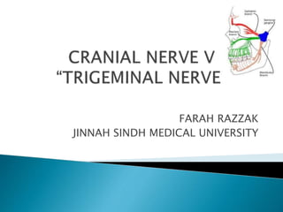
- 1. FARAH RAZZAK JINNAH SINDH MEDICAL UNIVERSITY
- 2. Trigeminal nerve is the largest cranial nerve and contains both sensory and motor fibers. It is sensory nerve to the greater part of the head and motor nerve to several muscles including muscles of mastication.
- 3. MAIN SENSORY NUCLEUS: Lies in posterior part of the pons lateral to motor nucleus. Continuous below the spinal nucleus. SPINAL NUCLEUS: Continuous superiorly with main sensory nucleus in the pons, Extends inferiorly through the whole length of the medulla oblongata and into the upper part of the spinal cord as far as the second cervical segment. MESENCEPHALIC NUCLEUS: Situated in the lateral part of the gray matter around the cerebral aqueduct. Extends inferiorly into the pons as far as the main sensory nucleus. MOTOR NUCLEUS: Situated in the pons medial to main sensory nucleus.
- 4. The sensations of pain ,temperature, touch and pressure from the skin of the face and mucous membrane travel along axons whose cell bodies are situated in TRIGEMINAL SENSORY GANGLION. Central processes of these cells form the large sensory root of the trigeminal nerve. They then divide into (a) ascending branches which terminate in the sensory nucleus. (b) descending branches terminate in spinal nucleus. The sensations of touch and pressure are conveyed by nerve fibers that terminate in the sensory nucleus . The sensations of pain and temperature pass to the spinal nucleus. The sensory fibers from the ophthalmic division terminate in the inferior part of spinal nucleus. Sensory fibers from maxillary division terminates into the middle part of spinal nucleus And that of mandibular division terminates into superior part of spinal nucleus. Proprioceptive impulses from the muscles of mastication and from the facial and the extraocular muscles are carried by the fibers in the sensory root of trigeminal nerve that have by passed the trigeminal ganglion. These fibers arises from the unipolar cells of the mesencephalic nucleus.
- 5. The axons of the main sensory nucleus and spinal nucleus and the central processes of the cells in the mesencephalic nucleus now crosses the medial plane and now ascend as the trigeminal lemniscus to terminate on the nerve cells of the ventral posteromedial nucleus of the thalamus. The axons of these cells now travel through the internal capsule to the postcentral gyrus.
- 6. The cells of the motor nucleus give rise to the axons that form the motor root . The motor nucleus supplies the muscles of mastication Tensor tympani Tensor veil palatine Mylohyoid Anterior belly of the Digastric muscle
- 7. Leaves the anterior aspect of pons as a small motor and large sensory root. Nerve passes forward out of the posterior cranial fossa and rest on the apex of the petrous part of the temporal bone in the middle cranial fossa. The large sensory now expands to form TRIGEMINAL GANGLION enclosed in within a pouch of dura mater called as “MECKEL CAVE”
- 8. 1.OPTHALMIC NERVE: Leaves the skull through superior orbital fissure to enter the orbital cavity. It has further branches: (a) Nasocilliary (b)Anterior ethmoid (c)Posterior ethmoid (d)Infratrochlear (e)Supraorbital (f) Supratrochlear (g)Lacrimal
- 9. 2.MAXILLARY NERVE: Leaves the skull through foramen rotunda it has further divisions In the cranium Middle meningeal nerve in the meninges From the pterygopalatine fossa Infraorbital nerve through Infraorbital canal Zygomatic nerve (zygomaticotemporal nerve, zygomaticofacial nerve) through Inferior orbital fissure Nasal Branches (nasopalatine) through Sphenopalatine foramen Superior alveolar nerves (Posterior superior alveolar nerve, Middle superior alveolar nerve, Anterior superior alveolar nerve) Palatine nerves (Greater palatine nerve, Lesser palatine nerve), including the Nasopalatine nerve Pharyngeal nerve In the infraorbital canal Anterior superior alveolar nerve Infraorbital nerve On the face Inferior palpebral nerve Superior labial nerve Lateral nasal nerve
- 10. 3.MANDIBULAR NERVE: Contains both sensory and motor fibers Leave the skull through foramen ovule (a)Nerve to mylohyoid (b)Auric temporal (c)Lingual (d)Mental (e)Buccal (g) Inferior alveolar