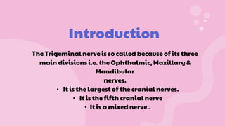
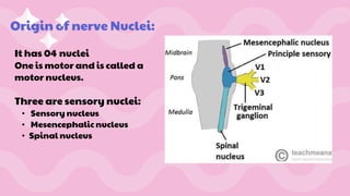
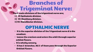
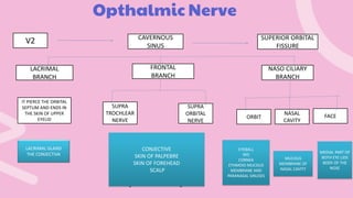
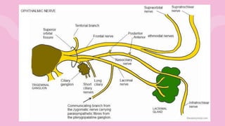
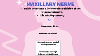
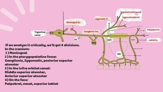
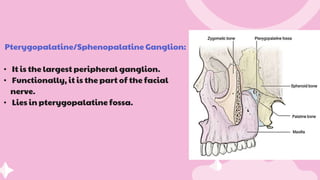
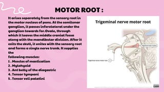
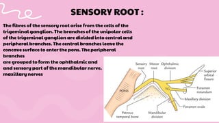
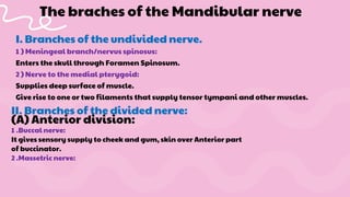
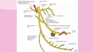
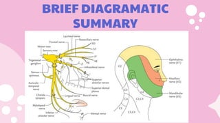
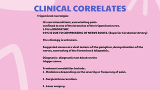
TRIGEMINAL NERVE
Presented
by :
Manahil
Mushtaq
COURSE OF
CONTENTS:
• Introduction
• Origin Of nerve
• Branches of trigeminal nerve
• Course of nerve
• Structures supplied
• Clinical correlates
Introduction
The Trigeminal nerve is so called because of its three
main divisions i.e. the Ophthalmic, Maxillary &
Mandibular
nerves.
• It is the largest of the cranial nerves.
• It is the fifth cranial nerve
• It is a mixed nerve..
Origin of nerve Nuclei:
It has 04 nuclei
One is motor and is called a
motor nucleus.
Three are sensory nuclei:
• Sensory nucleus
• Mesencephalic nucleus
• Spinal nucleus
Trigeminal ganglion:
Shape: Cresentric or Semilunar in shape.
Location: Located lateral to the Cavernous Sinus in the petrous part
of Temporal Bone.
Blood Supply: Through the ganglionic branches of ICA (Internal
Carotid Artery) and acessory meningeal arteries, which enter
through Foramen Ovale
Branches of
Trigeminal Nerve:
The 3 main divisions of the trigeminal nerve are;
1 ) VI Opthalmic division
2 ) V2 Maxillary division.
3 ) V3 Mandibular division.
OPTHALMIC NERVE
• It is the superior division of the Trigeminal nerve & is the
smallest.
• leaves the cranium and enters the orbit through superior
orbital fissure.
. It is wholly sensory.
• It has 3 branches. All 3 of them pass through the Superior
Orbital Fissure.
Opthalmic Nerve
V1
CAVERNOUS
SINUS
V2
LACRIMAL
BRANCH
NASO CILIARY
BRANCH
IT PIERCE THE ORBITAL
SEPTUM AND ENDS IN
THE SKIN OF UPPER
EYELID
SUPERIOR ORBITAL
FISSURE
FRONTAL
BRANCH
CONJECTIVE
SKIN OF PALPEBRE
SKIN OF FOREHEAD
SCALP
FACE
NASAL
CAVITY
SUPRA
ORBITAL
NERVE
ORBIT
SUPRA
TROCHLEAR
NERVE
LACRIMAL GLAND
THE CONJECTIVA
MUCOUS
MEMBRANE OF
NASAL CAVITY
MEDIAL PART OF
BOTH EYE LIDS
BODY OF THE
NOSE
EYEBALL
IRIS
CORNEA
ETHMOID MUCOUS
MEMBRANE AND
PARANASAL SINUSES
MAXILLARY NERVE
• This is the second & intermediate division of the
trigeminal nerve.
• It is wholly sensory.
V2
Cavernous Sinus
Foramen Rotundum
Crosses the upper part of
pterygopalatine
enters orbit through
inferior orbital fissure
If we analyse it critically, we'll get 4 divisions.
In the cranium:
1 ) Meningeal
2 ) In the pterygopalatine fossa:
Ganglionic, Zygomatic, posterior superior
alveolar
3 ) In the infra orbital canal:
Middle superior alveolar,
Anterior superior alveolar
4) On the face:
Palpebral, nasal, superior labial
Pterygopalatine/Sphenopalatine Ganglion:
• It is the largest peripheral ganglion.
• Functionally, it is the part of the facial
nerve.
• Lies in pterygopalatine fossa.
The branches of the Pterygopalatine ganglion are
I) Orbital branches:
II) Palatine branches:
1 . Anterior/greater palatine nerve
2 .Middle palatine nerve
3 .Posterior palatine nerve
III.Nasal branches:
1 . Posterior superior lateral nerve
2 .Nasopalatine/Sphenopalatine nerve
IV.Pharyngeal branch:
It is the third & largest
division of the
trigeminal nerve.
It is made up of 2 roots:
1 ) Sensory Root
2 ) Motor Root
MANDIBULAR Nerve:
V3
Cavernous Sinus
Foraman Ovale
It arises separately from the sensory root in
the motor nucleus of pons. At the semilunar
ganglion, it passes inferolateral under the
ganglion towards for.Ovale, through
which it leaves the middle cranial fossa
along with the mandibular division. After it
exits the skull, it unites with the sensory root
and forms a single nerve trunk. It supplies
the
following muscles:
MOTOR ROOT :
1 . Muscles of mastication
2 . Mylohyoid
3 . Ant belly of the diagastric
4. Tensor tympani
5 . Tensor veli palatini
The fibres of the sensory root arise from the cells of the
trigeminal ganglion. The branches of the unipolar cells
of the trigeminal ganglion are divided into central and
peripheral branches. The central branches leave the
concave surface to enter the pons. The peripheral
branches
are grouped to form the ophthalmic and
and sensory part of the mandibular nerve.
maxillary nerves
SENSORY ROOT :
I. Branches of the undivided nerve.
1 ) Meningeal branch/nervus spinosus:
Enters the skull through Foramen Spinosum.
2 ) Nerve to the medial pterygoid:
Supplies deep surface of muscle.
Give rise to one or two filaments that supply tensor tympani and other muscles.
The braches of the Mandibular nerve
II. Branches of the divided nerve:
(A) Anterior division:
1 .Buccal nerve:
It gives sensory supply to cheek and gum, skin over Anterior part
of buccinator.
2 .Massetric nerve:
It supplies to masseter muscle and also gives branch to TMJ.
3 . Deep temporal nerve:
They passes above the head of lateral pterygoid and sink deep into Temporalis
muscle.
4. Nerve of the lateral-pterygoid:
The braches of the Mandibular nerve
They are a couple. One supplying each muscle head.
(B) Posterior division:
1 . Auriculotemporal nerve
2 .Lingual nerve
3 .Inferior alveolar nerv
The auriculotemporal nerve arises by a medial & lateral roots that enclircle the
MMA & unite behind it just below the foramen spinosum.
Branches of the Auriculotemporal nerve:
1 . Parotid branches----secretomotor, vasomotor.
2 . Articular branches - to the TMJ.
3 . Auricular branches---to the skin of the helix & tragus.
4. Meatal branches----- Meatus of the tymphanic membrane
5 . Terminal branches---- scalp over the temporal region
The auriculotemporal nerve
Location:
It lies between the ramus of the mandible & the muscle in the
pterygomandibular space.
It then passes deep to reach the side of the tongue.
Supply:
It gives off sensory fibres to the tonsil & the mucous
Lingual nerve
Communication of the facial
with the lingal nerve:
Communication of the facial with the lingal nerve:
As the lingual nerve passes medially to the lateral
pterygoid, it is joined from behind by the chorda
tympani. This nerve conveys secretory fibres from the
facial nerve. The parasympathetic secretory fibres control
the submandibular & sublingual salivary glands.
membrane of the posterior part of the oral cavity.
Inferior Alveolar Nerve.
It is the largest terminal branch of the posterior division
of the mandibular nerve.
Supply:
to the mandibular teeth, foramena of the teeth to supply
mainly the pulp as well as the periodontium.
Branches of the nerve :-
1 . Mental nerve:
It supplies to the skin of the chin & the
mucous membrane as well as the skin of the lower lip
.
2 . Incisive branch:
Continues anteriorly from the mental
nerve in the body of the mandible to form the incisive
plexus & supplies the canine & incisors.
3 . Mylohyoid nerve: it sUpplies the mylohyoid muscle as well
as the anterior belly of the digastric.
BRIEF DIAGRAMATIC
SUMMARY
CLINICAL CORRELATES
It is an intermittant, excruciating pain
confined to one of the branches of the trigeminal nerve.
1 0% is IDIOPATHIC
90% IS DUE TO COMPRESSING OF NERVE ROUTE. (Superior Cerebellar Artery)
The etiology is unknown.
Suggested causes are viral lesions of the ganglion, demyelination of the
nerves, narrowing of the foramina & Idiopathic.
Diagnosis: diagnostic test block on the
trigger zones.
Treatment modalities include..
1 . Medicines depending on the severity or frequency of pain.
2 . Surgical Innervention.
3 . Laser surgery.
Trigeminal nueralgia:
Diagram
Thank You !

cranial nerve 5 detailed decription and presentation

  • 1.
  • 2.
    COURSE OF CONTENTS: • Introduction •Origin Of nerve • Branches of trigeminal nerve • Course of nerve • Structures supplied • Clinical correlates
  • 3.
    Introduction The Trigeminal nerveis so called because of its three main divisions i.e. the Ophthalmic, Maxillary & Mandibular nerves. • It is the largest of the cranial nerves. • It is the fifth cranial nerve • It is a mixed nerve..
  • 4.
    Origin of nerveNuclei: It has 04 nuclei One is motor and is called a motor nucleus. Three are sensory nuclei: • Sensory nucleus • Mesencephalic nucleus • Spinal nucleus
  • 5.
    Trigeminal ganglion: Shape: Cresentricor Semilunar in shape. Location: Located lateral to the Cavernous Sinus in the petrous part of Temporal Bone. Blood Supply: Through the ganglionic branches of ICA (Internal Carotid Artery) and acessory meningeal arteries, which enter through Foramen Ovale
  • 6.
    Branches of Trigeminal Nerve: The3 main divisions of the trigeminal nerve are; 1 ) VI Opthalmic division 2 ) V2 Maxillary division. 3 ) V3 Mandibular division. OPTHALMIC NERVE • It is the superior division of the Trigeminal nerve & is the smallest. • leaves the cranium and enters the orbit through superior orbital fissure. . It is wholly sensory. • It has 3 branches. All 3 of them pass through the Superior Orbital Fissure.
  • 7.
    Opthalmic Nerve V1 CAVERNOUS SINUS V2 LACRIMAL BRANCH NASO CILIARY BRANCH ITPIERCE THE ORBITAL SEPTUM AND ENDS IN THE SKIN OF UPPER EYELID SUPERIOR ORBITAL FISSURE FRONTAL BRANCH CONJECTIVE SKIN OF PALPEBRE SKIN OF FOREHEAD SCALP FACE NASAL CAVITY SUPRA ORBITAL NERVE ORBIT SUPRA TROCHLEAR NERVE LACRIMAL GLAND THE CONJECTIVA MUCOUS MEMBRANE OF NASAL CAVITY MEDIAL PART OF BOTH EYE LIDS BODY OF THE NOSE EYEBALL IRIS CORNEA ETHMOID MUCOUS MEMBRANE AND PARANASAL SINUSES
  • 9.
    MAXILLARY NERVE • Thisis the second & intermediate division of the trigeminal nerve. • It is wholly sensory. V2 Cavernous Sinus Foramen Rotundum Crosses the upper part of pterygopalatine enters orbit through inferior orbital fissure
  • 10.
    If we analyseit critically, we'll get 4 divisions. In the cranium: 1 ) Meningeal 2 ) In the pterygopalatine fossa: Ganglionic, Zygomatic, posterior superior alveolar 3 ) In the infra orbital canal: Middle superior alveolar, Anterior superior alveolar 4) On the face: Palpebral, nasal, superior labial
  • 11.
    Pterygopalatine/Sphenopalatine Ganglion: • Itis the largest peripheral ganglion. • Functionally, it is the part of the facial nerve. • Lies in pterygopalatine fossa.
  • 12.
    The branches ofthe Pterygopalatine ganglion are I) Orbital branches: II) Palatine branches: 1 . Anterior/greater palatine nerve 2 .Middle palatine nerve 3 .Posterior palatine nerve III.Nasal branches: 1 . Posterior superior lateral nerve 2 .Nasopalatine/Sphenopalatine nerve IV.Pharyngeal branch:
  • 13.
    It is thethird & largest division of the trigeminal nerve. It is made up of 2 roots: 1 ) Sensory Root 2 ) Motor Root MANDIBULAR Nerve: V3 Cavernous Sinus Foraman Ovale
  • 14.
    It arises separatelyfrom the sensory root in the motor nucleus of pons. At the semilunar ganglion, it passes inferolateral under the ganglion towards for.Ovale, through which it leaves the middle cranial fossa along with the mandibular division. After it exits the skull, it unites with the sensory root and forms a single nerve trunk. It supplies the following muscles: MOTOR ROOT : 1 . Muscles of mastication 2 . Mylohyoid 3 . Ant belly of the diagastric 4. Tensor tympani 5 . Tensor veli palatini
  • 15.
    The fibres ofthe sensory root arise from the cells of the trigeminal ganglion. The branches of the unipolar cells of the trigeminal ganglion are divided into central and peripheral branches. The central branches leave the concave surface to enter the pons. The peripheral branches are grouped to form the ophthalmic and and sensory part of the mandibular nerve. maxillary nerves SENSORY ROOT :
  • 16.
    I. Branches ofthe undivided nerve. 1 ) Meningeal branch/nervus spinosus: Enters the skull through Foramen Spinosum. 2 ) Nerve to the medial pterygoid: Supplies deep surface of muscle. Give rise to one or two filaments that supply tensor tympani and other muscles. The braches of the Mandibular nerve II. Branches of the divided nerve: (A) Anterior division: 1 .Buccal nerve: It gives sensory supply to cheek and gum, skin over Anterior part of buccinator. 2 .Massetric nerve:
  • 17.
    It supplies tomasseter muscle and also gives branch to TMJ. 3 . Deep temporal nerve: They passes above the head of lateral pterygoid and sink deep into Temporalis muscle. 4. Nerve of the lateral-pterygoid: The braches of the Mandibular nerve They are a couple. One supplying each muscle head. (B) Posterior division: 1 . Auriculotemporal nerve 2 .Lingual nerve 3 .Inferior alveolar nerv
  • 18.
    The auriculotemporal nervearises by a medial & lateral roots that enclircle the MMA & unite behind it just below the foramen spinosum. Branches of the Auriculotemporal nerve: 1 . Parotid branches----secretomotor, vasomotor. 2 . Articular branches - to the TMJ. 3 . Auricular branches---to the skin of the helix & tragus. 4. Meatal branches----- Meatus of the tymphanic membrane 5 . Terminal branches---- scalp over the temporal region The auriculotemporal nerve
  • 19.
    Location: It lies betweenthe ramus of the mandible & the muscle in the pterygomandibular space. It then passes deep to reach the side of the tongue. Supply: It gives off sensory fibres to the tonsil & the mucous Lingual nerve
  • 20.
    Communication of thefacial with the lingal nerve: Communication of the facial with the lingal nerve: As the lingual nerve passes medially to the lateral pterygoid, it is joined from behind by the chorda tympani. This nerve conveys secretory fibres from the facial nerve. The parasympathetic secretory fibres control the submandibular & sublingual salivary glands. membrane of the posterior part of the oral cavity.
  • 21.
    Inferior Alveolar Nerve. Itis the largest terminal branch of the posterior division of the mandibular nerve. Supply: to the mandibular teeth, foramena of the teeth to supply mainly the pulp as well as the periodontium. Branches of the nerve :- 1 . Mental nerve: It supplies to the skin of the chin & the mucous membrane as well as the skin of the lower lip . 2 . Incisive branch: Continues anteriorly from the mental nerve in the body of the mandible to form the incisive plexus & supplies the canine & incisors. 3 . Mylohyoid nerve: it sUpplies the mylohyoid muscle as well as the anterior belly of the digastric.
  • 23.
  • 24.
    CLINICAL CORRELATES It isan intermittant, excruciating pain confined to one of the branches of the trigeminal nerve. 1 0% is IDIOPATHIC 90% IS DUE TO COMPRESSING OF NERVE ROUTE. (Superior Cerebellar Artery) The etiology is unknown. Suggested causes are viral lesions of the ganglion, demyelination of the nerves, narrowing of the foramina & Idiopathic. Diagnosis: diagnostic test block on the trigger zones. Treatment modalities include.. 1 . Medicines depending on the severity or frequency of pain. 2 . Surgical Innervention. 3 . Laser surgery. Trigeminal nueralgia:
  • 25.
  • 27.