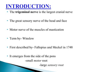
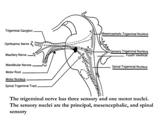
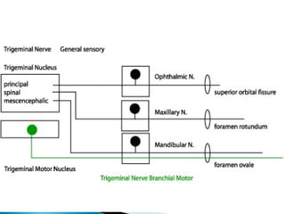
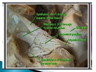
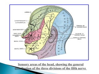
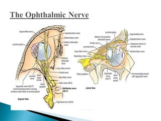
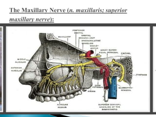
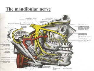
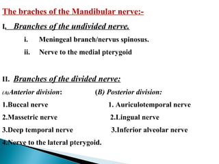
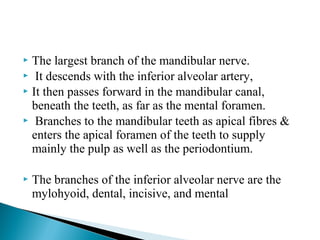
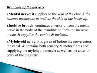
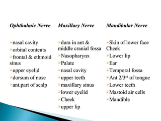
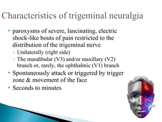
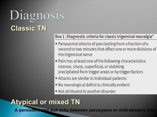
ODMORNIN
 The trigeminal nerve is the largest cranial nerve
 The great sensory nerve of the head and face
 Motor nerve of the muscles of mastication
 Term by- Winslow
 First described by- Fallopius and Meckel in 1748
 It emerges from the side of the pons
-small motor root
-large sensory root
The trigeminal nerve has three sensory and one motor nuclei.
The sensory nuclei are the principal, mesencephalic, and spinal
sensory
 Arise from the motor nucleus within pons and
medulla.
 At the semilunar ganglion motor root passes in a
lateral and inferior direction
 Leaves the middle cranial fossa through the foramen
ovale
 Just after leaving the skull it unites with sensory root
to form a single nerve trunk.
 Arise from the semilunar ganglion which lies in a
cavity of the dura mater near the apex of the petrous
temporal bone.
 They pass backward below the superior petrosal sinus
and tentorium cerebelli, and, entering the pons, divide
into upper and lower roots.
 Occupies a cavity called cavum Meckelii
 Crescent-shaped, with its convexity directed forward:
medially, in relation with the internal carotid artery
and the posterior part of the cavernous sinus.
 The greater superficial petrosal nerve lies underneath
the ganglion.
 The ophthalmic, maxillary, and mandibular nerves
proceed from its convex border.
Sensory areas of the head, showing the general
distribution of the three divisions of the fifth nerve
Ganglia associated with Trigeminal nerve
 ciliary ganglion - ophthalmic nerve
 sphenopalatine ganglion - maxillary nerve
 Otic ganglia - mandibular nerve
 submaxillary ganglia - mandibular nerve
 Smallest of the three divisions of the trigeminal,
 Arises from the upper part of the semilunar ganglion which
passes forward along the lateral wall of the cavernous sinus,
below the oculomotor and trochlear nerves;
 Just before entering the orbit, through the superior orbital
fissure, it divides into three branches, lacrimal, frontal, and
nasociliary
 the smallest of the three branches of the ophthalmic
 It enters the lacrimal gland and gives off several filaments,
which supply the gland and the conjunctiva.
 The largest branch of the ophthalmic
 It enters the orbit through the superior orbital fissure,
and runs forward between the Levator palpebrae
superioris and the periosteum
 Midway between the apex and base of the orbit it
divides into two branches, supratrochlear and
supraorbital.
It supplies the skin of the lower part of the forehead
close to the middle line and the conjunctiva and skin of
the upper eyelid.
The supraorbital nerve (n. supraorbitalis):
It exit the orbit through the supraorbital notch
supply the upper eyelid, conjuctiva, and forehead.
 It supplies internal nasal branches to the mucous membrane of
the front part of the septum and lateral wall of the nasal cavity.
 Finally, it emerges as the external nasal branch and supplies
the skin of the ala and apex of the nose.  
 
 Branches:
◦ the long root of the ciliary ganglion
◦ the long ciliary
◦ the ethmoidal nerves
Enters the postero-superior angle of the ciliary ganglion
It is sometimes joined by a filament from the cavernous plexus
of the sympathetic chain.
The long ciliary nerves (nn. ciliares longi):
The long ciliary nerves are supposed to contain sympathetic
fibers from the superior cervical ganglion to the Dilator pupillæ
muscle.
 Is given off from the nasociliary just before it enters the
anterior ethmoidal foramen.
 Passes to the medial angle of the eye, and supplies the skin of
the eyelids and side of the nose, the conjunctiva, lacrimal sac.
The ethmoidal branches (nn. ethmoidales):
 Supply the ethmoidal sinus
 The posterior branch leaves the orbital cavity through the
posterior ethmoidal foramen and gives some filaments to the
sphenoidal sinus.
 second division of the trigeminal, is a sensory nerve
 It is intermediate, both in position and size, between
the ophthalmic and mandibular
 It begins at the middle of the semilunar ganglion as a
flattened plexiform band
 It leaves the skull through the foramen rotundum,
where it becomes more cylindrical in form
 In the Cranium:
◦ Middle meningeal.
 In the Pterygopalatine Fossa:
◦ Zygomatic
◦ Sphenopalatine
◦ Posterior superior alveolar.
 In the Infraorbital Canal:
◦ Anterior superior alveolar
◦ Middle superior alveolar.
 On the Face:
◦ Inferior palpebral
◦ External nasal
◦ Superior labial
 Given off from the maxillary nerve directly after its
origin from the semilunar ganglion
 It accompanies the middle meningeal artery
 Supplies the dura mater
 Arises in the pterygopalatine fossa
 Enters the orbit by the inferior orbital fissure
 Divides into two branches, zygomaticotemporal and
zygomaticofacial.
 Originates from maxillary nerve in pterygopalatine
fossa ,through the pterygopalatine fissure enter the
infratemporal fossa and goes to the posterior surface
of maxilla through small alveolar foramen.
 Supply mucous membrane of maxillary sinus , molar
tooth, except mb root of 1st
molar, and adjacent buccal
gingiva.
 Orbital branch – supply orbital wall, sphenoidal and
ethmoidal sinus
 Greater palatine – supply mucosa and gland of hard palate
and adjacent gingiva almost as forward as
incisor teeth
 Lesser palatine –supply soft palate and tonsils
 Nasal branch –supply mucous membrane of superior and
middle concha, lining of post. ethmoidal
sinus and nasal septum…. Its largest br. is
nasopalatine that supplies mucosa,
gingiva, gland adjacent to incisor teeth
 Pharyngeal branch –supply mucosa and gland of
nasopharynx
 Given off from the nerve in the posterior part of the
infraorbital canal
 Runs downward and forward in a canal in the lateral
wall of the maxillary sinus
 supply the two premolar teeth and mesiobuccal root
of 1st
molar and adjacent buccal gingiva.
 Absent in 30% individuals
 Given off from the nerve just before its exit from the
infraorbital foramen
 It descends in a canal in the anterior wall of the
maxillary sinus
 Divides into branches which supply the incisor and
canine teeth, adjacent labial gingiva
 Supply the skin and conjunctiva of the lower eyelid,
joining at the lateral angle of the orbit with the facial
and zygomaticofacial nerves
 Supply the skin of the side of the nose and the septum
alaque nasi
 Join with the terminal twigs of the nasociliary nerve
 The largest and most numerous
 Distributed to the skin of the upper lip, the mucous
membrane of the mouth, and labial glands.
 They are joined immediately beneath the orbit, by
filaments from the facial nerve, and form the
infraorbital plexus.
The braches of the Mandibular nerve:-
I. Branches of the undivided nerve.
i. Meningeal branch/nervus spinosus.
ii. Nerve to the medial pterygoid
II. Branches of the divided nerve:
(A)Anterior division: (B) Posterior division:
1.Buccal nerve 1. Auriculotemporal nerve
2.Massetric nerve 2.Lingual nerve
3.Deep temporal nerve 3.Inferior alveolar nerve
4.Nerve to the lateral pterygoid.
 Enters the skull through the foramen spinosum with the
middle meningeal artery.
 two branches:
◦ anterior branch which accompany the main divisions of the
artery and supply the dura mater
◦ the posterior branch also supplies the mucous lining of the
mastoid cells;
The Internal Pterygoid Nerve
A slender branch, which enters the deep surface of the
muscle
It crosses the mandibular notch with the masseteric artery,
to the deep surface of the Masseter, in which it ramifies
nearly as far as its anterior border
The Deep Temporal Nerves
Two in number, anterior and posterior.
Laterally above the lateral pterygoid musle, curve around
infratemporal crest and ascend to temporal fossa and
supplies temporalis muscle
 The buccinator nerve supplies the skin over the
Buccinator, and the mucous membrane lining its inner
surface and adjacent oral mucosa and buccal gingiva of
lower molar.  
 Generally arises by two roots, between which the
middle meningeal artery ascends.
 It runs backward beneath the Pterygoideus
externus to the medial side of the neck of the
mandible.
 It supplies parotid gland, skin over the meatus,
and tympanic membrane, post. Portion of TMJ
and skin over temporal region
 This is the smallest of the posterior division.
 It passes medial to the lateral pterygoid & it lies between the
ramus of the mandible & the muscle in the pterygomandibular
space.
 It gives off sensory fibers to the tonsil & the mucous
membrane of the posterior part of the oral cavity.
 In the pterygomandibular space, it lies parallel to the inferior
alveolar nerve, but medial & anterior to it.
 It then passes deep to reach the side of the tongue.
Here it lies in the lateral lingual sulcus against the
deep surface of the mandible on the medial side of
the roots of the third molar tooth where it is covered
only by mucous membrane of the gum.
 From here it passes on to the side of the tongue where
it crosses the styloglossus & runs on the lateral
surface of the hyoglossus & deep to the mylohyoid in
close relation to the deep part of the submandibular
gland &its duct.
 Its branches of communication are with the facial
(through the chorda tympani), the inferior alveolar
and hypoglossal nerves, and the submaxillary
ganglion.
 Its branches of distribution
◦ Sublingual gland
◦ The lingual - mucous membrane
◦ The lingual portion of the gums
◦ The terminal filaments communicate, at the tip of the
tongue, with the hypoglossal nerve
 The largest branch of the mandibular nerve.
 It descends with the inferior alveolar artery,
 It then passes forward in the mandibular canal,
beneath the teeth, as far as the mental foramen.
 Branches to the mandibular teeth as apical fibres &
enters the apical foramen of the teeth to supply
mainly the pulp as well as the periodontium.
 The branches of the inferior alveolar nerve are the
mylohyoid, dental, incisive, and mental
Branches of the nerve :-
1.Mental nerve: it supplies to the skin of the chin & the
mucous membrane as well as the skin of the lower lip.
2.Incisive branch: continues anteriorly from the mental
nerve in the body of the mandible to form the incisive
plexus & supplies the canine & incisors.
3.Mylohyoid nerve: it is given of before the nerve enters
the canal & contains both sensory & motor fibres and
supplying the mylohyoid muscle as well as the anterior
belly of the digastric.
Ophthalmic NerveOphthalmic Nerve Maxillary NerveMaxillary Nerve Mandibular NerveMandibular Nerve
nasal cavitynasal cavity
orbital contentsorbital contents
frontal & ethmoidfrontal & ethmoid
sinussinus
upper eyelidupper eyelid
dorsum of nosedorsum of nose
ant.part of scalpant.part of scalp
dura in ant &dura in ant &
middle cranial fossamiddle cranial fossa
NasopharynxNasopharynx
PalatePalate
nasal cavitynasal cavity
upper teethupper teeth
maxillary sinusmaxillary sinus
lower eyelidlower eyelid
CheekCheek
upper lipupper lip
Skin of lower faceSkin of lower face
CheekCheek
Lower lipLower lip
EarEar
Temporal fossaTemporal fossa
Ant 2/3Ant 2/3rdrd
of tongueof tongue
Lower teethLower teeth
Mastoid air cellsMastoid air cells
MandibleMandible
APPLIED
ANATOMY
Maxillofacial procedures that run the risk of injury
to one of the peripheral branch of trigeminal nerve:
 Inferior alveolar nerve: mandibular
impacted 3rd
molar,endosteal implant placement,visor
osteotomy, mandibular osteotomies,cysts and tumor
removal,genioplasties and ostomyelitis.
 Lingual nerve:
3rd
molar removal, excision of sublingual gland,
instrumentation of floor of mouth ,sulcoplasties of
lingual vestibule, tumor removal and mandibular
ramus osteotomies
 Infraorbital nerve:
lefort-II & III level ostotomies,caldwell-luc
procedures,orbital osteotomies, fractures of mid face
and orbits
 Neuralgia
◦ Unexplained peripheral nerve pain
neuralgia (TN)
 Fothergill’s disease
 Tic douloureux (painful jerking)
 Mean age: 30--50 y/o
 Female predominance (male : female = 2:3)
 paroxysms of severe, lancinating, electric
shock-like bouts of pain restricted to the
distribution of the trigeminal nerve
◦ Unilaterally (right side)
◦ The mandibular (V3) and/or maxillary (V2)
branch or, rarely, the ophthalmic (V1) branch
 Spontaneously attack or triggered by trigger
zone & movement of the face
 Seconds to minutes
 Uncertain
◦ Traumatic compression of the trigeminal nerve by neoplastic
(cerebellopontine angle tumor) or vascular anomalies
◦ Infectious agents
 Human herpes simplex virus (HSV)
◦ Demyelinating conditions
 Multiple sclerosis (MS)
Classic TNClassic TN
Atypical or mixed TNAtypical or mixed TN
A persistent and dull ache between paroxysms or mild sensory loss
 Medical treatment
◦ Carbamazepine (Tegretol) – first line
◦ Oxcarbazepine
◦ Gabapentin (Neurontin)
◦ Lamotrigine
◦ Baclofen
◦ Phenytoin
◦ Clonazepam
◦ Valproate
◦ Mexiletine
◦ Topiramate
Second line
Others
 Surgical treatment
◦ Gasserian ganglion-level procedures
 Microvascular decompression (MVD)
 Ablative treatments
 Radiofrequency thermocoagulation (RFT)
 Glycerol rhizolysis (GR)
 Balloon compression (BC)
◦ Peripheral procedures
 Peripheral neurectomy
 Cryotherapy (cryonanlgesia)
 Alcohol block
Neuro-
destructive
procedure
Surgical
decompress
 Sudden onset of parasympathetic dysarythmia ,
sympathetic hypotension, apnea or gastric
hypermotility during stimulation of the sensory
branches of the trigeminal nerve.
 TCR may occurs during defferent cranio-facial
surgeries.
 Trigeminal sensory nerve endings send neuronal
signals via gasserian ganglion to sensory nucleus
forming the afferent pathway.
 Which continues along the short internuclial nerve
fibers in the reticular formation to connect with the
efferent pathway in the motor nucleus of vagus
nerve.
 Lesion in middle cranial fossa especially in the region
between trigeminal ganglion and ICA.
 Usual etiologies: tumor, aneurysm, trauma and
infection.
 Characterized by occulo-sympathetic paresis and
trigeminal involvement.
 Other cranial N involvement 4 and 6 also present
 Lesions located at the apex of the temporal bone.
 Trauma, osteitis, and otitis media.
 Involve ophthalmic division of trigeminal and nearby
abducent N.
 Pain and sensory disturbance in the distribution of
trigeminal N, lateral rectus palsy and occulo-
sympathetic paresis
 Trauma, Tumor, carotid aneurysm, and carotid
cavernous fistula and infection.
 Ophthalmic, maxillary and abducent and trochlear N
involvement.
 Total ophthalmoplegia and occulomotor palsy if lesion
proceeds from the sella.
 Occulo-sympathetic paresis rarely.
 Involves 3,4,6 and ophthalmic division of trigeminal N.
 Complete ophthalmoplegia, pain and sensory
disturbance in the distribution of ophthalmic division.
 Occulo-sympathetic paresis.
Mental nerve nueralgia.
 Due to resorption of the lower alveolar ridge, the
borders of the denture flange may compress on the
mental nerve, causing pain. Radiologically, the
foramen can be seen at the level of the surface of the
ridge. Shifting the foramen down is the treatment of
choice.
 Similar pain is felt due to narrowing of the
foramen. Decompession of the nerve by carefully
enlargening the foramen is the treatment of choice.
Causalgia:
 It is a term applied to severe pain which arises after injury
to or sectioning of a peripheral sensory nerve.
Clinical Features:
It usually follows extraction of a multirooted tooth particularly
when extraction is difficult or traumatic. The pain arises within
few days to several weeks after the extraction and has a typical
burning quality. The pain develops at the site of the injury and
is evoked by application of heat or cold..
Treatment:
Injection of procaine, alcohol
Surgical curettage of bone
Resection of nerves
Crocodile Tears:
 In this patient exhibits profuse lacrimation during eating
especially hot or spicy food.
It generally follows facial paralysis, either of Bells palsy type
or a result of herpes zoster, head injury or intra cranial operative
trauma.
 Whenever an autonomic nerve degenerates from injury or
disease any closely adjacent normal autonomic fibers will give
out sprouts which can connect up with appropriate cholinergic
or adrenergic endings thus a salivary lacrimal reflex arc is
established resulting in crocodile tears.
Treatment:
 Intracranial division of Auriculotemporal nerve.
Auriculo Temporal Syndrome (Frey's Syndrome
Gustatory Sweating)
It is an unusual phenomenon which arises as a result of
damage to the auriculo- temporal nerve and subsequent
reinnervation of the sweat glands by parasympathetic
salivary fibers.
Etiology:
 The syndrome follows some surgery such as
removal of a parotid tumor or the ramus of the mandible,
TMJ or a parotitis.
Trans-axonal excitation rather than actual anatomic
misdirection of fibers
Clinical Features:
 The patient typically exhibits flushing and sweating
of the involved side of the face chiefly in the temporal
area, during eating especially spicy or sour ones.
Profuse sweating may be provoked by the
administration of pilocarpine or eliminated by the
administration of atropine or procaine block.
Treatment:
 Intracranial division of Auriculotemporal nerve
Trigeminal nerve

Trigeminal nerve

  • 1.
  • 4.
     The trigeminalnerve is the largest cranial nerve  The great sensory nerve of the head and face  Motor nerve of the muscles of mastication  Term by- Winslow  First described by- Fallopius and Meckel in 1748  It emerges from the side of the pons -small motor root -large sensory root
  • 5.
    The trigeminal nervehas three sensory and one motor nuclei. The sensory nuclei are the principal, mesencephalic, and spinal sensory
  • 8.
     Arise fromthe motor nucleus within pons and medulla.  At the semilunar ganglion motor root passes in a lateral and inferior direction  Leaves the middle cranial fossa through the foramen ovale  Just after leaving the skull it unites with sensory root to form a single nerve trunk.
  • 9.
     Arise fromthe semilunar ganglion which lies in a cavity of the dura mater near the apex of the petrous temporal bone.  They pass backward below the superior petrosal sinus and tentorium cerebelli, and, entering the pons, divide into upper and lower roots.
  • 10.
     Occupies acavity called cavum Meckelii  Crescent-shaped, with its convexity directed forward: medially, in relation with the internal carotid artery and the posterior part of the cavernous sinus.  The greater superficial petrosal nerve lies underneath the ganglion.  The ophthalmic, maxillary, and mandibular nerves proceed from its convex border.
  • 12.
    Sensory areas ofthe head, showing the general distribution of the three divisions of the fifth nerve
  • 13.
    Ganglia associated withTrigeminal nerve  ciliary ganglion - ophthalmic nerve  sphenopalatine ganglion - maxillary nerve  Otic ganglia - mandibular nerve  submaxillary ganglia - mandibular nerve
  • 15.
     Smallest ofthe three divisions of the trigeminal,  Arises from the upper part of the semilunar ganglion which passes forward along the lateral wall of the cavernous sinus, below the oculomotor and trochlear nerves;  Just before entering the orbit, through the superior orbital fissure, it divides into three branches, lacrimal, frontal, and nasociliary
  • 16.
     the smallestof the three branches of the ophthalmic  It enters the lacrimal gland and gives off several filaments, which supply the gland and the conjunctiva.
  • 17.
     The largestbranch of the ophthalmic  It enters the orbit through the superior orbital fissure, and runs forward between the Levator palpebrae superioris and the periosteum  Midway between the apex and base of the orbit it divides into two branches, supratrochlear and supraorbital.
  • 18.
    It supplies theskin of the lower part of the forehead close to the middle line and the conjunctiva and skin of the upper eyelid. The supraorbital nerve (n. supraorbitalis): It exit the orbit through the supraorbital notch supply the upper eyelid, conjuctiva, and forehead.
  • 20.
     It suppliesinternal nasal branches to the mucous membrane of the front part of the septum and lateral wall of the nasal cavity.  Finally, it emerges as the external nasal branch and supplies the skin of the ala and apex of the nose.      Branches: ◦ the long root of the ciliary ganglion ◦ the long ciliary ◦ the ethmoidal nerves
  • 21.
    Enters the postero-superiorangle of the ciliary ganglion It is sometimes joined by a filament from the cavernous plexus of the sympathetic chain. The long ciliary nerves (nn. ciliares longi): The long ciliary nerves are supposed to contain sympathetic fibers from the superior cervical ganglion to the Dilator pupillæ muscle.
  • 22.
     Is givenoff from the nasociliary just before it enters the anterior ethmoidal foramen.  Passes to the medial angle of the eye, and supplies the skin of the eyelids and side of the nose, the conjunctiva, lacrimal sac. The ethmoidal branches (nn. ethmoidales):  Supply the ethmoidal sinus  The posterior branch leaves the orbital cavity through the posterior ethmoidal foramen and gives some filaments to the sphenoidal sinus.
  • 24.
     second divisionof the trigeminal, is a sensory nerve  It is intermediate, both in position and size, between the ophthalmic and mandibular  It begins at the middle of the semilunar ganglion as a flattened plexiform band  It leaves the skull through the foramen rotundum, where it becomes more cylindrical in form
  • 25.
     In theCranium: ◦ Middle meningeal.  In the Pterygopalatine Fossa: ◦ Zygomatic ◦ Sphenopalatine ◦ Posterior superior alveolar.  In the Infraorbital Canal: ◦ Anterior superior alveolar ◦ Middle superior alveolar.  On the Face: ◦ Inferior palpebral ◦ External nasal ◦ Superior labial
  • 26.
     Given offfrom the maxillary nerve directly after its origin from the semilunar ganglion  It accompanies the middle meningeal artery  Supplies the dura mater
  • 27.
     Arises inthe pterygopalatine fossa  Enters the orbit by the inferior orbital fissure  Divides into two branches, zygomaticotemporal and zygomaticofacial.
  • 28.
     Originates frommaxillary nerve in pterygopalatine fossa ,through the pterygopalatine fissure enter the infratemporal fossa and goes to the posterior surface of maxilla through small alveolar foramen.  Supply mucous membrane of maxillary sinus , molar tooth, except mb root of 1st molar, and adjacent buccal gingiva.
  • 29.
     Orbital branch– supply orbital wall, sphenoidal and ethmoidal sinus  Greater palatine – supply mucosa and gland of hard palate and adjacent gingiva almost as forward as incisor teeth  Lesser palatine –supply soft palate and tonsils  Nasal branch –supply mucous membrane of superior and middle concha, lining of post. ethmoidal sinus and nasal septum…. Its largest br. is nasopalatine that supplies mucosa, gingiva, gland adjacent to incisor teeth  Pharyngeal branch –supply mucosa and gland of nasopharynx
  • 30.
     Given offfrom the nerve in the posterior part of the infraorbital canal  Runs downward and forward in a canal in the lateral wall of the maxillary sinus  supply the two premolar teeth and mesiobuccal root of 1st molar and adjacent buccal gingiva.  Absent in 30% individuals
  • 31.
     Given offfrom the nerve just before its exit from the infraorbital foramen  It descends in a canal in the anterior wall of the maxillary sinus  Divides into branches which supply the incisor and canine teeth, adjacent labial gingiva
  • 32.
     Supply theskin and conjunctiva of the lower eyelid, joining at the lateral angle of the orbit with the facial and zygomaticofacial nerves
  • 33.
     Supply theskin of the side of the nose and the septum alaque nasi  Join with the terminal twigs of the nasociliary nerve
  • 34.
     The largestand most numerous  Distributed to the skin of the upper lip, the mucous membrane of the mouth, and labial glands.  They are joined immediately beneath the orbit, by filaments from the facial nerve, and form the infraorbital plexus.
  • 36.
    The braches ofthe Mandibular nerve:- I. Branches of the undivided nerve. i. Meningeal branch/nervus spinosus. ii. Nerve to the medial pterygoid II. Branches of the divided nerve: (A)Anterior division: (B) Posterior division: 1.Buccal nerve 1. Auriculotemporal nerve 2.Massetric nerve 2.Lingual nerve 3.Deep temporal nerve 3.Inferior alveolar nerve 4.Nerve to the lateral pterygoid.
  • 37.
     Enters theskull through the foramen spinosum with the middle meningeal artery.  two branches: ◦ anterior branch which accompany the main divisions of the artery and supply the dura mater ◦ the posterior branch also supplies the mucous lining of the mastoid cells; The Internal Pterygoid Nerve A slender branch, which enters the deep surface of the muscle
  • 38.
    It crosses themandibular notch with the masseteric artery, to the deep surface of the Masseter, in which it ramifies nearly as far as its anterior border The Deep Temporal Nerves Two in number, anterior and posterior. Laterally above the lateral pterygoid musle, curve around infratemporal crest and ascend to temporal fossa and supplies temporalis muscle
  • 39.
     The buccinatornerve supplies the skin over the Buccinator, and the mucous membrane lining its inner surface and adjacent oral mucosa and buccal gingiva of lower molar.  
  • 40.
     Generally arisesby two roots, between which the middle meningeal artery ascends.  It runs backward beneath the Pterygoideus externus to the medial side of the neck of the mandible.  It supplies parotid gland, skin over the meatus, and tympanic membrane, post. Portion of TMJ and skin over temporal region
  • 41.
     This isthe smallest of the posterior division.  It passes medial to the lateral pterygoid & it lies between the ramus of the mandible & the muscle in the pterygomandibular space.  It gives off sensory fibers to the tonsil & the mucous membrane of the posterior part of the oral cavity.  In the pterygomandibular space, it lies parallel to the inferior alveolar nerve, but medial & anterior to it.
  • 42.
     It thenpasses deep to reach the side of the tongue. Here it lies in the lateral lingual sulcus against the deep surface of the mandible on the medial side of the roots of the third molar tooth where it is covered only by mucous membrane of the gum.  From here it passes on to the side of the tongue where it crosses the styloglossus & runs on the lateral surface of the hyoglossus & deep to the mylohyoid in close relation to the deep part of the submandibular gland &its duct.
  • 43.
     Its branchesof communication are with the facial (through the chorda tympani), the inferior alveolar and hypoglossal nerves, and the submaxillary ganglion.
  • 44.
     Its branchesof distribution ◦ Sublingual gland ◦ The lingual - mucous membrane ◦ The lingual portion of the gums ◦ The terminal filaments communicate, at the tip of the tongue, with the hypoglossal nerve
  • 46.
     The largestbranch of the mandibular nerve.  It descends with the inferior alveolar artery,  It then passes forward in the mandibular canal, beneath the teeth, as far as the mental foramen.  Branches to the mandibular teeth as apical fibres & enters the apical foramen of the teeth to supply mainly the pulp as well as the periodontium.  The branches of the inferior alveolar nerve are the mylohyoid, dental, incisive, and mental
  • 47.
    Branches of the nerve :- 1.Mental nerve: itsupplies to the skin of the chin & the mucous membrane as well as the skin of the lower lip. 2.Incisive branch: continues anteriorly from the mental nerve in the body of the mandible to form the incisive plexus & supplies the canine & incisors. 3.Mylohyoid nerve: it is given of before the nerve enters the canal & contains both sensory & motor fibres and supplying the mylohyoid muscle as well as the anterior belly of the digastric.
  • 48.
    Ophthalmic NerveOphthalmic Nerve Maxillary NerveMaxillary Nerve Mandibular NerveMandibular Nerve nasalcavitynasal cavity orbital contentsorbital contents frontal & ethmoidfrontal & ethmoid sinussinus upper eyelidupper eyelid dorsum of nosedorsum of nose ant.part of scalpant.part of scalp dura in ant &dura in ant & middle cranial fossamiddle cranial fossa NasopharynxNasopharynx PalatePalate nasal cavitynasal cavity upper teethupper teeth maxillary sinusmaxillary sinus lower eyelidlower eyelid CheekCheek upper lipupper lip Skin of lower faceSkin of lower face CheekCheek Lower lipLower lip EarEar Temporal fossaTemporal fossa Ant 2/3Ant 2/3rdrd of tongueof tongue Lower teethLower teeth Mastoid air cellsMastoid air cells MandibleMandible
  • 49.
  • 50.
    Maxillofacial procedures thatrun the risk of injury to one of the peripheral branch of trigeminal nerve:  Inferior alveolar nerve: mandibular impacted 3rd molar,endosteal implant placement,visor osteotomy, mandibular osteotomies,cysts and tumor removal,genioplasties and ostomyelitis.
  • 51.
     Lingual nerve: 3rd molarremoval, excision of sublingual gland, instrumentation of floor of mouth ,sulcoplasties of lingual vestibule, tumor removal and mandibular ramus osteotomies  Infraorbital nerve: lefort-II & III level ostotomies,caldwell-luc procedures,orbital osteotomies, fractures of mid face and orbits
  • 52.
     Neuralgia ◦ Unexplainedperipheral nerve pain neuralgia (TN)  Fothergill’s disease  Tic douloureux (painful jerking)  Mean age: 30--50 y/o  Female predominance (male : female = 2:3)
  • 53.
     paroxysms ofsevere, lancinating, electric shock-like bouts of pain restricted to the distribution of the trigeminal nerve ◦ Unilaterally (right side) ◦ The mandibular (V3) and/or maxillary (V2) branch or, rarely, the ophthalmic (V1) branch  Spontaneously attack or triggered by trigger zone & movement of the face  Seconds to minutes
  • 55.
     Uncertain ◦ Traumaticcompression of the trigeminal nerve by neoplastic (cerebellopontine angle tumor) or vascular anomalies ◦ Infectious agents  Human herpes simplex virus (HSV) ◦ Demyelinating conditions  Multiple sclerosis (MS)
  • 56.
    Classic TNClassic TN Atypicalor mixed TNAtypical or mixed TN A persistent and dull ache between paroxysms or mild sensory loss
  • 57.
     Medical treatment ◦Carbamazepine (Tegretol) – first line ◦ Oxcarbazepine ◦ Gabapentin (Neurontin) ◦ Lamotrigine ◦ Baclofen ◦ Phenytoin ◦ Clonazepam ◦ Valproate ◦ Mexiletine ◦ Topiramate Second line Others
  • 58.
     Surgical treatment ◦Gasserian ganglion-level procedures  Microvascular decompression (MVD)  Ablative treatments  Radiofrequency thermocoagulation (RFT)  Glycerol rhizolysis (GR)  Balloon compression (BC) ◦ Peripheral procedures  Peripheral neurectomy  Cryotherapy (cryonanlgesia)  Alcohol block Neuro- destructive procedure Surgical decompress
  • 59.
     Sudden onsetof parasympathetic dysarythmia , sympathetic hypotension, apnea or gastric hypermotility during stimulation of the sensory branches of the trigeminal nerve.  TCR may occurs during defferent cranio-facial surgeries.
  • 60.
     Trigeminal sensorynerve endings send neuronal signals via gasserian ganglion to sensory nucleus forming the afferent pathway.  Which continues along the short internuclial nerve fibers in the reticular formation to connect with the efferent pathway in the motor nucleus of vagus nerve.
  • 61.
     Lesion inmiddle cranial fossa especially in the region between trigeminal ganglion and ICA.  Usual etiologies: tumor, aneurysm, trauma and infection.  Characterized by occulo-sympathetic paresis and trigeminal involvement.  Other cranial N involvement 4 and 6 also present
  • 62.
     Lesions locatedat the apex of the temporal bone.  Trauma, osteitis, and otitis media.  Involve ophthalmic division of trigeminal and nearby abducent N.  Pain and sensory disturbance in the distribution of trigeminal N, lateral rectus palsy and occulo- sympathetic paresis
  • 63.
     Trauma, Tumor,carotid aneurysm, and carotid cavernous fistula and infection.  Ophthalmic, maxillary and abducent and trochlear N involvement.  Total ophthalmoplegia and occulomotor palsy if lesion proceeds from the sella.  Occulo-sympathetic paresis rarely.
  • 64.
     Involves 3,4,6and ophthalmic division of trigeminal N.  Complete ophthalmoplegia, pain and sensory disturbance in the distribution of ophthalmic division.  Occulo-sympathetic paresis.
  • 65.
    Mental nerve nueralgia. Due to resorption of the lower alveolar ridge, the borders of the denture flange may compress on the mental nerve, causing pain. Radiologically, the foramen can be seen at the level of the surface of the ridge. Shifting the foramen down is the treatment of choice.  Similar pain is felt due to narrowing of the foramen. Decompession of the nerve by carefully enlargening the foramen is the treatment of choice.
  • 66.
    Causalgia:  It isa term applied to severe pain which arises after injury to or sectioning of a peripheral sensory nerve. Clinical Features: It usually follows extraction of a multirooted tooth particularly when extraction is difficult or traumatic. The pain arises within few days to several weeks after the extraction and has a typical burning quality. The pain develops at the site of the injury and is evoked by application of heat or cold.. Treatment: Injection of procaine, alcohol Surgical curettage of bone Resection of nerves
  • 67.
    Crocodile Tears:  Inthis patient exhibits profuse lacrimation during eating especially hot or spicy food. It generally follows facial paralysis, either of Bells palsy type or a result of herpes zoster, head injury or intra cranial operative trauma.  Whenever an autonomic nerve degenerates from injury or disease any closely adjacent normal autonomic fibers will give out sprouts which can connect up with appropriate cholinergic or adrenergic endings thus a salivary lacrimal reflex arc is established resulting in crocodile tears. Treatment:  Intracranial division of Auriculotemporal nerve.
  • 68.
    Auriculo Temporal Syndrome(Frey's Syndrome Gustatory Sweating) It is an unusual phenomenon which arises as a result of damage to the auriculo- temporal nerve and subsequent reinnervation of the sweat glands by parasympathetic salivary fibers.
  • 69.
    Etiology:  The syndromefollows some surgery such as removal of a parotid tumor or the ramus of the mandible, TMJ or a parotitis. Trans-axonal excitation rather than actual anatomic misdirection of fibers
  • 70.
    Clinical Features:  Thepatient typically exhibits flushing and sweating of the involved side of the face chiefly in the temporal area, during eating especially spicy or sour ones. Profuse sweating may be provoked by the administration of pilocarpine or eliminated by the administration of atropine or procaine block. Treatment:  Intracranial division of Auriculotemporal nerve

Editor's Notes

  • #53  During an attack of TN, the sufferer will almost always remain still and refrain from speech or movement of the face, so as not to trigger further attacks of pain. The face may contort into a painful wince. Early descriptions of TN confused these sudden attacks with seizures, leading to the term tic doloureux or neuralgia epileptiforme. TN attacks rarely occur when the sufferer is asleep, but may be worsened or alleviated by leaning or lying in a specific position.
  • #54 brief paroxysms of "shock-like" pain in 1 or more divisions of the trigeminal nerve (cranial nerve V). The pain most often occurs in "machine-gun-like" volleys, lasting a few seconds to a minute, and recurring frequently for weeks at a time. The pain can be so intense that it may precipitate facial spasms or wincing; hence the term "tic douloureux." It typically occurs unilaterally, but in 4% of patients -- of whom, most have underlying multiple sclerosis -- it is bilateral. Patients may also experience a dull ache between the paroxysms of pain.
  • #55 The pain is provoked by touching certain trigger zones or by other stimuli such as cold wind, talking, brushing the teeth, chewing, shaving or washing the face. Trigger zones most commonly are located on the cheek, lip, nose, or buccal mucosa
  • #57 Image: CT, MRI, MRA Try and error: carbamazepine