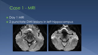
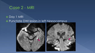
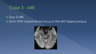
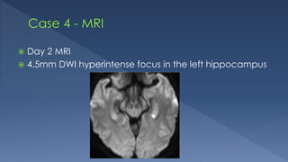
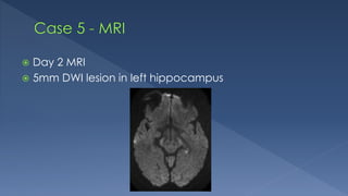
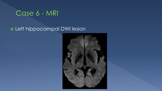
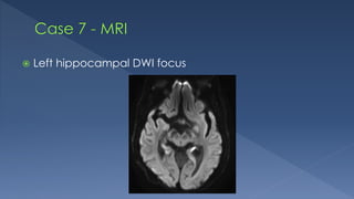
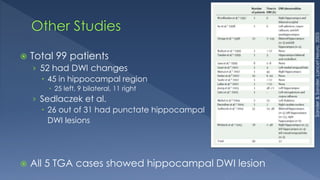
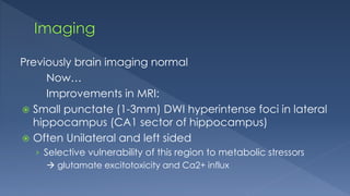
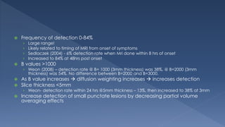
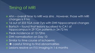
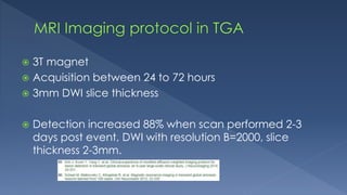
 Self-limiting antegrade amnesia
› In absence of other causes
 Witnessed
 Antegrade amnesia
› Unable to form new memories
› Perserveration
 “Broken record”
› Sometimes also retrograde
 No other cognitive impairment or altered consciousness
› Otherwise, alert and well
 Duration of episode resolves within 24hrs
› 1-10 hrs, average 6hrs
 No other neurological deficit/epileptic features/head trauma
› Diagnoses of exclusion
 Precipitating event
 No concensus
 Theories include:
› Vascular dysfunction
 Arterial or Venous
› Paroxysmal neuronal discharge/Epileptic phenomena
 Self propagating wave of neuronal depolarisation
 Nothing
› Self resolving
 Exclude other causes
› Diagnosis
 treatment
› Prognosis
DDx Clinical Findings MRI findings
Transient epileptic
amnesia
<1hr, multiple
attacks at time of
presentation
Increased T2/FLAIR in
hippocampus, thalamus and
cortex
TIA/CVA Amnesia in absence
of other focal
neurodeficits rare
DWI in vascular territories
Wernickes
encephalopathy
More global
amnesia and
inattention
Symmetrical increased T2/FLAIR
in mammillary bodies, medial
thalami, tectal plate and
periaqueductal area
tectal region (white arrows), periaqueductal area
(black arrowheads), and mamillary bodies (white
arrowheads
TGA Antegrade amnesia
<24hrs
DWI punctate (1-3mm) foci in
hippocampus, uni/bilateral
DDx
Transient epileptic
amnesia
TIA/CVA
Wernickes
encephalopathy
TGA
 Fn
› Involved in learning & memory
 Part of mesial temporal lobe
› Below temporal horn of lateral ventricles
› Seahorse
› Made up of dentate gyrus, C1-4.
 Blood supply:
› PCA
 hippocampal arteries
› AChA
 Branch of ICA
 5 TGA cases presented to the Calvary Hospital
› Between March 2013 to February 2015
 All had MRI findings typical of TGA
 61 yo male
 No significant PMHx
 Acute confusion and amnesia
› Repetitive questioning
 Alert
 Ix:
› CTB: NAD
› LP: NAD
 Day 1 MRI
 2 punctate DWI lesions in left hippocampus
 66yo male
 PMHx: T2 DM, hypertension and hypercholesterolaemia
 Acute onset of amnesia and confusion
 Alert
 Repetitive questioning
 CTB: NAD
 Day 1 MRI
 Punctate DWI lesion in left hippocampus
 62yo female
 PMHx: Meniere’s disease, migraine and hypertension
 Sudden onset of anterograde and retrograde amnesia
 Nausea and vomiting, worse than usual Meniere’s
 Alert
 CTB: NAD
 Day 2 MRI
 5mm DWI hyperintense focus in the left hippocampus
 63yo female
 Sudden onset confusion and amnesia at work
 PMHx: NAD
 Alert
› No memory of days events
 CTB: NAD
 Day 2 MRI
 4.5mm DWI hyperintense focus in the left hippocampus
 64yo female
 PMHx: OA
 Amnesic events at the gym and whilst doing errands
 Day 2 MRI
 5mm DWI lesion in left hippocampus
 Left hippocampal DWI lesion
 81yo female
 PMHx: AF, AV replacement
 Acute confusion and dysphasia
› Resolved next day
 Acute left hippocampal infarction
 Left hippocampal DWI focus
 78yo male
 PMHx: EtOH, COPD
 Recurrent episodes of decreased levels of
consciousness
› Staring and not responding
› Over last few months
› Lasts 10mins
 Followed by 2-3 hrs of fatigue
 Complex partial seizures
 Cases demonstrating DWI focus in hippocampus
› BUT not TGA clinically
Hippocampal DWI Lesions ≠ TGA
 Total 99 patients
› 52 had DWI changes
 45 in hippocampal region
 25 left, 9 bilateral, 11 right
› Sedlaczek et al.
 26 out of 31 had punctate hippocampal
DWI lesions
 All 5 TGA cases showed hippocampal DWI lesion
Sander&Sander,LancetNeuro.2005
 Small case series
 Reflective of literature
 Diagnosis to consider
 Review area
 Clinical diagnosis
“Clinical correlation is recommended”
 Dr Yash Gawarikar
 Dr Alexander Lam
 Dr Brett Jones
 Dr Yun Tae Hwang
 Consistent with other studies
 MRI findings supports clinical diagnosis
› Treatment and prognosis
 100% MRI detection rate
› Why?
 Optimised protocol
 t = 24-72 hrs
 b = 2000
 3mm thick slices
Previously brain imaging normal
Now…
Improvements in MRI:
 Small punctate (1-3mm) DWI hyperintense foci in lateral
hippocampus (CA1 sector of hippocampus)
 Often Unilateral and left sided
› Selective vulnerability of this region to metabolic stressors
 glutamate excitotoxicity and Ca2+ influx
 MR-spectroscopy of hippocampal DWI lesion
› Lactate peak  further evidence for CA1 neuronal dysfuction
 No abnormality in vessels on MRA
 Dy/dx with Wernicke encephalopathy
 DWI in medial thalami, mammillary bodies,
periaqueductal region, tectal plate
 Frequency of detection 0-84%
› Large range!
› Likely related to timing of MRI from onset of symptoms
› Sedlaczek (2004) - 6% detection rate when Mri done within 8 hrs of onset
› Increased to 84% at 48hrs post onset
 B values >1000
› Weon (2008) – detection rate @ B= 1000 (3mm thickness) was 38%, @ B=2000 (3mm
thickness) was 54%. No difference between B=2000 and B=3000.
 As B value increases  diffusion weighting increases  increases detection
 Slice thickness <5mm
› Weon- detection rate within 24 hrs @5mm thickness – 13%, then increased to 38% at 3mm
 Increase detection of small punctate lesions by decreasing partial volume
averaging effects
 Ahn – overall time to MRI was 6hrs . However, those with MRI
changes is 9 hrs
 16 out of 203 TGA over 7yrs with DWI hippocampal changes
 Bartsch – found that lesions localised to CA1 of
hippocampus in 29 TGA patients in 24-72 hrs
 Peak incidence at 12-72hrs
 DWI normalisation on Day 10
 Similar to time course of ischaemic
  careful timing to find abnormalities
 Lesions resolve on F/U imaging in 1-6 months
 3T magnet
 Acquisition between 24 to 72 hours
 3mm DWI slice thickness
 Detection increased 88% when scan performed 2-3
days post event, DWI with resolution B=2000, slice
thickness 2-3mm.

Transient Global Amnesia

  • 2.
     Self-limiting antegradeamnesia › In absence of other causes
  • 3.
     Witnessed  Antegradeamnesia › Unable to form new memories › Perserveration  “Broken record” › Sometimes also retrograde  No other cognitive impairment or altered consciousness › Otherwise, alert and well  Duration of episode resolves within 24hrs › 1-10 hrs, average 6hrs  No other neurological deficit/epileptic features/head trauma › Diagnoses of exclusion  Precipitating event
  • 4.
     No concensus Theories include: › Vascular dysfunction  Arterial or Venous › Paroxysmal neuronal discharge/Epileptic phenomena  Self propagating wave of neuronal depolarisation
  • 5.
  • 6.
     Exclude othercauses › Diagnosis  treatment › Prognosis
  • 7.
    DDx Clinical FindingsMRI findings Transient epileptic amnesia <1hr, multiple attacks at time of presentation Increased T2/FLAIR in hippocampus, thalamus and cortex TIA/CVA Amnesia in absence of other focal neurodeficits rare DWI in vascular territories Wernickes encephalopathy More global amnesia and inattention Symmetrical increased T2/FLAIR in mammillary bodies, medial thalami, tectal plate and periaqueductal area tectal region (white arrows), periaqueductal area (black arrowheads), and mamillary bodies (white arrowheads TGA Antegrade amnesia <24hrs DWI punctate (1-3mm) foci in hippocampus, uni/bilateral DDx Transient epileptic amnesia TIA/CVA Wernickes encephalopathy TGA
  • 8.
     Fn › Involvedin learning & memory  Part of mesial temporal lobe › Below temporal horn of lateral ventricles › Seahorse › Made up of dentate gyrus, C1-4.
  • 9.
     Blood supply: ›PCA  hippocampal arteries › AChA  Branch of ICA
  • 10.
     5 TGAcases presented to the Calvary Hospital › Between March 2013 to February 2015  All had MRI findings typical of TGA
  • 11.
     61 yomale  No significant PMHx  Acute confusion and amnesia › Repetitive questioning  Alert  Ix: › CTB: NAD › LP: NAD
  • 12.
     Day 1MRI  2 punctate DWI lesions in left hippocampus
  • 13.
     66yo male PMHx: T2 DM, hypertension and hypercholesterolaemia  Acute onset of amnesia and confusion  Alert  Repetitive questioning  CTB: NAD
  • 14.
     Day 1MRI  Punctate DWI lesion in left hippocampus
  • 15.
     62yo female PMHx: Meniere’s disease, migraine and hypertension  Sudden onset of anterograde and retrograde amnesia  Nausea and vomiting, worse than usual Meniere’s  Alert  CTB: NAD
  • 16.
     Day 2MRI  5mm DWI hyperintense focus in the left hippocampus
  • 17.
     63yo female Sudden onset confusion and amnesia at work  PMHx: NAD  Alert › No memory of days events  CTB: NAD
  • 18.
     Day 2MRI  4.5mm DWI hyperintense focus in the left hippocampus
  • 19.
     64yo female PMHx: OA  Amnesic events at the gym and whilst doing errands
  • 20.
     Day 2MRI  5mm DWI lesion in left hippocampus
  • 21.
  • 22.
     81yo female PMHx: AF, AV replacement  Acute confusion and dysphasia › Resolved next day  Acute left hippocampal infarction
  • 23.
  • 24.
     78yo male PMHx: EtOH, COPD  Recurrent episodes of decreased levels of consciousness › Staring and not responding › Over last few months › Lasts 10mins  Followed by 2-3 hrs of fatigue  Complex partial seizures
  • 25.
     Cases demonstratingDWI focus in hippocampus › BUT not TGA clinically
  • 26.
  • 27.
     Total 99patients › 52 had DWI changes  45 in hippocampal region  25 left, 9 bilateral, 11 right › Sedlaczek et al.  26 out of 31 had punctate hippocampal DWI lesions  All 5 TGA cases showed hippocampal DWI lesion Sander&Sander,LancetNeuro.2005
  • 28.
     Small caseseries  Reflective of literature  Diagnosis to consider  Review area  Clinical diagnosis “Clinical correlation is recommended”
  • 29.
     Dr YashGawarikar  Dr Alexander Lam  Dr Brett Jones  Dr Yun Tae Hwang
  • 34.
     Consistent withother studies  MRI findings supports clinical diagnosis › Treatment and prognosis  100% MRI detection rate › Why?  Optimised protocol  t = 24-72 hrs  b = 2000  3mm thick slices
  • 37.
    Previously brain imagingnormal Now… Improvements in MRI:  Small punctate (1-3mm) DWI hyperintense foci in lateral hippocampus (CA1 sector of hippocampus)  Often Unilateral and left sided › Selective vulnerability of this region to metabolic stressors  glutamate excitotoxicity and Ca2+ influx
  • 38.
     MR-spectroscopy ofhippocampal DWI lesion › Lactate peak  further evidence for CA1 neuronal dysfuction  No abnormality in vessels on MRA  Dy/dx with Wernicke encephalopathy  DWI in medial thalami, mammillary bodies, periaqueductal region, tectal plate
  • 39.
     Frequency ofdetection 0-84% › Large range! › Likely related to timing of MRI from onset of symptoms › Sedlaczek (2004) - 6% detection rate when Mri done within 8 hrs of onset › Increased to 84% at 48hrs post onset  B values >1000 › Weon (2008) – detection rate @ B= 1000 (3mm thickness) was 38%, @ B=2000 (3mm thickness) was 54%. No difference between B=2000 and B=3000.  As B value increases  diffusion weighting increases  increases detection  Slice thickness <5mm › Weon- detection rate within 24 hrs @5mm thickness – 13%, then increased to 38% at 3mm  Increase detection of small punctate lesions by decreasing partial volume averaging effects
  • 40.
     Ahn –overall time to MRI was 6hrs . However, those with MRI changes is 9 hrs  16 out of 203 TGA over 7yrs with DWI hippocampal changes  Bartsch – found that lesions localised to CA1 of hippocampus in 29 TGA patients in 24-72 hrs  Peak incidence at 12-72hrs  DWI normalisation on Day 10  Similar to time course of ischaemic   careful timing to find abnormalities  Lesions resolve on F/U imaging in 1-6 months
  • 41.
     3T magnet Acquisition between 24 to 72 hours  3mm DWI slice thickness  Detection increased 88% when scan performed 2-3 days post event, DWI with resolution B=2000, slice thickness 2-3mm.

Editor's Notes

  • #3 Constellation of symptoms
  • #4 Perserveration = Repetitive questioning In bold is are the Caplan and Hodges diagnostic criteria ??Diagnosis of exclusion
  • #5 Ischaemia or venous congestion Abnormal firing of neurones
  • #7 So if we do nothing for TGA, why is imaging important? Because TGA more of a diagnoses of exclusion, imaging is used to diagnose/exclude other causes of amnesia
  • #8 The other causes of amnesia include Clinical and Mri findings are different in terms of distribution of signal.
  • #10 Hippocampus vulnerable to ischemia. As hippocampal artery supplies an internal anastomosis forming a link between an upper and a lower artery  watershed area = “hypoxia-susceptible sector of Sommer” Hippocampus vulnerable to excitotoxic mechanisms. Stressful events  overexcite hippocampus  glutamate release  triggers spreading depression and the functional ablation of the hippocampus
  • #11 5 clinically diagnosed cases of TGA presented to Calvary hospital between 2014-2015 All had similar findings on MRI, which I will present to you.
  • #26 During this period there were DWI hippocampal lesions, that were clinically NOT TGA
  • #27 These last 2 cases were to demonstrate that not all hippocampal DWI lesions are TGA Clinical ___ important
  • #28 Our small case series of 5 patients, how does this compare with other studies? Sander and Sander collated case reports and case series , with a combined total of 99 patients. 52 had DWI changes , of which 45 were located hippocampus. Reason why some studies had no MRI changes: timing. TGA usual present by 24 -72hr DWI abnormalities highly sensitive for ischaemia but not specific
  • #29 Now that we know what is out there in the literature, we need to be mindful of the implications and limitations of our case series We do have a small sample size, however our findings are reflective of the literature as discussed prior. In radiology, TGA is a diagnosis to keep in mind. And we know that the high yield review area is in the hippocampus. However, ultimately TGA is a clinical diagnosis – and that clinical correlation is recommended.
  • #35 Our small casse series is Increases detection rates
  • #42 Can this protocol pick up all the DDx?? Since TGA is diagnosis of exclusion, this protocol is able to exclude other DDx… Increase sensitivity /detection rate