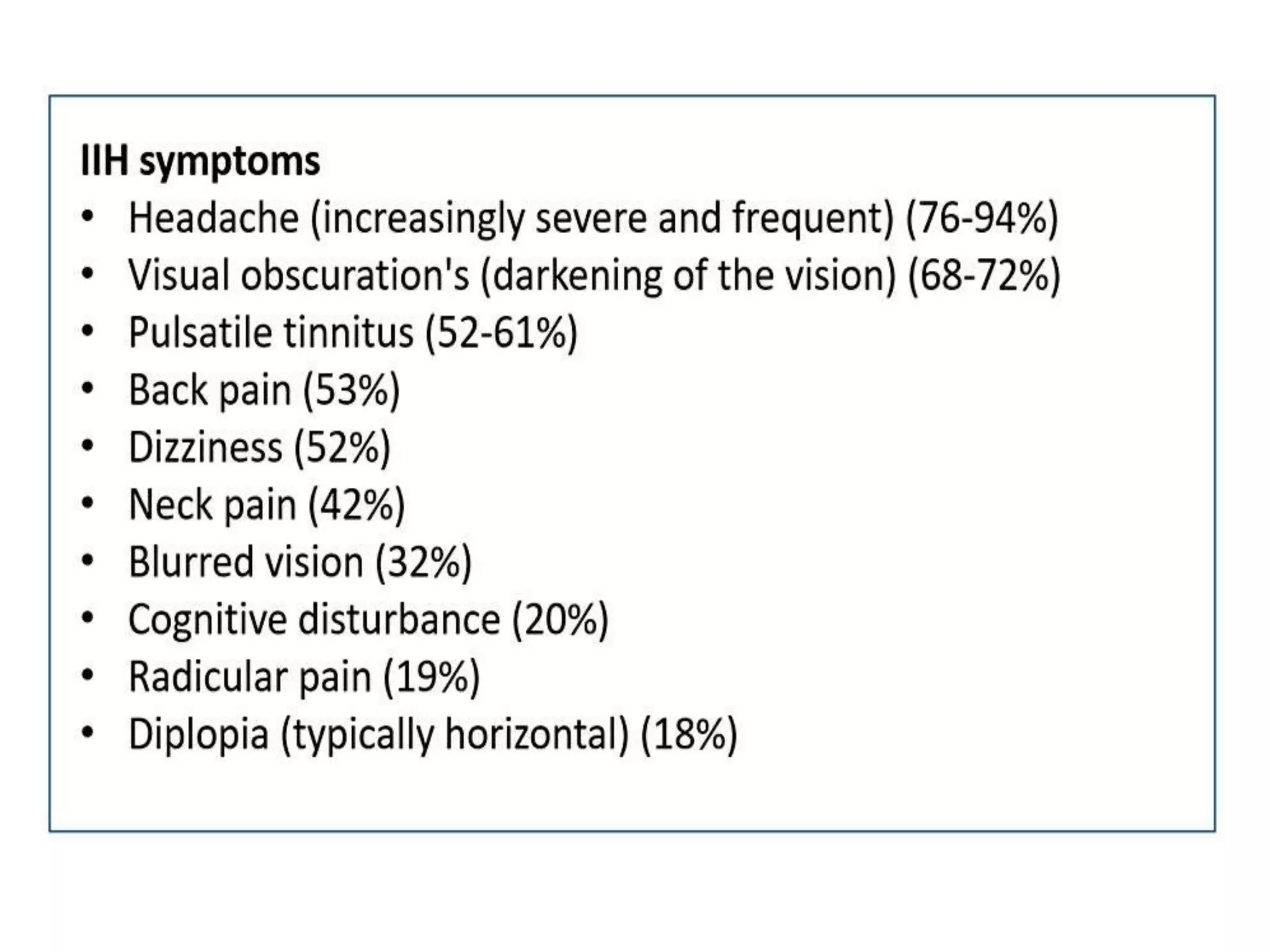
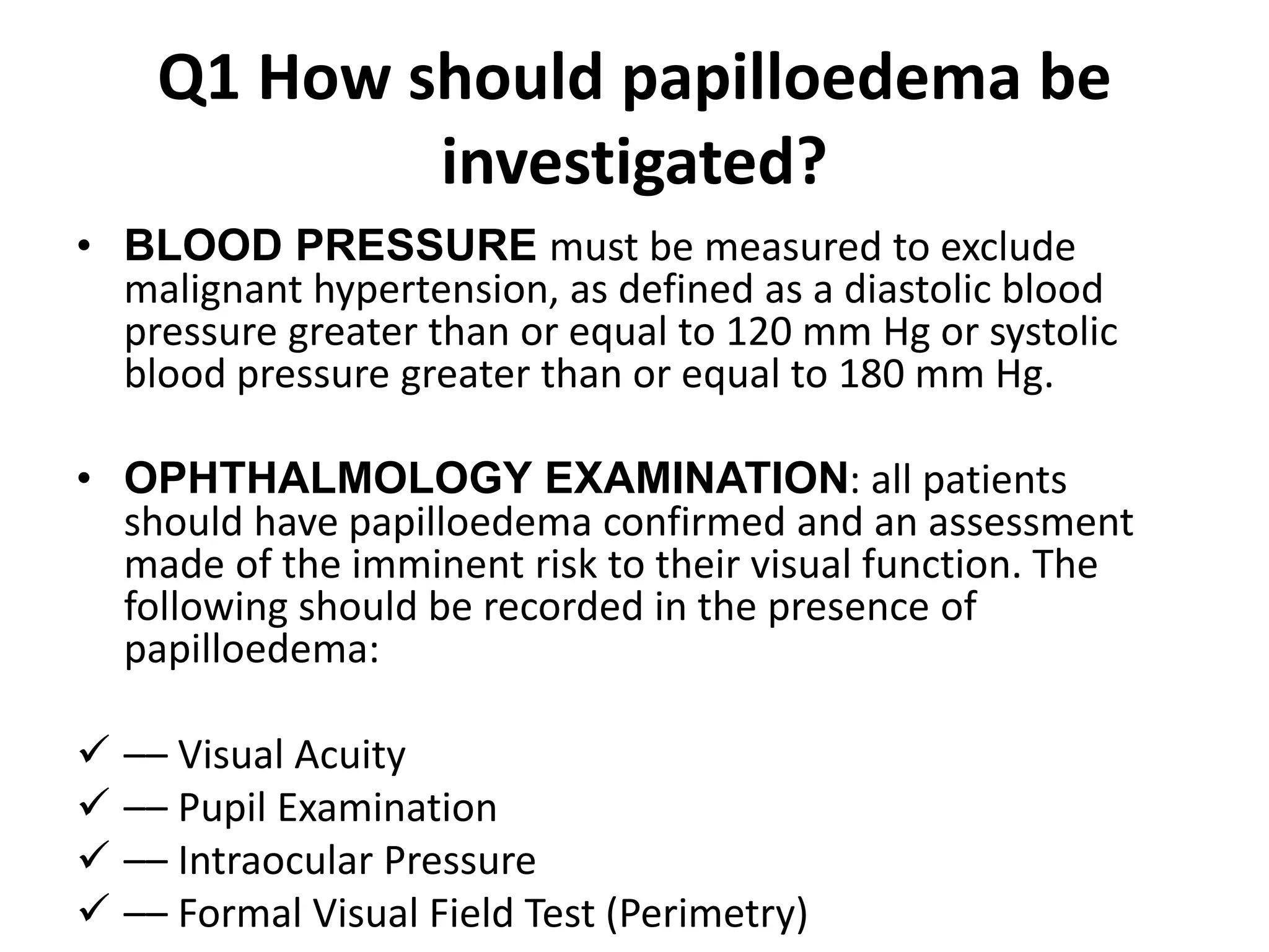
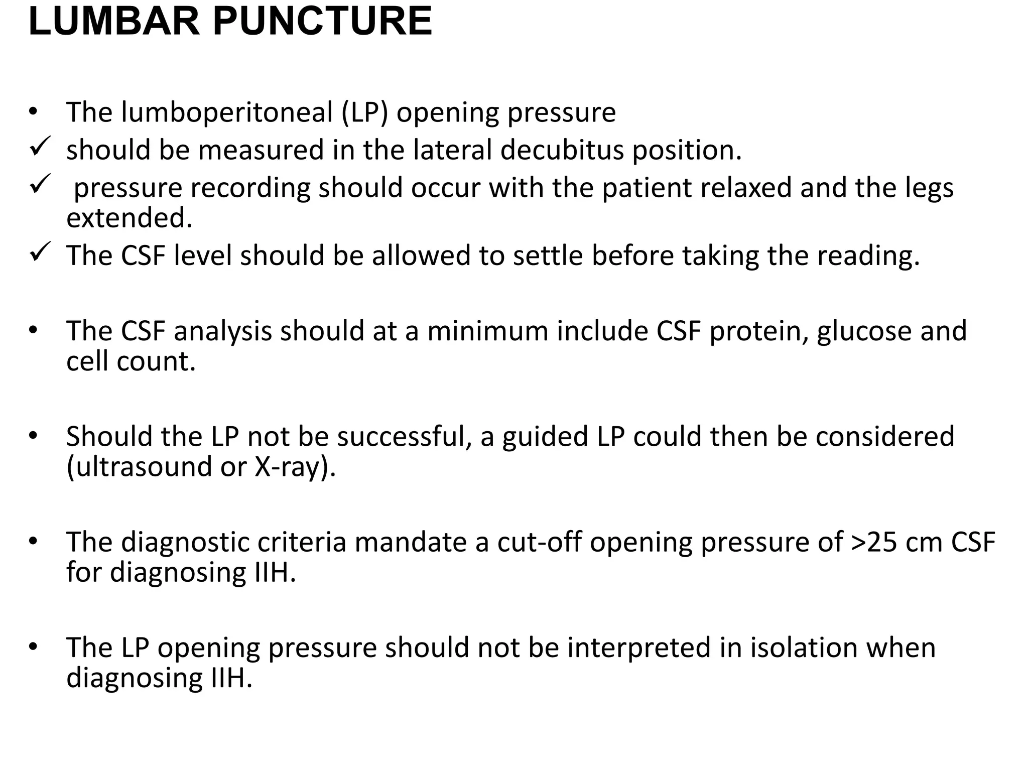
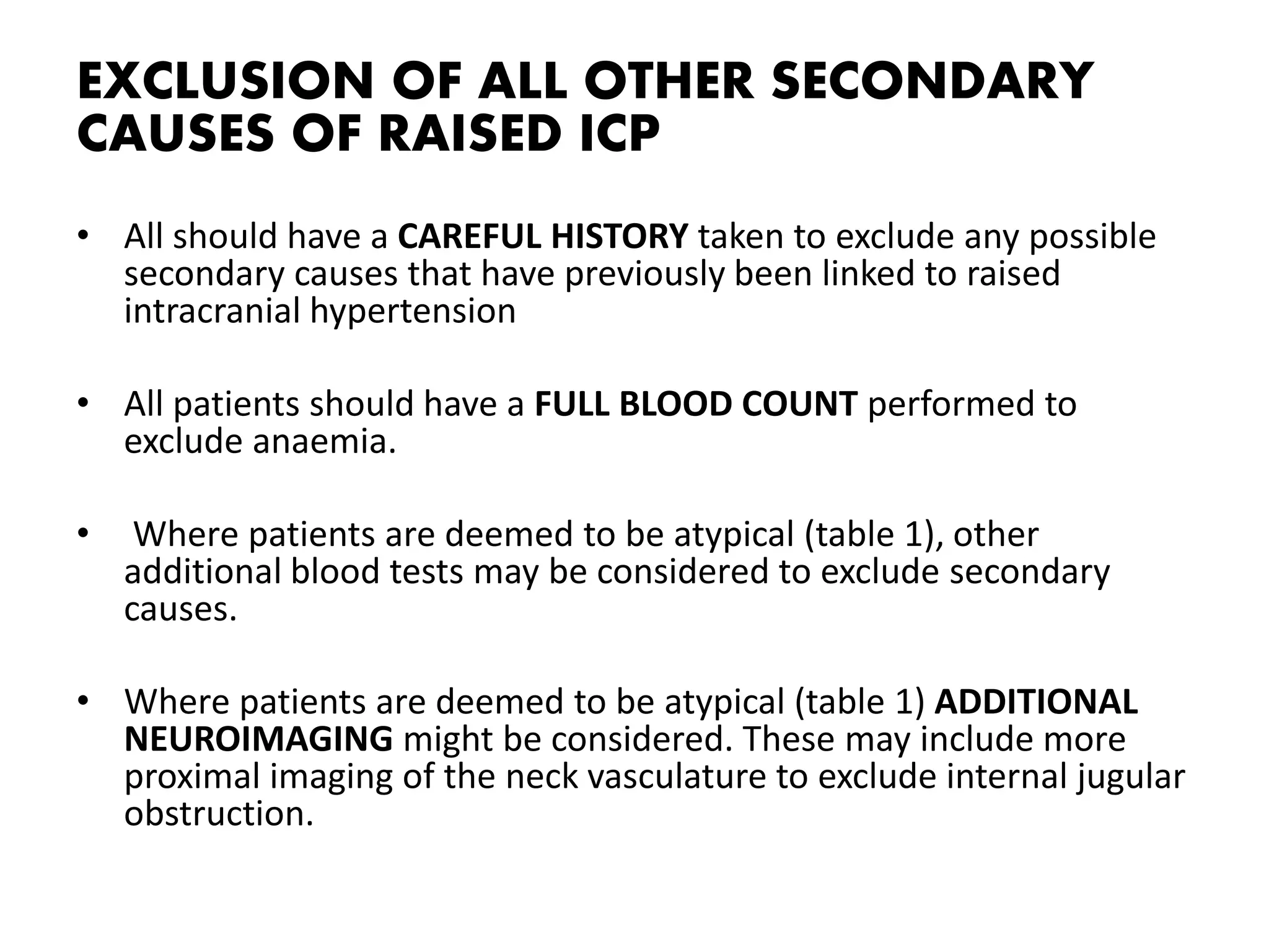
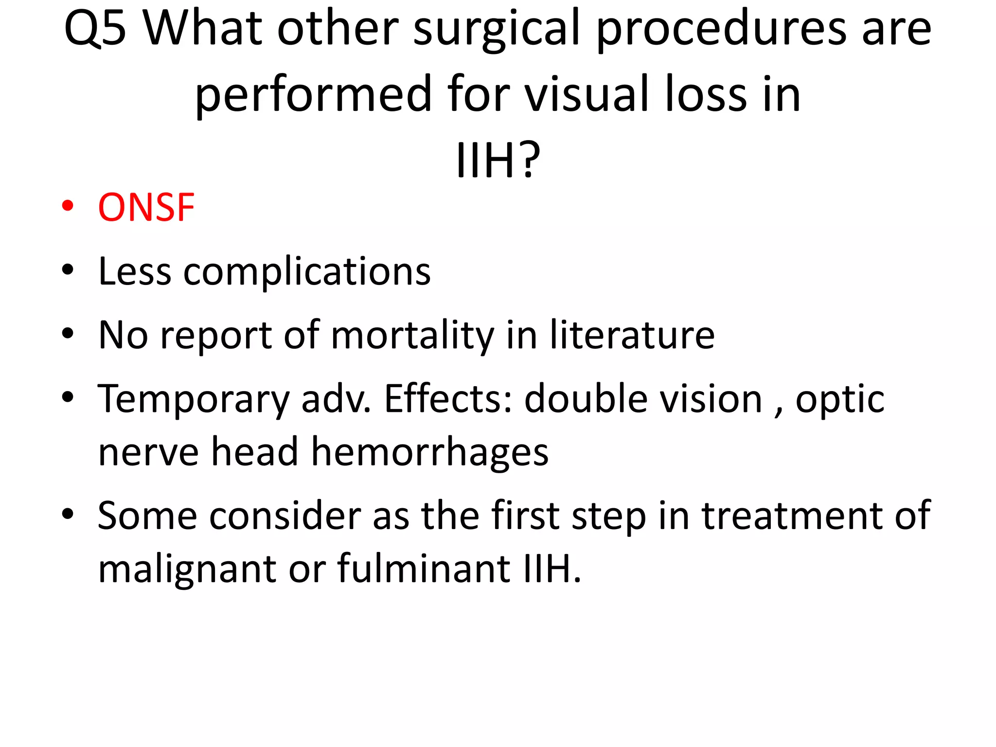
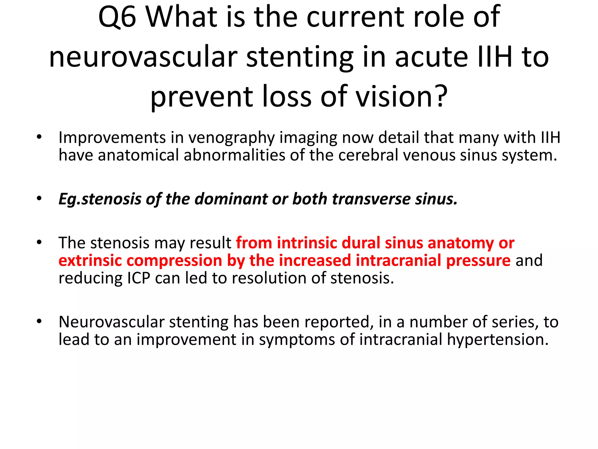
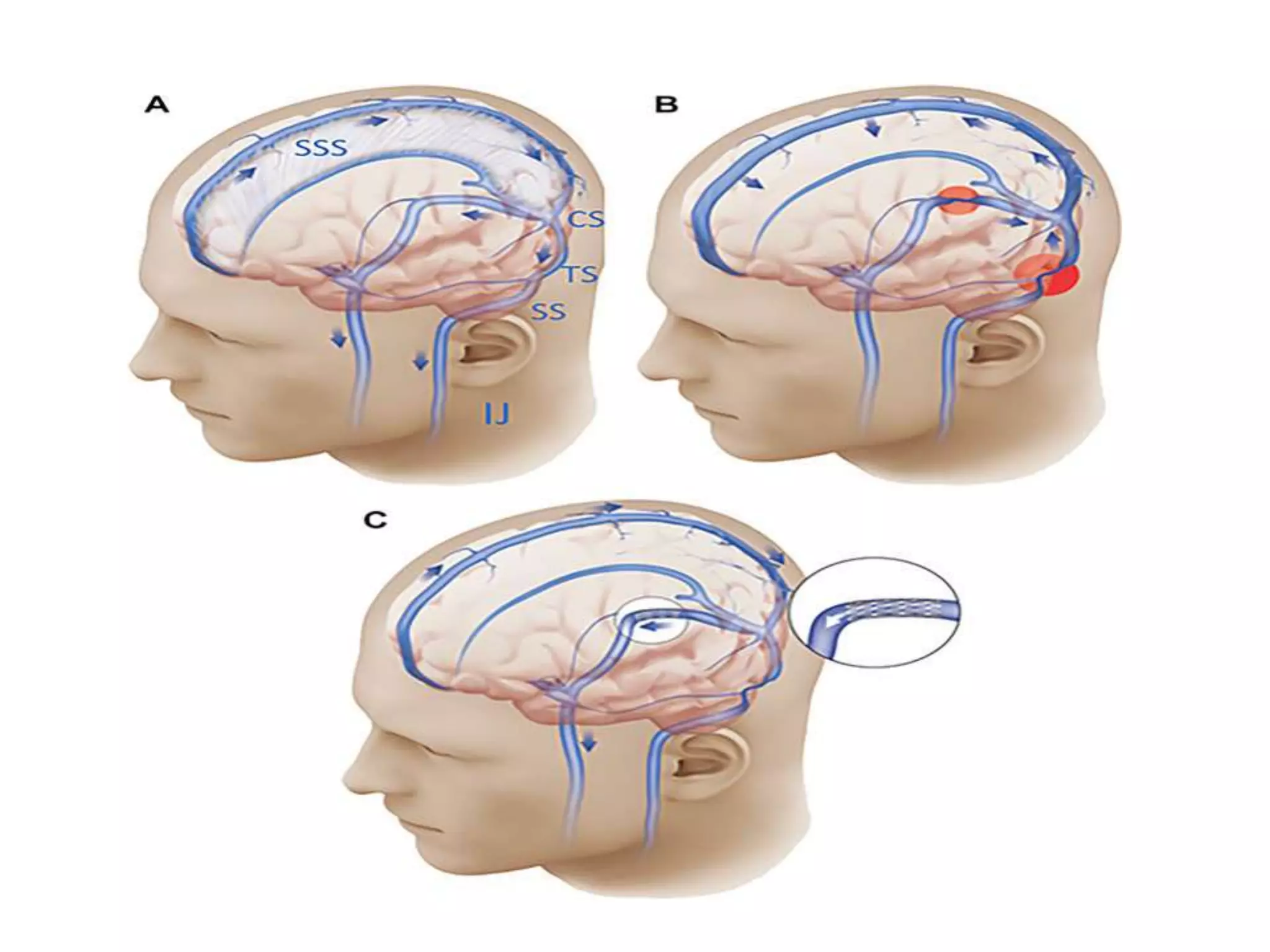
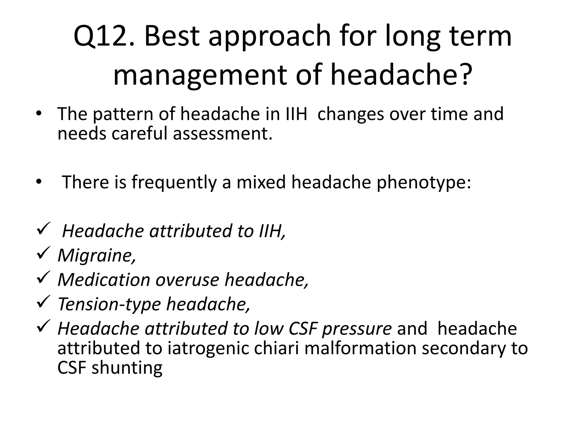
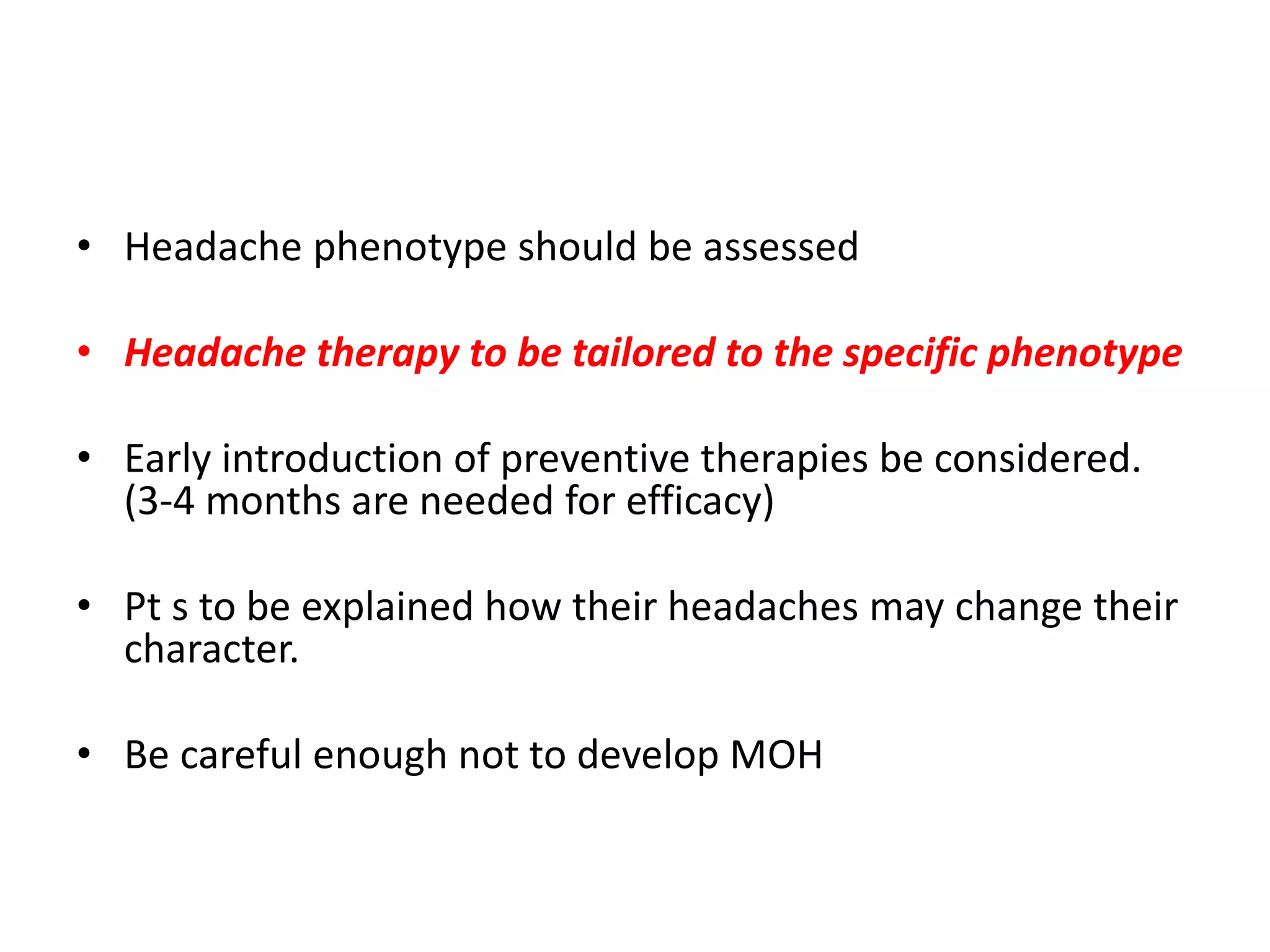
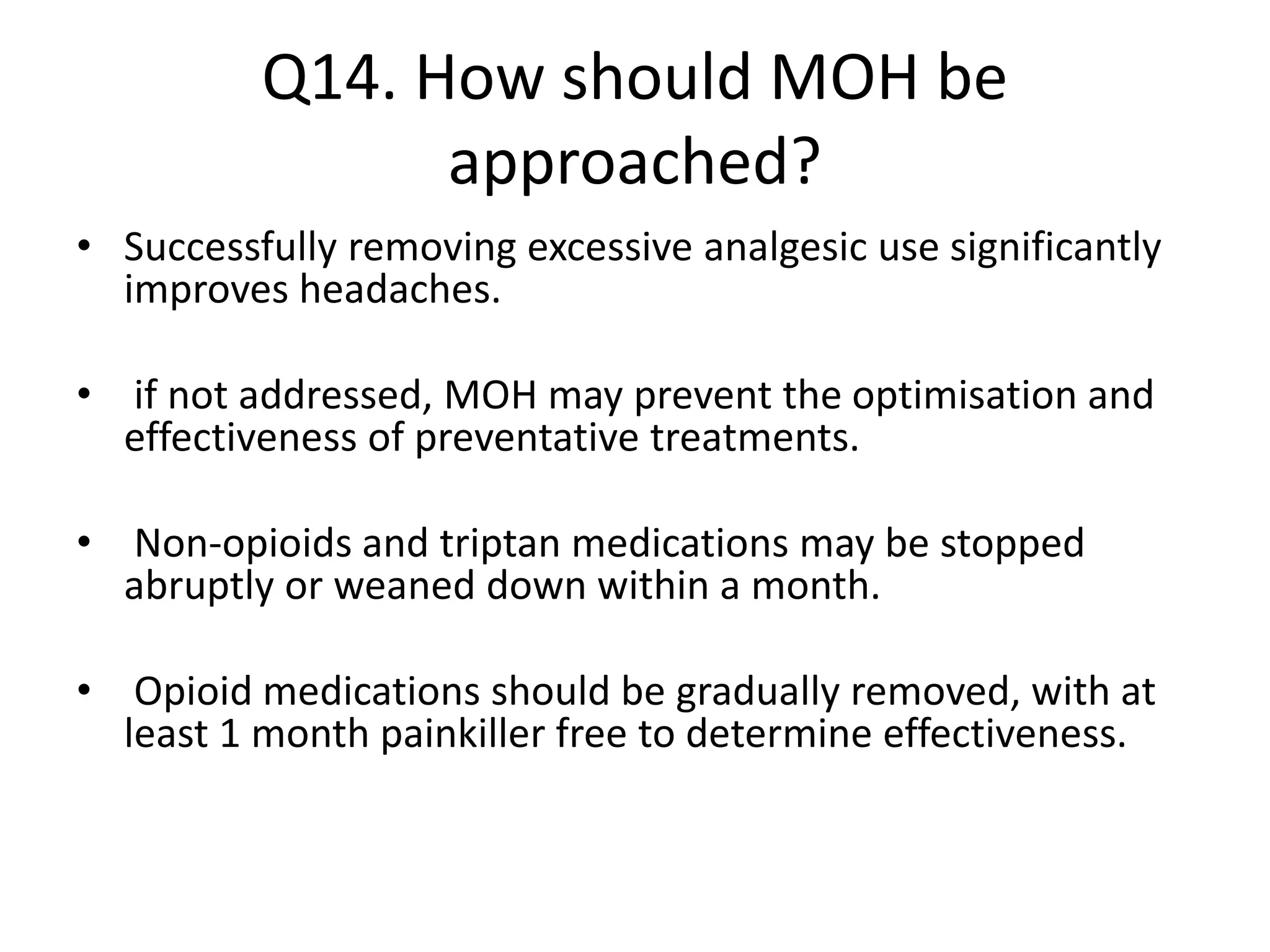
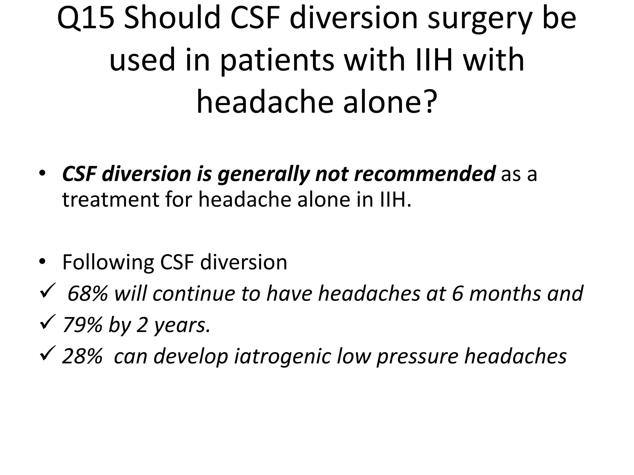
6. Q1 How should papilloedema be
investigated?
• BLOOD PRESSURE must be measured to exclude
malignant hypertension, as defined as a diastolic blood
pressure greater than or equal to 120 mm Hg or systolic
blood pressure greater than or equal to 180 mm Hg.
• OPHTHALMOLOGY EXAMINATION: all patients
should have papilloedema confirmed and an assessment
made of the imminent risk to their visual function. The
following should be recorded in the presence of
papilloedema:
–– Visual Acuity
–– Pupil Examination
–– Intraocular Pressure
–– Formal Visual Field Test (Perimetry)
7. NEUROLOGICAL EXAMINATION
• Record cranial nerve examination. Where IIH is suspected, typically
there should be no cranial nerve involvement other than sixth
cranial nerve palsy/palsies.
• Should other cranial nerves and/or other pathological findings be
involved, an alternative diagnosis should be considered.
NEUROIMAGING
• Urgent MRI brain within 24 hours; if unavailable within 24 hours,
then urgent CT brain with subsequent MRI brain if no lesion
identified.
• There should be no evidence of hydrocephalus, mass, structural,
vascular lesion and no abnormal meningeal enhancement.
• CT or MR venography is mandatory to exclude cerebral sinus
thrombosis within 24 hours.
• Characteristics of raised intracranial pressure may be seen on
neuroimaging (box 1)
8. LUMBAR PUNCTURE
• The lumboperitoneal (LP) opening pressure
should be measured in the lateral decubitus position.
pressure recording should occur with the patient relaxed and the legs
extended.
The CSF level should be allowed to settle before taking the reading.
• The CSF analysis should at a minimum include CSF protein, glucose and
cell count.
• Should the LP not be successful, a guided LP could then be considered
(ultrasound or X-ray).
• The diagnostic criteria mandate a cut-off opening pressure of >25 cm CSF
for diagnosing IIH.
• The LP opening pressure should not be interpreted in isolation when
diagnosing IIH.
9.
10.
11.
12. EXCLUSION OF ALL OTHER SECONDARY
CAUSES OF RAISED ICP
• All should have a CAREFUL HISTORY taken to exclude any possible
secondary causes that have previously been linked to raised
intracranial hypertension
• All patients should have a FULL BLOOD COUNT performed to
exclude anaemia.
• Where patients are deemed to be atypical (table 1), other
additional blood tests may be considered to exclude secondary
causes.
• Where patients are deemed to be atypical (table 1) ADDITIONAL
NEUROIMAGING might be considered. These may include more
proximal imaging of the neck vasculature to exclude internal jugular
obstruction.
13. Management principles
• For optimal management of patients with IIH, there must be clear
communication between clinicians for seamless joint care between
the various specialties (figure 3).
• Weight loss reduces ICP and has been shown to be effective in
improving papilloedema and headaches.
• The main principles of management of IIH are:
1. To treat the underlying disease
2. To protect the vision
3. To minimise the headache morbidity.
Twenty-three questions were formulated to cover the three principle
domains of management in IIH
14. Primary principle for IIH management: modify the
underlying
disease through weight loss
Q2 What is the best way to modify the disease to induce remission?
Weight loss is the only disease-modifying therapy in typical IIH.
• Once definite IIH is diagnosed, all patients with a BMI >30 kg/m2 should be
counselled about weight management at the earliest opportunity.
•
• The amount of weight loss required to put the disease into remission is not known.
• up to 15% of weight loss was required to put IIH into remission in one cohort.
• Patients should be referred to a community weight management programme or a
hospital-based weight programme.
15. • There is an increasing role for bariatric surgery
for sustained weight loss and for use in IIH.
• For those who are not obese secondary
causes should be revisited , and the role of
weight gain/loss remains uncertain.
16. Second principle for IIH management:
protect the vision
Q3 How should IIH be treated when there is imminent risk of visual loss?
Where there is evidence of declining visual function, the acute management to
preserve vision is surgical.
• A temporary measure of a lumbar drain could be useful to protect the vision while
planning urgent surgical treatment.
• There is evidence that many of the surgical procedures, such as CSF diversion and
optic nerve sheath fenestration (ONSF), work well in the short term.
• While they are working, the underlying disease should be modified with weight
loss
In the absence of high class evidence, we do not recommend the use of corticosteroids
for fulminant IIH at this time, and indeed a prolonged treatment course of
corticosteroids would not be recommended due to weight gain.
17. Q4 What is currently the best surgical procedure for
visual loss in IIH?
• In the UK, the preferred surgical procedure is neurosurgical CSF diversion
• Ventriculoperitoneal (VP) should be the preferred CSF diversion procedure for
visual deterioration in IIH, due to lower reported revisions per patient.
• An LP shunt could also be used.
.
• Adjustable valves with antigravity or antisiphon devices should be considered for
use to reduce the risk of low pressure headaches.
• A mortality rate although low but does exist.
• Complications of shunts include abdominal pain, shunt obstruction, migration and
infection, low pressure headaches, subdural haematoma and tonsillar herniation
18. Q5 What other surgical procedures are
performed for visual loss in
IIH?
• ONSF
• Less complications
• No report of mortality in literature
• Temporary adv. Effects: double vision , optic
nerve head hemorrhages
• Some consider as the first step in treatment of
malignant or fulminant IIH.
19. Q6 What is the current role of
neurovascular stenting in acute IIH to
prevent loss of vision?
• Improvements in venography imaging now detail that many with IIH
have anatomical abnormalities of the cerebral venous sinus system.
• Eg.stenosis of the dominant or both transverse sinus.
• The stenosis may result from intrinsic dural sinus anatomy or
extrinsic compression by the increased intracranial pressure and
reducing ICP can led to resolution of stenosis.
• Neurovascular stenting has been reported, in a number of series, to
lead to an improvement in symptoms of intracranial hypertension.
20.
21. Overall improvement of symptoms was noted in 16 patients (88.9%)
postprocedure, with 1 patient (5.6%) requiring shunt placement and 2 other
patients (11.1%) requiring postprocedural LP to monitor intracranial pressure
to determine candidacy for further
surgical interventions to treat residual symptoms.
22. After treatment, >75% exhibit an improvement in headaches,
~50% improvement in tinnitus, and ~50 % improvement in
ophthalmologic testing.
23. • Demerits of neurovascular stenting: long term anti-
thrombotic treament.
• The role of neurovascular stenting in IIH to preserve
rapidly deteriorating vision is not yet established, as
there is a lack of quality data in this area.
• It may be useful for highly selected patients with IIH
with venous sinus stenosis with an elevated pressure
gradient and elevated ICP in whom traditional
therapies have not worked.
24. Q7 What is the role of serial lumbar
punctures in IIH?
• The relief from a LP is typically short lived as CSF is
secreted from the choroid plexus at a rate of
25mL/hour and consequently the volume removed in a
so-called therapeutic tap is rapidly replaced.
Serial lumbar punctures are not recommended for
management of IIH.
• Despite the relief of headache in nearly three quarters
of patients, LPs are associated with significant anxiety
in many patients and can led to acute and chronic back
pain in some patients
26. The IIHTT also reported improved quality of life
outcomes at 6 months with acetazolamide.
27. Failed to show a treatment effect. Importantly, 48%
discontinued acetazolamide due to adverse effects.
28. • IT CONCLUDED:
‘the two included RCTs showed modest benefits
for acetazolamide for some outcomes, there is
insufficient evidence to recommend or reject
the efficacy of this intervention, or any other
treatments currently available, for treating
people with IIH’.
29. Q9. How should acetazolamide be
prescribed?
The IIHTT used a maximal dose of 4 g daily, with 44% o participants
achieving 4 g/day, and the majority toleratin 1 g/day.47 Ball et all identified
that 48% discontinued a mean doses of 1.5 g due to side effects.
A popular starting dose of acetazolamide is 250–500 mg twice a day, with
the majority of clinicians titrating the daily dose up.
Patients should be warned of the adverse side effects which include increase
risk of diarrhoea, dysgeusia, fatigue, nausea, paraesthesia tinnitus,
vomiting, depression and rarely renal stones.
There is no consensus over the use of normal release an modified release
acetazolamide.
30. Q10. What are the drugs helpful in IIH?
• Topiramate has carbonic anhydrase activity and can suppress appetite.
• There is evidence of efficacy of topiramate in treating migraine.
• may have a role with weekly dose escalation from 25 mg to 50 mg bd.
• caution: women must be informed that it can reduce the efficacy of the
contraceptive pill/oral contraceptives and other hormonal contraceptives.
• women must be counselled regarding side effects (including depression and
cognitive slowing) and potential teratogenetic risks.
Uncertainties
• The role of other diuretics such as furosemide, amiloride and coamilofruse
are not certain but are used by some as alternative therapies.
31. Q11. What is the best way to manage
headache in newly diagnosed IIH?
• Pt. to be informed about the risk of painkiller overuse.
• Simple analgesics in the first few weeks. (NSAIDs or paracetamol)
Indomethacin also reduces ICP
• Opioids not to be prescribed
• Greater occipital nerve blocks may be considered (evidence lacking)
• Acetazolamide alone is not helpful
• Repeated or solo LPs for this purpose are not recommended
32. Q12. Best approach for long term
management of headache?
• The pattern of headache in IIH changes over time and
needs careful assessment.
• There is frequently a mixed headache phenotype:
Headache attributed to IIH,
Migraine,
Medication overuse headache,
Tension-type headache,
Headache attributed to low CSF pressure and headache
attributed to iatrogenic chiari malformation secondary to
CSF shunting
33. • Headache phenotype should be assessed
• Headache therapy to be tailored to the specific phenotype
• Early introduction of preventive therapies be considered.
(3-4 months are needed for efficacy)
• Pt s to be explained how their headaches may change their
character.
• Be careful enough not to develop MOH
34. Q13. what therapeutic strategies are
helpful for headache in IIH?
• Migrainous phenotype is the commonest (68%)
• Triptan ± NSAID/paracetamol + antiemetic
• Limit: 2 days per week / 10 days per month
• Migraine preventatives be tried( useful in IIH in ocular remission)
• Caution: drugs causing weight gain( b blocker, TCA, sod.
Valproate,flunarizine)
• Caution for depression: flunarizine, topiramate, b blocker
• Topiramate: migraine, wt. loss
but depression , cognition , decreased OCP efficacy and teratogenicity
are the downsides
35. • venlafaxine for depression. (weight neutral)
• Preventatives for a minimum duration of 3m for a trial.
Lifestyle modification:
limit caffeine intake
Ensure regular meals and
adequate hydration,
Exercise programme and
sleep hygiene.
Behavioural and stress management techniques can be implemented such as yoga,
cognitive–behavioural therapy and mindfulness.
36. Q14. How should MOH be
approached?
• Successfully removing excessive analgesic use significantly
improves headaches.
• if not addressed, MOH may prevent the optimisation and
effectiveness of preventative treatments.
• Non-opioids and triptan medications may be stopped
abruptly or weaned down within a month.
• Opioid medications should be gradually removed, with at
least 1 month painkiller free to determine effectiveness.
37. Q15 Should CSF diversion surgery be
used in patients with IIH with
headache alone?
• CSF diversion is generally not recommended as a
treatment for headache alone in IIH.
• Following CSF diversion
68% will continue to have headaches at 6 months and
79% by 2 years.
28% can develop iatrogenic low pressure headaches
38. Q16 Should neurovascular stenting be
used in patients with IIH with
headache alone?
• Currently its not recommended as a treatment
for headache in IIH
39. MANAGEMENT OF HEADACHE IN
SHUNTED PATIENTS IN IIH
• Q17 How should an acute exacerbation of
headache be investigated in those who are
already shunted?
40.
41. Q18 How should an acute exacerbation of headache be
treated in those who are already shunted?
• In shunted patients with deteriorating headaches, low
pressure headache and shunt over drainage should be
considered.
• In established overdrainage or low CSF pressure,
consideration should be given to the valve settings or tying
the shunt off.
• In the absence of shunt over drainage headache
management should follow the section above
• Consider medication overuse headache as a cause of acute
exacerbation in shunted patients.
42. Q19 Other chronic problems to be
dealt with?
• Pts with IIH have higher incidence of anxiety &
depression , and lower quality of life
• Sleep apnea
• PCOS may coexist
• Cognitive dysfunction may coexist
43. Q20 Pregnant pts. In IIH?
• Acetazolamide : limited evidence to make any
safety recommendations.
• Perinatal exposure in rodents have shown
teratogenicity.
• Topiramate : teratogenic
• If pregnancy takes place on topiramate , it is to be
reduced and stopped as soon as possible.
44. Q 21. acute exacerbation of IIH with
imminent risk to vision?
• Serial lumbar punctures as a temporary
measure may be considered till long term
measures as diversion or ONSF is available.
45. Q22. management of IIHWOP?
• without papilledema the risk of vision loss has
not been identified
• Headache is the principal morbidity
• Counsel about weight management
• Surgical management not to be considered
routinely
46. Q23. how should we monitor & follow
up these patients?
• All pts with papilledema should have the
following
1. visual acuity
2. pupil examination
3. formal visual field assessment
4. dilated fundal examination to grade the
papilloedema.
5. BMI calculation.
47. • In pts without papilledema :
Time to time assessment of headache character,
severity, frequency & frequency of analgesic
use.
Periodic monitoring of serum electrolytes is recommended
Decreases weight
MOH(use of simple analgesics on more
than 15 days per month or opioids, combined preparations
or triptan medication on greater than 10 days per month for
more than 3 months)
And that the drug which had once worked may not work later.
Raised ICP is settling and papillledema has resolved