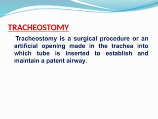
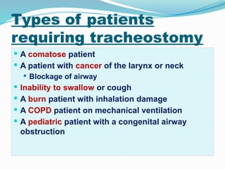
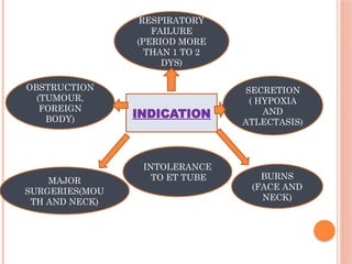
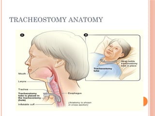
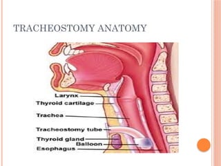
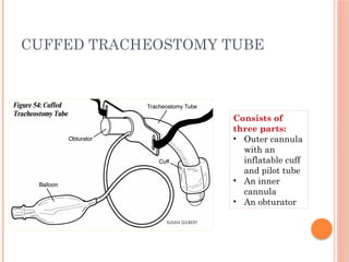
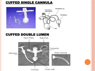
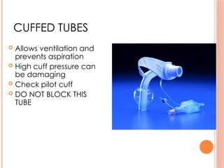
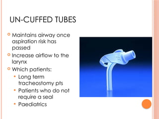
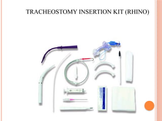
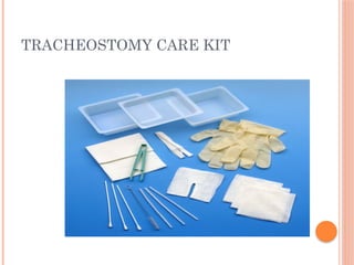
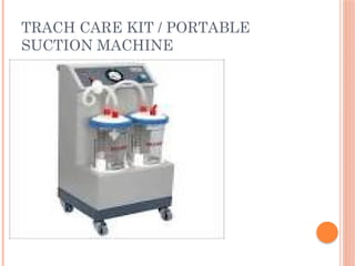
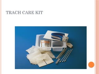
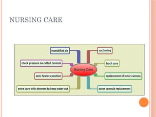
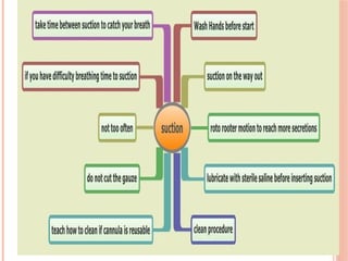
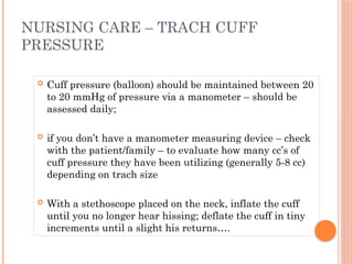
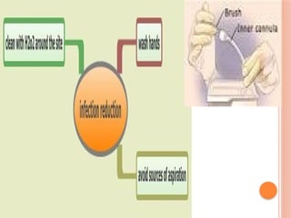
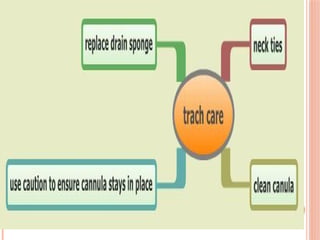
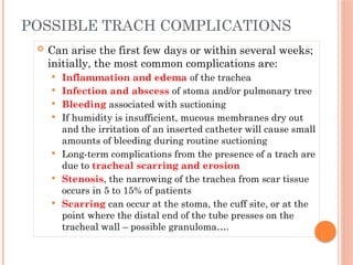
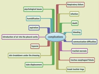
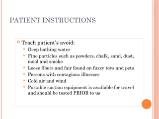
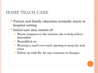
TRACHEOSTOMY
Tracheostomy is a surgical procedure or an
artificial opening made in the trachea into
which tube is inserted to establish and
maintain a patent airway.
Types of patients
requiring tracheostomy
 A comatose patient
 A patient with cancer of the larynx or neck
 Blockage of airway
 Inability to swallow or cough
 A burn patient with inhalation damage
 A COPD patient on mechanical ventilation
 A pediatric patient with a congenital airway
obstruction
INDICATION
OBSTRUCTION
(TUMOUR,
FOREIGN
BODY)
MAJOR
SURGERIES(MOU
TH AND NECK)
BURNS
(FACE AND
NECK)
RESPIRATORY
FAILURE
(PERIOD MORE
THAN 1 TO 2
DYS)
SECRETION
( HYPOXIA
AND
ATLECTASIS)
INTOLERANCE
TO ET TUBE
TRACHEOSTOMY ANATOMY
LANDMARKS
TRACHEOSTOMY TUBE TYPES
CUFFED TRACHEOSTOMY TUBE
Consists of
three parts:
• Outer cannula
with an
inflatable cuff
and pilot tube
• An inner
cannula
• An obturator
UNCUFFED SINGLE CANNULA
UNCUFFED DOUBLE CANNULA
CUFFED SINGLE CANNULA
CUFFED DOUBLE LUMEN
CUFFED TUBES
 Allows ventilation and
prevents aspiration
 High cuff pressure can
be damaging
 Check pilot cuff
 DO NOT BLOCK THIS
TUBE
UN-CUFFED TUBES
 Maintains airway once
aspiration risk has
passed
 Increase airflow to the
larynx
 Which patients:
 Long term
tracheostomy pts
 Patients who do not
require a seal
 Paediatrics
FENESTRATED TUBE
 Increases airflow to larynx/
vocalisation
 Cuffed or un-cuffed
 These are used for weaning
 Enables phonation (speaking)
 The fenestrated tube can be
used as such if the patient is
tolerating the cuff down
 To suction always use the non
fenestrated inner tube for
suctioning
 Fenestrated are the only tubes
(when inner fenestrated tube
insitu and cuff is down) that can
be intentionally occluded
CARING FOR THE PATIENT
 Having a tracheostomy can be very traumatic
and many patients find it difficult to adjust.
 Patients with a new tracheostomy will need lots
of support, reassurance and education
TRACHEOSTOMY INSERTION KIT (RHINO)
TRACHEOSTOMY CARE KIT
TRACH CARE KIT / PORTABLE
SUCTION MACHINE
TRACH CARE KIT
NURSING CARE
CARING FOR THE PATIENT
 Having a tracheostomy can be very traumatic
and many patients find it difficult to adjust.
 Patients with a new tracheostomy will need lots
of support, reassurance and education
 Must conduct a thorough assessment of patient at
the start of visit
 Observe for signs of hypoxia, infection, excessive secretions,
pain, etc.
 Examine trach tube, any attached tubing and equipment, as
well as stoma site
 Observe for redness, purulent drainage, and abnormal
bleeding around the stoma – note the amount, color,
consistency, and odor of secretions
 Auscultate breath sounds
 Ensure that appropriate emergency trach supplies and CPR
equipment is at bedside
 Be aware of when and why the trach was inserted , how it was
performed, the type and size of tube inserted
NURSING CARE
CARE OF THE PATIENT WITH A
TRACHEOSTOMY
 SAFETY FIRST
 CARE OF THE STOMA
 COMMUNICATION
 PSYCHOLOGICAL
 NUTRITION
 INFECTION CONTROL
SAFETY FIRST
WHEN CARING FOR A PATIENT WITH A
TRACHEOSTOMY YOU MUST ENSURE THAT:-
 THERE ARE SPARE TRACHEOSTOMIES
AVAILABLE CLOSE BY 1 THE SAME SIZE
AND THE OTHER A SIZE SMALLER
 A TRACHEAL DILITATION KIT IS CLOSE
BY
 SUCTION EQUIPMENT IS AVAILABLE
 DIFFERENT SIZE SUCTION CATHETERS
AVAILABLE
 OXYGEN IS AVAILABLE
 EMERGENCY EQUIPMENT IS AVAILABLE
INCLUDING A RESUSCITATION BAG AND
MASK AND DEFIBRILLATOR AND
EMERGENCY DRUGS
 Many trach patients have acute or chronic disease that
predispose to stagnation of secretions
 Frequent repositioning, deep breathing and coughing,
chest physiotherapy, postural drainage, oral and
parenteral hydration and supplemental humidification
all help to thin and mobilize secretions
 Tubing from an external moisture source accumulates
moisture and will need frequent draining – ensure the
tubing is positioned LOWER than the patient to avoid
aspiration risk!
NURSING CARE: MOBILIZING
SECRETIONS
Having a tracheostomy tube, however, by-
passes these mechanisms so humidification
must be provided to keep secretions thin
and to avoid mucus plugs
TRACHEOSTOMY HUMIDIFICATION
 Heat moisture exchanger
HUMIDIFICATION EXAMPLES
 Dyspnea: Flared nostrils, chest retractions
and/or prolonged wheezing
 Noisy breathing
 Cyanosis and clammy skin
 Restlessness and agitation
 Copious secretions; moist cough
 Low oxygen saturation
 Increased peak inspiratory pressure on
mechanical ventilator
INDICATIONS FOR SUCTIONING
 Necessary for all trach patients to remove secretions and
assess for airway patency
 Acute care patients need to be assessed every two hours
(teach family members)…
 Routinely done 2x / day, but more often if needed –
particularly a newly placed tracheostomy or when there is
infection present
 Suctioning activates psychological and physiological
reflexes that make the experience both uncomfortable and
frightening
NURSING CARE - SUCTIONING
 Selection of the appropriate size suction
catheter is vital in reducing the risk of
trauma during suctioning
 Divide the internal diameter of the
tracheostomy by two, and multiply the
answer by three to obtain the French gauge
suction catheter:
 Size 8 tracheostomy tube (patient); (8mm/2) x 3 =
12; therefore, a size 12F gauge catheter is
suitable for suctioning
SELECTING A SUCTION CATHETER
 PPE – (mask, goggles, gloves)
 Bottle of normal saline
 Appropriately sized suction catheter
 Trach care kit
 Disposable inner cannula
 02 source – connected to patient (suction
equipment) regulator set at 80-120 mmHg
 Ambu bag to ventilate patient prior to suctioning
if appropriate
GATHERING EQUIPMENT FOR
SUCTIONING – OPEN SYSTEM
 Place patient in semi-fowler’s position
 Select appropriate sized suction catheter
 Hyper oxygenate BEFORE each suction pass (exceptions to hyper
oxygenation are children and patients with long-term
tracheostomies)
 Insert catheter to a pre-measured depth or (to point of resistance if
deep suctioning)
 Apply suction on withdrawal while slowly removing suction catheter
 Limit suctioning to 5 seconds for pre-measured depth and 10-15
seconds for deep suctioning
 Use suction pressure between 80 – 120 mmHg
 Limit suctioning to 3 passes and discontinue if HR drops by 20;
increases by 40, produces arrhythmias, or decreases 02 < 90%
PROCEDURE FOR SUCTIONING
CLOSING SUCTIONING SYSTEM –
BALLARD SUCTIONING
 Cuff pressure (balloon) should be maintained between 20
to 20 mmHg of pressure via a manometer – should be
assessed daily;
 if you don’t have a manometer measuring device – check
with the patient/family – to evaluate how many cc’s of
cuff pressure they have been utilizing (generally 5-8 cc)
depending on trach size
 With a stethoscope placed on the neck, inflate the cuff
until you no longer hear hissing; deflate the cuff in tiny
increments until a slight his returns….
NURSING CARE – TRACH CUFF
PRESSURE
 Assess and evaluate how the cuff is working
 Periodically relieve pressure on the trachea
 Let secretions above the cuff drain down so you
can suction them
DEFLATING AND INFLATING THE
CUFF IS A WAY TO:
NURSING CARE: CARE OF THE STOMA /
INFECTION CONTROL
 THE STOMA HAS TO BE
CARED FOR CAREFULLY
 IT NEEDS TO BE CLEANED
AND INSPECTED 2-3
TIMES A DAY
 IT SHOULD BE CLEANED
USING ASEPTIC
TECHNIQUE AND
APPROPRIATE DRESSINGS
APPLIED TO AID HEALING
 ONCE TUBE IS REMOVED
THE STOMA WILL CLOSE
SPONTANEOUSLY OVER A
FEW DAYS
 The majority of trach tubes have inner cannulas
that require cleaning one to three times daily
unless they are disposable
 Use sterile technique to clean the reusable
cannula with ½ strength hydrogen peroxide and
normal saline or just NS
 Reinsert and lock back into place within a 15
minute time frame
MAINTENANCE OF THE INNER
CANNULA
 To lower the risk of a new trach tube accidentally
dislodging, ties are usually not changed within
the first 24 HOURS FOLLOWING INSERTION;
thereafter, ties are generally changed daily
 To lower the risk of accidental decannulation (the
trach tube coming out) the tie changes should be
performed by two people or with new ties secured
BEFORE old ties are removed.
TRACHEOSTOMY TIES
 Trach tubes, (both single cannula type and the outer
cannula of a universal type) are changed one to four
weeks (check physicians order – consult if needed)
 Silicon tubes can crack and tear; soft PVC tubes can
stiffen with age and metal tubes can develop cracks
 When a patient has had a tracheostomy for several
months, the stoma is well formed and tube changes
can be done safely on a monthly basis using a clean
technique; the initial tube change is usually
performed by MD
NURSING CARE: CHANGING THE
TRACH TUBE
 Assess the stoma for the infection and skin
breakdown
 Clean stoma with Q-tip moistened with NS;
avoid using hydrogen peroxide unless infection
present (as it can impair healing) –
 Dressings around the stoma are changed when
excessive exudate is present .
NURSING CARE: TRACH SITE CARE
AND DRESSING CHANGES
 A tracheostomy WILL NOT prevent a patient from eating –
although some patients may have concurrent swallowing
problems that may need evaluation by an otolaryngologist or
speech pathologist
 Patients may have poor appetite because of disease
progression or reaction to copious secretions; suctioning
PRIOR to meals is helpful
 Inability to speak is anxiety-provoking for most patients –
you will need to evaluate alternative methods of
communication for your patient until long-term speaking
solutions are initiated
NURSING CARE: NUTRITION AND
COMMUNICATION
 Can arise the first few days or within several weeks;
initially, the most common complications are:
 Inflammation and edema of the trachea
 Infection and abscess of stoma and/or pulmonary tree
 Bleeding associated with suctioning
 If humidity is insufficient, mucous membranes dry out
and the irritation of an inserted catheter will cause small
amounts of bleeding during routine suctioning
 Long-term complications from the presence of a trach are
due to tracheal scarring and erosion
 Stenosis, the narrowing of the trachea from scar tissue
occurs in 5 to 15% of patients
 Scarring can occur at the stoma, the cuff site, or at the
point where the distal end of the tube presses on the
tracheal wall – possible granuloma….
POSSIBLE TRACH COMPLICATIONS
 Trach patient’s avoid:
 Deep bathing water
 Fine particles such as powders, chalk, sand, dust,
mold and smoke
 Loose fibers and fair found on fuzzy toys and pets
 Persons with contagious illnesses
 Cold air and wind
 Portable suction equipment is available for travel
and should be tested PRIOR to us
PATIENT INSTRUCTIONS
 Patient and family education normally starts in
hospital setting
 Initial care may consist of:
 Warm compress to the incision site to help relieve
discomfort
 Humidified air
 Wearing a scarf over trach opening to keep dry and
clean
 Follow up with Dr. for any concerns or changes
HOME TRACH CARE
TRACHEOSTOMY TUBE CARE powerpoint presentation

TRACHEOSTOMY TUBE CARE powerpoint presentation

  • 1.
    TRACHEOSTOMY Tracheostomy is asurgical procedure or an artificial opening made in the trachea into which tube is inserted to establish and maintain a patent airway.
  • 2.
    Types of patients requiringtracheostomy  A comatose patient  A patient with cancer of the larynx or neck  Blockage of airway  Inability to swallow or cough  A burn patient with inhalation damage  A COPD patient on mechanical ventilation  A pediatric patient with a congenital airway obstruction
  • 3.
    INDICATION OBSTRUCTION (TUMOUR, FOREIGN BODY) MAJOR SURGERIES(MOU TH AND NECK) BURNS (FACEAND NECK) RESPIRATORY FAILURE (PERIOD MORE THAN 1 TO 2 DYS) SECRETION ( HYPOXIA AND ATLECTASIS) INTOLERANCE TO ET TUBE
  • 4.
  • 5.
  • 6.
  • 7.
  • 9.
    CUFFED TRACHEOSTOMY TUBE Consistsof three parts: • Outer cannula with an inflatable cuff and pilot tube • An inner cannula • An obturator
  • 10.
  • 11.
  • 12.
    CUFFED TUBES  Allowsventilation and prevents aspiration  High cuff pressure can be damaging  Check pilot cuff  DO NOT BLOCK THIS TUBE
  • 13.
    UN-CUFFED TUBES  Maintainsairway once aspiration risk has passed  Increase airflow to the larynx  Which patients:  Long term tracheostomy pts  Patients who do not require a seal  Paediatrics
  • 14.
    FENESTRATED TUBE  Increasesairflow to larynx/ vocalisation  Cuffed or un-cuffed  These are used for weaning  Enables phonation (speaking)  The fenestrated tube can be used as such if the patient is tolerating the cuff down  To suction always use the non fenestrated inner tube for suctioning  Fenestrated are the only tubes (when inner fenestrated tube insitu and cuff is down) that can be intentionally occluded
  • 15.
    CARING FOR THEPATIENT  Having a tracheostomy can be very traumatic and many patients find it difficult to adjust.  Patients with a new tracheostomy will need lots of support, reassurance and education
  • 16.
  • 17.
  • 18.
    TRACH CARE KIT/ PORTABLE SUCTION MACHINE
  • 19.
  • 20.
  • 21.
    CARING FOR THEPATIENT  Having a tracheostomy can be very traumatic and many patients find it difficult to adjust.  Patients with a new tracheostomy will need lots of support, reassurance and education
  • 22.
     Must conducta thorough assessment of patient at the start of visit  Observe for signs of hypoxia, infection, excessive secretions, pain, etc.  Examine trach tube, any attached tubing and equipment, as well as stoma site  Observe for redness, purulent drainage, and abnormal bleeding around the stoma – note the amount, color, consistency, and odor of secretions  Auscultate breath sounds  Ensure that appropriate emergency trach supplies and CPR equipment is at bedside  Be aware of when and why the trach was inserted , how it was performed, the type and size of tube inserted NURSING CARE
  • 23.
    CARE OF THEPATIENT WITH A TRACHEOSTOMY  SAFETY FIRST  CARE OF THE STOMA  COMMUNICATION  PSYCHOLOGICAL  NUTRITION  INFECTION CONTROL
  • 24.
    SAFETY FIRST WHEN CARINGFOR A PATIENT WITH A TRACHEOSTOMY YOU MUST ENSURE THAT:-  THERE ARE SPARE TRACHEOSTOMIES AVAILABLE CLOSE BY 1 THE SAME SIZE AND THE OTHER A SIZE SMALLER  A TRACHEAL DILITATION KIT IS CLOSE BY  SUCTION EQUIPMENT IS AVAILABLE  DIFFERENT SIZE SUCTION CATHETERS AVAILABLE  OXYGEN IS AVAILABLE  EMERGENCY EQUIPMENT IS AVAILABLE INCLUDING A RESUSCITATION BAG AND MASK AND DEFIBRILLATOR AND EMERGENCY DRUGS
  • 25.
     Many trachpatients have acute or chronic disease that predispose to stagnation of secretions  Frequent repositioning, deep breathing and coughing, chest physiotherapy, postural drainage, oral and parenteral hydration and supplemental humidification all help to thin and mobilize secretions  Tubing from an external moisture source accumulates moisture and will need frequent draining – ensure the tubing is positioned LOWER than the patient to avoid aspiration risk! NURSING CARE: MOBILIZING SECRETIONS
  • 26.
    Having a tracheostomytube, however, by- passes these mechanisms so humidification must be provided to keep secretions thin and to avoid mucus plugs TRACHEOSTOMY HUMIDIFICATION
  • 27.
     Heat moistureexchanger HUMIDIFICATION EXAMPLES
  • 28.
     Dyspnea: Flarednostrils, chest retractions and/or prolonged wheezing  Noisy breathing  Cyanosis and clammy skin  Restlessness and agitation  Copious secretions; moist cough  Low oxygen saturation  Increased peak inspiratory pressure on mechanical ventilator INDICATIONS FOR SUCTIONING
  • 29.
     Necessary forall trach patients to remove secretions and assess for airway patency  Acute care patients need to be assessed every two hours (teach family members)…  Routinely done 2x / day, but more often if needed – particularly a newly placed tracheostomy or when there is infection present  Suctioning activates psychological and physiological reflexes that make the experience both uncomfortable and frightening NURSING CARE - SUCTIONING
  • 30.
     Selection ofthe appropriate size suction catheter is vital in reducing the risk of trauma during suctioning  Divide the internal diameter of the tracheostomy by two, and multiply the answer by three to obtain the French gauge suction catheter:  Size 8 tracheostomy tube (patient); (8mm/2) x 3 = 12; therefore, a size 12F gauge catheter is suitable for suctioning SELECTING A SUCTION CATHETER
  • 31.
     PPE –(mask, goggles, gloves)  Bottle of normal saline  Appropriately sized suction catheter  Trach care kit  Disposable inner cannula  02 source – connected to patient (suction equipment) regulator set at 80-120 mmHg  Ambu bag to ventilate patient prior to suctioning if appropriate GATHERING EQUIPMENT FOR SUCTIONING – OPEN SYSTEM
  • 32.
     Place patientin semi-fowler’s position  Select appropriate sized suction catheter  Hyper oxygenate BEFORE each suction pass (exceptions to hyper oxygenation are children and patients with long-term tracheostomies)  Insert catheter to a pre-measured depth or (to point of resistance if deep suctioning)  Apply suction on withdrawal while slowly removing suction catheter  Limit suctioning to 5 seconds for pre-measured depth and 10-15 seconds for deep suctioning  Use suction pressure between 80 – 120 mmHg  Limit suctioning to 3 passes and discontinue if HR drops by 20; increases by 40, produces arrhythmias, or decreases 02 < 90% PROCEDURE FOR SUCTIONING
  • 35.
    CLOSING SUCTIONING SYSTEM– BALLARD SUCTIONING
  • 36.
     Cuff pressure(balloon) should be maintained between 20 to 20 mmHg of pressure via a manometer – should be assessed daily;  if you don’t have a manometer measuring device – check with the patient/family – to evaluate how many cc’s of cuff pressure they have been utilizing (generally 5-8 cc) depending on trach size  With a stethoscope placed on the neck, inflate the cuff until you no longer hear hissing; deflate the cuff in tiny increments until a slight his returns…. NURSING CARE – TRACH CUFF PRESSURE
  • 37.
     Assess andevaluate how the cuff is working  Periodically relieve pressure on the trachea  Let secretions above the cuff drain down so you can suction them DEFLATING AND INFLATING THE CUFF IS A WAY TO:
  • 38.
    NURSING CARE: CAREOF THE STOMA / INFECTION CONTROL  THE STOMA HAS TO BE CARED FOR CAREFULLY  IT NEEDS TO BE CLEANED AND INSPECTED 2-3 TIMES A DAY  IT SHOULD BE CLEANED USING ASEPTIC TECHNIQUE AND APPROPRIATE DRESSINGS APPLIED TO AID HEALING  ONCE TUBE IS REMOVED THE STOMA WILL CLOSE SPONTANEOUSLY OVER A FEW DAYS
  • 39.
     The majorityof trach tubes have inner cannulas that require cleaning one to three times daily unless they are disposable  Use sterile technique to clean the reusable cannula with ½ strength hydrogen peroxide and normal saline or just NS  Reinsert and lock back into place within a 15 minute time frame MAINTENANCE OF THE INNER CANNULA
  • 42.
     To lowerthe risk of a new trach tube accidentally dislodging, ties are usually not changed within the first 24 HOURS FOLLOWING INSERTION; thereafter, ties are generally changed daily  To lower the risk of accidental decannulation (the trach tube coming out) the tie changes should be performed by two people or with new ties secured BEFORE old ties are removed. TRACHEOSTOMY TIES
  • 43.
     Trach tubes,(both single cannula type and the outer cannula of a universal type) are changed one to four weeks (check physicians order – consult if needed)  Silicon tubes can crack and tear; soft PVC tubes can stiffen with age and metal tubes can develop cracks  When a patient has had a tracheostomy for several months, the stoma is well formed and tube changes can be done safely on a monthly basis using a clean technique; the initial tube change is usually performed by MD NURSING CARE: CHANGING THE TRACH TUBE
  • 44.
     Assess thestoma for the infection and skin breakdown  Clean stoma with Q-tip moistened with NS; avoid using hydrogen peroxide unless infection present (as it can impair healing) –  Dressings around the stoma are changed when excessive exudate is present . NURSING CARE: TRACH SITE CARE AND DRESSING CHANGES
  • 45.
     A tracheostomyWILL NOT prevent a patient from eating – although some patients may have concurrent swallowing problems that may need evaluation by an otolaryngologist or speech pathologist  Patients may have poor appetite because of disease progression or reaction to copious secretions; suctioning PRIOR to meals is helpful  Inability to speak is anxiety-provoking for most patients – you will need to evaluate alternative methods of communication for your patient until long-term speaking solutions are initiated NURSING CARE: NUTRITION AND COMMUNICATION
  • 46.
     Can arisethe first few days or within several weeks; initially, the most common complications are:  Inflammation and edema of the trachea  Infection and abscess of stoma and/or pulmonary tree  Bleeding associated with suctioning  If humidity is insufficient, mucous membranes dry out and the irritation of an inserted catheter will cause small amounts of bleeding during routine suctioning  Long-term complications from the presence of a trach are due to tracheal scarring and erosion  Stenosis, the narrowing of the trachea from scar tissue occurs in 5 to 15% of patients  Scarring can occur at the stoma, the cuff site, or at the point where the distal end of the tube presses on the tracheal wall – possible granuloma…. POSSIBLE TRACH COMPLICATIONS
  • 48.
     Trach patient’savoid:  Deep bathing water  Fine particles such as powders, chalk, sand, dust, mold and smoke  Loose fibers and fair found on fuzzy toys and pets  Persons with contagious illnesses  Cold air and wind  Portable suction equipment is available for travel and should be tested PRIOR to us PATIENT INSTRUCTIONS
  • 49.
     Patient andfamily education normally starts in hospital setting  Initial care may consist of:  Warm compress to the incision site to help relieve discomfort  Humidified air  Wearing a scarf over trach opening to keep dry and clean  Follow up with Dr. for any concerns or changes HOME TRACH CARE

Editor's Notes

  • #4 The tracheostomy can be performed in the OR or at bedside under moderate sedation. The tracheostomy is usually formed between the second and third or third and fourth tracheal cartilages. Percutaneous dilatational tracheostomy (PCT or PDT) is done at the patient’s bedside, usually in the ICU setting. The procedure generally takes 15 minutes or less…bedside procedure (1/4 of patients) are contraindicated in a quarter of the patients – mostly due to anatomical irregularities or coagulation problems. We will watch a graphic video of this procedure….
  • #6 http://www.medclip.com/index.php?page=videos&section=view&vid_id=100883 http://www.youtube.com/watch?v=E8irjJ4yMMg (very graphic)
  • #9 Cuff trach tubes are generally used for patients who have swallowing difficulties or who are receiving mechanical ventilation. Disposable and reusable trach tubes are both available – and can be custom made if needed. The outer cannula has an inflated cuff that keeps the airway open. When inflated, this tube seals the airway and prevents aspiration of oral or gastric secretions. The cuff directs air through but not around the tube. It is commonly used when mechanical ventilation is required, to provide a closed airway system. The inner cannula of the cuffed tube has a universal adaptor to use with a ventilator and other respiratory equipment. The inner cannulas can be removed, cleaned, and reinserted, unless it is disposable. The inner cannula fits inside the outer cannula. It has a lock to keep it from being coughed out, and is easy to remove for cleaning. The obturator has a rounded tip for smoothly inserting the outer tube and avoiding trauma to the tracheal wall. It is important to keep the obturator near the beside in case of an emergency. It is used to insert the tracheostomy tube. The cuffed tube with disposable inner cannula is used to obtain a closed circuit for ventilation; cuff should be inflated when using with ventilator – just enough to allow for minimal airleak; should be deflated when/if a patient uses a speaking valve; cuff pressure needs to be checked twice daily; inner cannula is disposable. The same aforementioned items pertain to a cuffed tube with reusable inner cannula with the exception that the inner cannula is NOT disposable – it can be reused when cleaned properly and thoroughly.
  • #22 Please note: When a trach is inserted, the natural warming, humidification and filtering of inhaled air (from nares / mouth) is lost. Therefor it is essential to provide an alternate form of humidification. Many forms exist – see next slide…
  • #29 PC’s – severe hypoxia, cardiac arrhythmias, and even cardiac arrest when the airway is occluded by the catheter and air is simultaneously sucked out the lungs
  • #31 Closed system suctioning (see next slide pictorial) would be termed as: an enclosed suctioning system (ballard suctioning for example) – the suctioning mechanism is part of the ballard system (inside a plastic sleeve) – everything is attached – you would just hyper oxygenate the patient – insert bullet of saline if needed to loosen secretions, and then insert ballard into trach until you meet resistance; you would slowly come back with catheter while holding the suction button to remove secretions
  • #32 It’s important to note: suction mouth AFTER trach suctioning to remove secretions ABOVE CUFF. DO NOT CONTAMINATE the trach be going from mouth back to trach Reassess the patient’s condition after suctioning and recommence oxygen therapy as soon as possible, ideally within 10 seconds of completing suctioning.
  • #42 Various ties are available on the market today such as velcro tape and twill tape. Maintain two finger breaths between patient neck and ties for ease and comfort….
  • #43 Important: make sure your patient HAS NOT eaten or receiving tube feeding for at least ONE hour prior to trach tube change! For cuffed tubes, test the cuff by inflating and deflating before inserting it! Always use the trach obturator for a smooth guide to insertion!
  • #44 copy and paste URL to view…..
  • #45 Patients require an extra measure of sensitivity the first few days post-trach – be sensitive to the fact that they are adjusting and coping with choking sensations and pain issues
  • #46 Stenosis: fairly common complication of trach patients, but are not usually significant enough for surgical intervention unless it narrows the airway by more than 50% Ulceration and scarring: may occur with prolonged exposure to a trach tube; treatment may be: serial dilation; endoscopic excision; anterior cricoid split or laryngotracheoplasty (balloon open) Fistula formation: may take months to develop. The constant pressure from a poorly fitted trach tube, excessive cuff volume, and/or nasogastric feeding tube all contribute to tissue necrosis. A fistula can develop between the trachea and the esophagus or can grow into the wall containing a major artery Aspiration of gastric contents: is the consequence for one path of erosion; hemorrhage results from the other. If your patient is coughing and choking during meals, and trach cuff inflation requires increasing amounts of air, your patient may have a tracheal-esophageal fistula. A patient with a fistula should be NPO and evaluated for surgery Decannulation: trach tubes are discontinued surgically or through a transition process of intermittent trials; the trach tube is capped or plugged for lengthening periods of time until the patient can tolerate if for 24 hours; during these times, patient should be closely observed for respiratory distress; systematic downsizing of tube may also be used for the weaning process; always assess your patient’s risk for aspiration BEFORE removing tube; patient should be NPO at least 4 hours prior to removal; once tube is removed, an occlusive dressing should be placed over the remaining stoma to form a seal so that patient can breath normally through the mouth and nose; once removed, the stoma normally closes by itself, if not, minor surgery will be done to close it; patient should be inserted to apply gentle pressure over the stoma dressing when coughing or speaking to aid in the stoma closure; dressings need to be CDI.