Downloaded 56 times


![• American Association for Respiratory Care. (1992). AARC clinical practice
guideline. Humidification during mechanical ventilation. Respiratory Care,
37(8), 887-890. [AARC, 1992]
• Carroll, P. (2003). Improve your suctioning technique. [On-line version].
RNWeb. Retrieved May 22, 2007, from
http://www.rnweb.com/rnweb/article/ articleDetail.jsp?id=107341.
• Centers for Disease Control and Prevention (CDC). (2004). Guidelines for
preventing health-care associated pneumonia 2003: Recommendations of
CDC and the Healthcare Infection Control Practices Advisory Committee.
MMWR 2004, 53(RR03), 1-36. [CDC, 2004]
• Elizabeth, T. (1999). Evaluating suitability for tracheostomy decannulation:
A critical evaluation of two management protocols.
• Journal of Medical Speech Language Pathology, 7(4), 273-281. Griggs, A.
(1998). Tracheostomy: Suctioning and humidification. Nursing Standard,
13(2), 49-53, 55-56. Hooper, M. (1996). N](https://image.slidesharecdn.com/trachcare-200208054606/85/Tracheostomy-care-56-320.jpg)


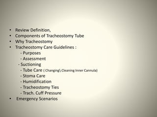
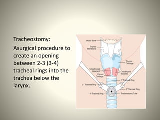
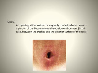
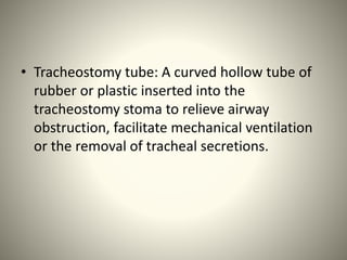
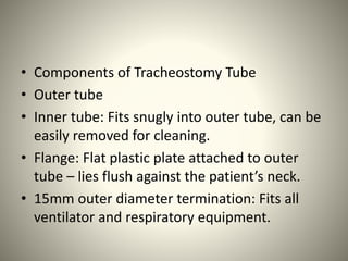
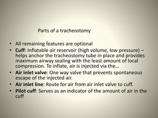
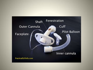
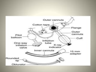
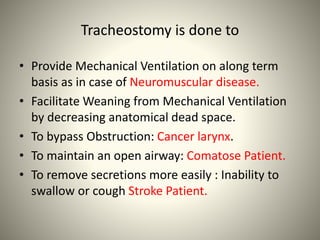
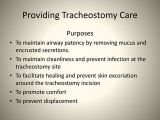
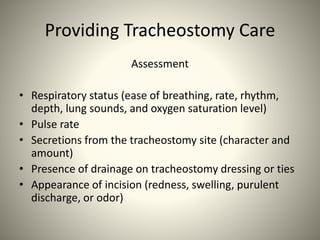
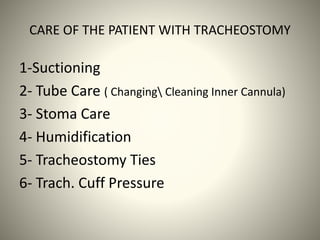
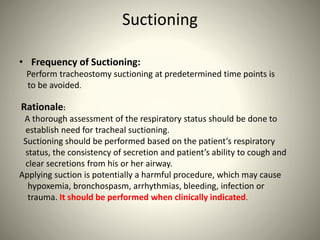
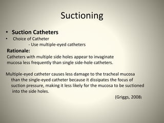
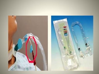
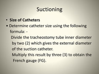
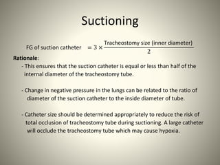
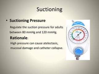
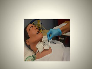
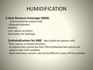
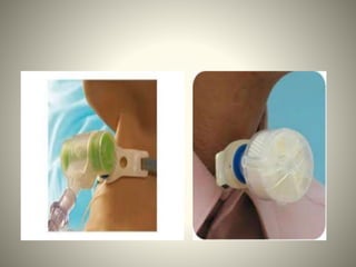
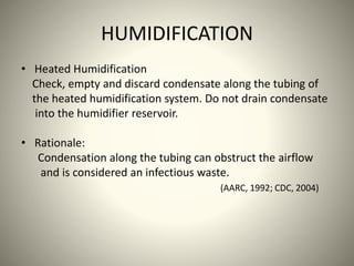
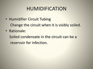
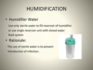
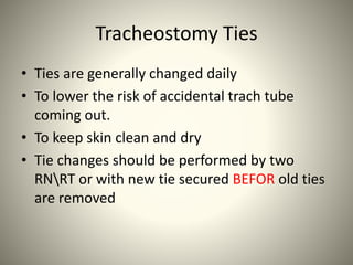
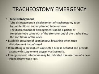
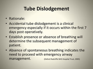
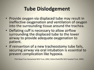
This document provides guidelines for tracheostomy care including components of a tracheostomy tube, purposes of tracheostomy, assessment of patients, suctioning procedures, inner cannula care, stoma care, humidification, and emergency scenarios. Key aspects of care include performing suctioning based on patient assessment rather than a set schedule, using aseptic technique, regulating suction pressure, preoxygenating patients, and changing dressings and tubes as needed to prevent infection and maintain an open airway. Various devices and methods are recommended for providing appropriate humidification.


