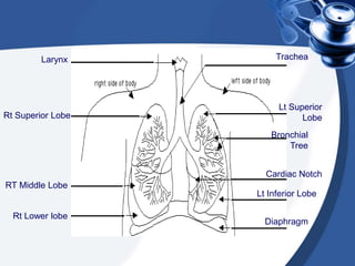
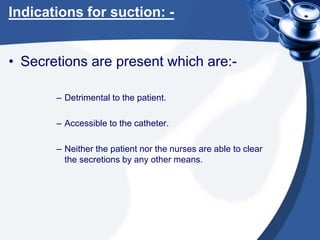
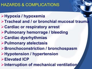
Suctioning
A Nursing Approach
Mr. Nestlee Sio Cabaccan RN,MSN
Aims
• To ensure the highest standards of
patient care through theoretical and
practical teaching of suction
techniques, together with safe and
effective use of suctioning equipment,
to nursing student.
Objectives
After This Session Candidates will
• Be familiar with the anatomy and physiology of
related structures and have an under standing on
nursing procedure.
• Be able to identify key features in assessing the
need of suction.
• Be able to state ways in reducing Mucosal trauma
and preventing Hypoxia Valsalva Maneuver.
• Be able to identify a safe value for negative
suctioning pressure and will be able to dismantle,
clean, set up and adjust suction machines
accordingly.
• It is anticipated that student will have the
opportunity to demonstrate safe suctioning
techniques to a competent student with the
supervision of the clinical instructor.
Content
1. Definition of suctioning
2. Brief history
3. Anatomy and Physiology
4. Purpose
5. Guidelines
Definition
• Suctioning is a method of removing
excessive secretions from the airway.
• May be applied to:
a. Oral
b. Nasopharyngeal
c. Tracheal passages
A Brief History Of Suction
• Airway suction was once described as a
“surprisingly simple technique” (Thompson,
1936) .
• In 1959, Boba et al studied the effects of
endotracheal suctioning in paralysed
patients. They reported that severe hypoxia
resulted from suctioning for one minute.
• Shumacker et al (1951), Keown (1960) and
Marx et al (1968) reported cardiac arrest
associated with endotracheal suction.
A Brief History Of Suction
• Rosen and Hillard (1962) stated that
deaths during suctioning procedures
have not been reported as often as
personal inquiries indicate that they
happen.
– “cardiac arrest may arise from the
stimulation of respiratory tract reflexes,”
• In 1984, Kergin et al., Using oximetry,
again reported reduction in blood
oxygen saturation during suctioning.
Anatomy And Physiology Of
Related Structures
Nose
Pharynx
Larynx
Trachea
Bronchi
Lungs –
alveoli
Larynx
Rt Superior Lobe
RT Middle Lobe
Rt Lower lobe
Lt Superior
Lobe
Bronchial
Tree
Cardiac Notch
Trachea
Lt Inferior Lobe
Diaphragm
Purpose
• To provide a patent airway by keeping
it clear of excessive secretions.
INDICATION
Therapeutic Diagnostic
NECESSARY EQUIPMENT
 Vaccum source with adjustable
regulator suction jar
 stethoscope
 Sterile gloves for open suctioning
method
 Clean gloves for closed suctioning
method
 Sterile catheter
 Clear protective goggles, apron & mask
 Sterile normal saline
 Bain’s circuit or ambu bag for
preoxygenate the patient
 Suction tray with hot water for flushing
The Vagus Nerves
• Have a more extensive distribution than any
other cranial nerves. The motor fibres supply
the smooth muscles and secretory glands of
the pharynx, larynx, trachea, heart,
oesophagus, stomach, intestines, pancreas,
gall bladder, bile ducts, spleen, kidneys,
ureter and blood vessels in the thoracic and
abdominal cavities.The sensory fibres
convey impulses from the lining membranes
of the same structures to the brain.
VAGUS NERVE
BRANCHES
OF THE
VAGUS
NERVE
Indications for suction: -
• Secretions are present which are:-
– Detrimental to the patient.
– Accessible to the catheter.
– Neither the patient nor the nurses are able to clear
the secretions by any other means.
HAZARDS & COMPLICATIONS
Hypoxia / hypoxemia
Tracheal and / or bronchial mucosal trauma
Cardiac or respiratory arrest
Pulmonary hemorrage / bleeding
Cardiac dysrhythmias
Pulmonary atelectasis
Bronchoconstriction / bronchospasm
Hypotension / hypertension
Elevated ICP
Interruption of mechanical ventilation
TYPES OF SUCTIONING
OPEN SUCTION CLOSED SUCTION
OPEN SUCTION SYSTEM:
Regularly using system in the
intubated patients.
CLOSED SUCTION SYSTEM:
 This is used to facilitate continuous
mechanical ventilation and oxygenation
during the suctioning.
 Closed suctioning is also indicated
when PEEP level above 10cmH2O.
MONITORING
The following should be monitored prior to,
during & after the procedure:
 Breath sounds
 Oxygen saturation
 RR & pattern
 Haemodynamic parameters (pulse rate, Blood
pressure)
 Cough effort
 ICP (If indicated and available)
 Sputum characteristics (colour, volume,
consistency & odor)
Choosing Correct Gauge
Catheter.
• E.G. tracheostomy tube size = 10.
• Multiply by three = 30.
• Divide by two = 15.
• Then choose the nearest, safest or most efficient gauge
catheter to that number i.e.
• For a size 10 tracheostomy tube, use a size 14 fg catheter.
• “It is essential to use the right size catheter for the lumen of
the tracheostomy tube:
• a 10FG catheter is appropriate for a size 6 tube,
• a 12FG catheter for a size 8 tube;
• a 14FG catheter for a size 10 tube,
• It is occasionally necessary to us a proportionately larger
diameter of catheter, especially if secretions are viscous, but
this must be done with care.” (Mallet 1985).
Choosing The Correct Amount
Of Negative Pressure.
• Suggestions for minimising the suction-induced hypoxemia
include, limiting the negative suction pressure, and the use
of hyper oxygenation.
• Negative suction pressure is also strongly associated with
trauma, which as we know leads to infection and increases
patient anxiety; the following article is included to
demonstrate this.
Achieving the correct depth of
insertion.
• Not introducing the catheter too deeply into the tracheo – bronchial
tree will reduce the likely hood of vagal stimulation, bronchospasm
and trauma. There is a degree of conflict within the research
(Kleiber 1986) with suggestions of efficient depths which range
from 1cm past the end of the tube to one cm past the carina.
• A general rule is proceed with the minimum amount of invasion,
the recommendation is to advance the catheter slowly until either a
cough reflex is initiated or resistance is felt upon
encountering either of these conditions, the nurse
should withdraw the catheter 1cm , apply suction and
withdraw the catheter.
• For patients with copious or tenacious secretions, who are showing
signs of ineffective airway clearance, deeper suctioning is
suggested. Care plans should include specific guidelines for
catheter insertion and should be updated routinely by the caregiver.
Individualisation of the care plan is essential.
Applying Suction Appropriately,
For Correct Amount Of Time.
• Insufflation of five litres of O2 down a sidearm during
endotracheal suction diminished the rate of decline of pao2
during suction of normal dog lungs. In patients with
respiratory insufficiency, the insufflation of O2 during
suction did not have any effect on the decreased pao2
seen during the endotracheal suction.
• The most effective way to prevent hypoxia during
endotracheal suction of patients with respiratory failure is to
hyperoxygenate for one minute with 100% O2 prior to
suction and limit suction to 15 seconds, (fell 1971).
• To err on the side of caution it is recommended that
suctioning is limited to 10 seconds only and that only 3 – 4
passes are completed in any one session.
Being Gentle.
• The airway mucosa is extremely sensitive to pressure and is
easily damaged. Chronic irritation can result in scar
formation, which may necessitate surgical intervention and
prolonged hospitalisation. Therefore, any suctioning of the
airway must be done with extreme gentleness.
• This again will reduce the likely hood of vagal stimulation,
bronchospasm and trauma and will greatly reduce patient
anxiety.
Patient Preparation
 Explain the procedure to the patient
(If patient is concious).
 The patient should receive hyper
oxygenation by the delivery of 100%
oxygen for >30 seconds prior to the
suctioning (Either with Bain’s circuit
or by increasing the FiO2 by
mechanical ventilator).
 Position the patient in supine
position.
 Auscultate the breath sounds.
PROCEDURE
Perform hand hygiene, wash
hands. It reduces
transmission of
microorganisms.
Turn on suction apparatus
and set vacuum regulator to
appropriate negative
pressure. For adult a pressure
of 100-120 mmHg, 80-
100/120Lmmhg for children &
60-80/1ooLmmhg for infants.
Continue…..
 Place the dominant thumb
over the control vent of the
suction port, applying
continuous or intermittent
suction for no more than 10
sec as you withdraw the
catheter into the sterile sleeve
of the closed suction device
 Repeat steps above if needed
 Clean suction catheter with
sterile saline until clear; being
careful not to instill solution
into the ETtube
 Suction oropharynx above
the artificial airway
 Wash hands
ASSESSMENT OF OUTCOME
Improvement in breath sounds.
Decreased peak inspiratory pressure;
Increased tidal volume delivery during
ventilation.
Improvement in arterial blood gas
values or saturation as reflected by
pulse oximetry. (SpO2)
Removal of pulmonary secretions.
CONTRAINDICATIONS
 Most contraindications are relative to the
patient's risk of developing adverse
reactions or worsening clinical condition as
result of the procedure.
 Suctioning is contraindicated when there is
fresh bleeding.
 When indicated, there is no absolute
contraindication to endotracheal suctioning
because the decision to abstain from
suctioning in order to avoid a possible
adverse reaction may, in fact, be lethal.
LIMITATIONS OF METHOD
Suctioning is potentially an harmful
procedure if carriedout improperly.
Suctioning should be done when
clinically necessary (not routinely).
The need for suctioning should be
assessed at least every 2hrs or more
frequently as need arises.

218935884-16744678-Safe-Suctioning.ppt

  • 1.
    Suctioning A Nursing Approach Mr.Nestlee Sio Cabaccan RN,MSN
  • 2.
    Aims • To ensurethe highest standards of patient care through theoretical and practical teaching of suction techniques, together with safe and effective use of suctioning equipment, to nursing student.
  • 3.
    Objectives After This SessionCandidates will • Be familiar with the anatomy and physiology of related structures and have an under standing on nursing procedure. • Be able to identify key features in assessing the need of suction. • Be able to state ways in reducing Mucosal trauma and preventing Hypoxia Valsalva Maneuver. • Be able to identify a safe value for negative suctioning pressure and will be able to dismantle, clean, set up and adjust suction machines accordingly. • It is anticipated that student will have the opportunity to demonstrate safe suctioning techniques to a competent student with the supervision of the clinical instructor.
  • 4.
    Content 1. Definition ofsuctioning 2. Brief history 3. Anatomy and Physiology 4. Purpose 5. Guidelines
  • 5.
    Definition • Suctioning isa method of removing excessive secretions from the airway. • May be applied to: a. Oral b. Nasopharyngeal c. Tracheal passages
  • 6.
    A Brief HistoryOf Suction • Airway suction was once described as a “surprisingly simple technique” (Thompson, 1936) . • In 1959, Boba et al studied the effects of endotracheal suctioning in paralysed patients. They reported that severe hypoxia resulted from suctioning for one minute. • Shumacker et al (1951), Keown (1960) and Marx et al (1968) reported cardiac arrest associated with endotracheal suction.
  • 7.
    A Brief HistoryOf Suction • Rosen and Hillard (1962) stated that deaths during suctioning procedures have not been reported as often as personal inquiries indicate that they happen. – “cardiac arrest may arise from the stimulation of respiratory tract reflexes,” • In 1984, Kergin et al., Using oximetry, again reported reduction in blood oxygen saturation during suctioning.
  • 8.
    Anatomy And PhysiologyOf Related Structures Nose Pharynx Larynx Trachea Bronchi Lungs – alveoli
  • 9.
    Larynx Rt Superior Lobe RTMiddle Lobe Rt Lower lobe Lt Superior Lobe Bronchial Tree Cardiac Notch Trachea Lt Inferior Lobe Diaphragm
  • 10.
    Purpose • To providea patent airway by keeping it clear of excessive secretions.
  • 11.
  • 12.
    NECESSARY EQUIPMENT  Vaccumsource with adjustable regulator suction jar  stethoscope  Sterile gloves for open suctioning method  Clean gloves for closed suctioning method  Sterile catheter  Clear protective goggles, apron & mask  Sterile normal saline  Bain’s circuit or ambu bag for preoxygenate the patient  Suction tray with hot water for flushing
  • 13.
    The Vagus Nerves •Have a more extensive distribution than any other cranial nerves. The motor fibres supply the smooth muscles and secretory glands of the pharynx, larynx, trachea, heart, oesophagus, stomach, intestines, pancreas, gall bladder, bile ducts, spleen, kidneys, ureter and blood vessels in the thoracic and abdominal cavities.The sensory fibres convey impulses from the lining membranes of the same structures to the brain.
  • 14.
  • 16.
    Indications for suction:- • Secretions are present which are:- – Detrimental to the patient. – Accessible to the catheter. – Neither the patient nor the nurses are able to clear the secretions by any other means.
  • 17.
    HAZARDS & COMPLICATIONS Hypoxia/ hypoxemia Tracheal and / or bronchial mucosal trauma Cardiac or respiratory arrest Pulmonary hemorrage / bleeding Cardiac dysrhythmias Pulmonary atelectasis Bronchoconstriction / bronchospasm Hypotension / hypertension Elevated ICP Interruption of mechanical ventilation
  • 18.
    TYPES OF SUCTIONING OPENSUCTION CLOSED SUCTION
  • 19.
    OPEN SUCTION SYSTEM: Regularlyusing system in the intubated patients. CLOSED SUCTION SYSTEM:  This is used to facilitate continuous mechanical ventilation and oxygenation during the suctioning.  Closed suctioning is also indicated when PEEP level above 10cmH2O.
  • 20.
    MONITORING The following shouldbe monitored prior to, during & after the procedure:  Breath sounds  Oxygen saturation  RR & pattern  Haemodynamic parameters (pulse rate, Blood pressure)  Cough effort  ICP (If indicated and available)  Sputum characteristics (colour, volume, consistency & odor)
  • 21.
    Choosing Correct Gauge Catheter. •E.G. tracheostomy tube size = 10. • Multiply by three = 30. • Divide by two = 15. • Then choose the nearest, safest or most efficient gauge catheter to that number i.e. • For a size 10 tracheostomy tube, use a size 14 fg catheter. • “It is essential to use the right size catheter for the lumen of the tracheostomy tube: • a 10FG catheter is appropriate for a size 6 tube, • a 12FG catheter for a size 8 tube; • a 14FG catheter for a size 10 tube, • It is occasionally necessary to us a proportionately larger diameter of catheter, especially if secretions are viscous, but this must be done with care.” (Mallet 1985).
  • 22.
    Choosing The CorrectAmount Of Negative Pressure. • Suggestions for minimising the suction-induced hypoxemia include, limiting the negative suction pressure, and the use of hyper oxygenation. • Negative suction pressure is also strongly associated with trauma, which as we know leads to infection and increases patient anxiety; the following article is included to demonstrate this.
  • 23.
    Achieving the correctdepth of insertion. • Not introducing the catheter too deeply into the tracheo – bronchial tree will reduce the likely hood of vagal stimulation, bronchospasm and trauma. There is a degree of conflict within the research (Kleiber 1986) with suggestions of efficient depths which range from 1cm past the end of the tube to one cm past the carina. • A general rule is proceed with the minimum amount of invasion, the recommendation is to advance the catheter slowly until either a cough reflex is initiated or resistance is felt upon encountering either of these conditions, the nurse should withdraw the catheter 1cm , apply suction and withdraw the catheter. • For patients with copious or tenacious secretions, who are showing signs of ineffective airway clearance, deeper suctioning is suggested. Care plans should include specific guidelines for catheter insertion and should be updated routinely by the caregiver. Individualisation of the care plan is essential.
  • 24.
    Applying Suction Appropriately, ForCorrect Amount Of Time. • Insufflation of five litres of O2 down a sidearm during endotracheal suction diminished the rate of decline of pao2 during suction of normal dog lungs. In patients with respiratory insufficiency, the insufflation of O2 during suction did not have any effect on the decreased pao2 seen during the endotracheal suction. • The most effective way to prevent hypoxia during endotracheal suction of patients with respiratory failure is to hyperoxygenate for one minute with 100% O2 prior to suction and limit suction to 15 seconds, (fell 1971). • To err on the side of caution it is recommended that suctioning is limited to 10 seconds only and that only 3 – 4 passes are completed in any one session.
  • 25.
    Being Gentle. • Theairway mucosa is extremely sensitive to pressure and is easily damaged. Chronic irritation can result in scar formation, which may necessitate surgical intervention and prolonged hospitalisation. Therefore, any suctioning of the airway must be done with extreme gentleness. • This again will reduce the likely hood of vagal stimulation, bronchospasm and trauma and will greatly reduce patient anxiety.
  • 26.
    Patient Preparation  Explainthe procedure to the patient (If patient is concious).  The patient should receive hyper oxygenation by the delivery of 100% oxygen for >30 seconds prior to the suctioning (Either with Bain’s circuit or by increasing the FiO2 by mechanical ventilator).  Position the patient in supine position.  Auscultate the breath sounds.
  • 27.
    PROCEDURE Perform hand hygiene,wash hands. It reduces transmission of microorganisms. Turn on suction apparatus and set vacuum regulator to appropriate negative pressure. For adult a pressure of 100-120 mmHg, 80- 100/120Lmmhg for children & 60-80/1ooLmmhg for infants.
  • 28.
    Continue…..  Place thedominant thumb over the control vent of the suction port, applying continuous or intermittent suction for no more than 10 sec as you withdraw the catheter into the sterile sleeve of the closed suction device  Repeat steps above if needed  Clean suction catheter with sterile saline until clear; being careful not to instill solution into the ETtube  Suction oropharynx above the artificial airway  Wash hands
  • 29.
    ASSESSMENT OF OUTCOME Improvementin breath sounds. Decreased peak inspiratory pressure; Increased tidal volume delivery during ventilation. Improvement in arterial blood gas values or saturation as reflected by pulse oximetry. (SpO2) Removal of pulmonary secretions.
  • 30.
    CONTRAINDICATIONS  Most contraindicationsare relative to the patient's risk of developing adverse reactions or worsening clinical condition as result of the procedure.  Suctioning is contraindicated when there is fresh bleeding.  When indicated, there is no absolute contraindication to endotracheal suctioning because the decision to abstain from suctioning in order to avoid a possible adverse reaction may, in fact, be lethal.
  • 31.
    LIMITATIONS OF METHOD Suctioningis potentially an harmful procedure if carriedout improperly. Suctioning should be done when clinically necessary (not routinely). The need for suctioning should be assessed at least every 2hrs or more frequently as need arises.