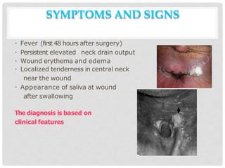
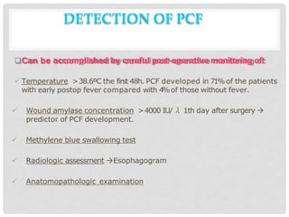
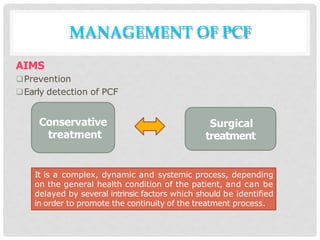
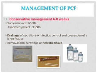
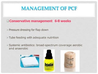
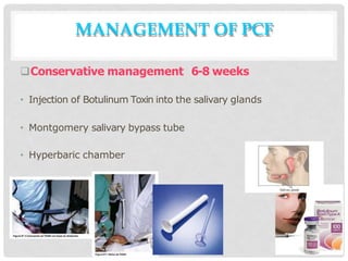
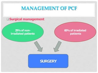
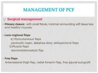
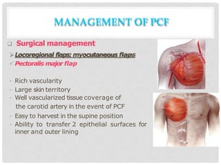
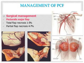
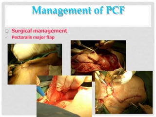
MANAGEMENT OF
PHARYNGOCUTANEOUS FISTULA
Elena Rizzo Riera
R1 ENT HUSE
DEFINITION
• Pharyngocutaneous fistula (PCF) is the communication of the
digestive tract with the cervical skin which originates the
appearance of saliva on the skin surface after swallowing
• At the level of the surgical incision
• Around the tracheostoma
• The continuous flow of saliva is the main factor involved in hinder
the closure of PCF.
INTRODUCTION
• The most common complication of oncologic laryngeal and
hypopharyngeal surgery.
Consequences:
 postoperative hospital stay
Late onset of swallowing
Reintervention
Death caused by carotid artery rupture
CLASSIFICATION
Classification based on SIZEof PCF
Type I  <2cm
Type I
I  2-4 cm
Type I
I
I  4-6 cm
Type IV  >6cm
CLASSIFICATION
• Guthrie classification (1974)
PCF with a horizontal diameter <8 mm
PCF with a diameter less than ¼of pharyngeal circumference
PCF less than the half of pharyngeal
circumference
PCF more than the half of pharyngeal
circumference
CLASSIFICATION
• Vilar Sancho classification
PCF in not-irradiated patients
PCF in irradiated patiens
a) Caused by dehiscence
c) Caused by loss of substance
CLASSIFICATION
Funk’s classification:
1. Pharyngocutaneous fistula
a)With RT,carotid artery involvement, or
microvascular anastomosis
b) Without RT, carotid artery affectation
or microvascular anastomosis
2. Pharyngostoma ( direct opening of the
p h a r y n x to the skin, o f t e n
accompanied by skin loss)
EPIDEMIOLOGY
• Incidence 5 – 65%
• M>F
• 50-70 years
• 7 -10 days after surgery
• Late onset: 153 days after the
procedure
Years Women Men Total
<40 1 0.7% 1 0.7 2 1.3
4-50 2 1.3% 21 13.3 23 14.5
51-60 4 2.5% 66 41.7 70 44.3
61-70 3 1.8% 44 27.8 47 29.7
>70 - - 16 10.2 16 10.2
Total 10 6.4 148 93..6 158 100
RISK FACTORS
Gastroesophageal reflux disease (GERD)
 71%of patients with laryngeal carcinoma have abnormal 24-hour pH studies
 Prophylactic use of Ranitidine (H2 receptor blocker) and Metoclopramide
(prokinetic) have decreased the incidence of PCF and the mean length of
hospital stay after total laryngectomy.
Malnutrition
 30-50%of incidence in patients with H&N cancers.
 The patients with >10% of weight loss in the 6 months prior to surgery were at
greatest risk (50%) for development of major postop complications.
Age
Vascular diseases related to smoking and alcohol drinking
Low preoperative Hemoglobin
Diabetes
Hepatopathy
Chronic obstructive pulmonary disease
Hypothyroidism
Immunosuppressive
therapy
Preoperative radiation
Chemotherapy
RISK FACTORS
RISK FACTORS
Positive surgical margins
Preoperative tracheostomy in emergency situations
Tumor stage and laryngeal site
 Supraglottic tumors have significantly more fistulas than glottic tumors.
Extension to the vallecula or the piriform fossa biggest excision of
pharyngeal mucosa  more tension.
Onset of oral feeding
Vomit
Infection
Cervical dissection
RISK FACTORS
Suture material
- Vicryl 3/0 >catgut
Vycril has greater tensile strength, less inflammatory reaction and a longer
half- life.
Suture tecnique
- Interrupted suture
- Extramucose suture
- Knots tied on the inside
- Tpharyngeal closure >vertical closure
SYMPTOMS AND SIGNS
• Fever (first 48 hours after surgery)
• Persistent elevated neck drain output
• Wound erythema and edema
• Localized tenderness in central neck
near the wound
• Appearance of saliva at wound
after swallowing
The diagnosis is based on
clinical features
DETECTION OF PCF
Can be accomplished by careful post-operative monitoring of:
 Temperature >38.6ºC the first 48h. PCF developed in 71%of the patients
with early postop fever compared with 4
%of those without fever.
 Wound amylase concentration >4000 IU/ λ 1th day after surgery 
predictor of PCF development.
 Methylene blue swallowing test
 Radiologic assessment Esophagogram
 Anatomopathologic examination
MANAGEMENT OF PCF
AIMS
Prevention
Early detection of PCF
Conservative
treatment
Surgical
treatment
It is a complex, dynamic and systemic process, depending
on the general health condition of the patient, and can be
delayed by several intrinsic factors which should be identified
in order to promote the continuity of the treatment process.
Prevention
• Surgical tecnique: hemostasis and manipulation. To avoid significant
crush injury to the tissues, to excise questionably viable mucosa, and
to avoid unnecessary tension on the pharyngeal closure.
Antibiotics: 50%of reduction of PCF incidence
Gastroesophageal reflux disease (GERD)
(ranitidine and metoclopramide 7 days)
Identification and management of comorbidities
Enteral nutrition: NGT or PEG for 10 days
MANAGEMENT OF PCF
 Conservative management 6-8 weeks
Successful rate: 60-80%
Irradiated patient: 35-50%
Drainage of secretions infection control and prevention of a
large fistula
Removal and curettage of necrotic tissue
MANAGEMENT OF PCF
Conservative management 6-8 weeks
Pressure dressing for flap down
Tube feeding with adequate nutrition
Systemic antibiotics: broad-spectrum coverage aerobic
and anaerobic
MANAGEMENT OF PCF
Conservative management 6-8 weeks
• Injection of Botulinum Toxin into the salivary glands
• Montgomery salivary bypass tube
• Hyperbaric chamber
MANAGEMENT OF PCF
Surgical management
25%of non-
irradiated patients
60%of irradiated
patients
SURGERY
MANAGEMENT OF PCF
 Surgical management
Primary closure: with small fistula, minimal sorrounding soft tissue loss
and healthy mucosa.
Loco-regional flaps
a) Myocutaneous flaps
pectoralis major, latissimus dorsi, deltopectoral flaps
b)Muscle flaps
sternocleidomastoid flap
Free flaps
Anterolateral thigh flap, radial forearm flap, free jejunal autograft
MANAGEMENT OF PCF
 Surgical management
Locoregional flaps: myocutaneous flaps
Pectoralis major flap
- Rich vascularity
- Large skin territory
- Well vascularized tissue coverage of
the carotid artery in the event of PCF
- Easy to harvest in the supine position
- Ability to transfer 2 epithelial surfaces for
inner and outer lining
MANAGEMENT OF PCF
MANAGEMENT OF PCF
 Surgical management
 Pectoralis major flap
 Total flap necrosis 1-3%
 Partial flap necrosis 4-7%
Management of PCF
 Surgical management
 Pectoralis major flap
CONCLUSIONS
Risk pre-surgery
Identification and treatment of comorbidities
Good surgical technique
Multidisciplinary approach
 post-operative hospital stay
Possible lethal complications
THANK YOU 

Pharyngocutaneous-fistula (1).pptx

  • 1.
  • 2.
    DEFINITION • Pharyngocutaneous fistula(PCF) is the communication of the digestive tract with the cervical skin which originates the appearance of saliva on the skin surface after swallowing • At the level of the surgical incision • Around the tracheostoma • The continuous flow of saliva is the main factor involved in hinder the closure of PCF.
  • 3.
    INTRODUCTION • The mostcommon complication of oncologic laryngeal and hypopharyngeal surgery. Consequences:  postoperative hospital stay Late onset of swallowing Reintervention Death caused by carotid artery rupture
  • 4.
    CLASSIFICATION Classification based onSIZEof PCF Type I  <2cm Type I I  2-4 cm Type I I I  4-6 cm Type IV  >6cm
  • 5.
    CLASSIFICATION • Guthrie classification(1974) PCF with a horizontal diameter <8 mm PCF with a diameter less than ¼of pharyngeal circumference PCF less than the half of pharyngeal circumference PCF more than the half of pharyngeal circumference
  • 6.
    CLASSIFICATION • Vilar Sanchoclassification PCF in not-irradiated patients PCF in irradiated patiens a) Caused by dehiscence c) Caused by loss of substance
  • 7.
    CLASSIFICATION Funk’s classification: 1. Pharyngocutaneousfistula a)With RT,carotid artery involvement, or microvascular anastomosis b) Without RT, carotid artery affectation or microvascular anastomosis 2. Pharyngostoma ( direct opening of the p h a r y n x to the skin, o f t e n accompanied by skin loss)
  • 8.
    EPIDEMIOLOGY • Incidence 5– 65% • M>F • 50-70 years • 7 -10 days after surgery • Late onset: 153 days after the procedure Years Women Men Total <40 1 0.7% 1 0.7 2 1.3 4-50 2 1.3% 21 13.3 23 14.5 51-60 4 2.5% 66 41.7 70 44.3 61-70 3 1.8% 44 27.8 47 29.7 >70 - - 16 10.2 16 10.2 Total 10 6.4 148 93..6 158 100
  • 9.
    RISK FACTORS Gastroesophageal refluxdisease (GERD)  71%of patients with laryngeal carcinoma have abnormal 24-hour pH studies  Prophylactic use of Ranitidine (H2 receptor blocker) and Metoclopramide (prokinetic) have decreased the incidence of PCF and the mean length of hospital stay after total laryngectomy. Malnutrition  30-50%of incidence in patients with H&N cancers.  The patients with >10% of weight loss in the 6 months prior to surgery were at greatest risk (50%) for development of major postop complications. Age Vascular diseases related to smoking and alcohol drinking
  • 10.
    Low preoperative Hemoglobin Diabetes Hepatopathy Chronicobstructive pulmonary disease Hypothyroidism Immunosuppressive therapy Preoperative radiation Chemotherapy RISK FACTORS
  • 11.
    RISK FACTORS Positive surgicalmargins Preoperative tracheostomy in emergency situations Tumor stage and laryngeal site  Supraglottic tumors have significantly more fistulas than glottic tumors. Extension to the vallecula or the piriform fossa biggest excision of pharyngeal mucosa  more tension. Onset of oral feeding Vomit Infection Cervical dissection
  • 12.
    RISK FACTORS Suture material -Vicryl 3/0 >catgut Vycril has greater tensile strength, less inflammatory reaction and a longer half- life. Suture tecnique - Interrupted suture - Extramucose suture - Knots tied on the inside - Tpharyngeal closure >vertical closure
  • 13.
    SYMPTOMS AND SIGNS •Fever (first 48 hours after surgery) • Persistent elevated neck drain output • Wound erythema and edema • Localized tenderness in central neck near the wound • Appearance of saliva at wound after swallowing The diagnosis is based on clinical features
  • 14.
    DETECTION OF PCF Canbe accomplished by careful post-operative monitoring of:  Temperature >38.6ºC the first 48h. PCF developed in 71%of the patients with early postop fever compared with 4 %of those without fever.  Wound amylase concentration >4000 IU/ λ 1th day after surgery  predictor of PCF development.  Methylene blue swallowing test  Radiologic assessment Esophagogram  Anatomopathologic examination
  • 15.
    MANAGEMENT OF PCF AIMS Prevention Earlydetection of PCF Conservative treatment Surgical treatment It is a complex, dynamic and systemic process, depending on the general health condition of the patient, and can be delayed by several intrinsic factors which should be identified in order to promote the continuity of the treatment process.
  • 16.
    Prevention • Surgical tecnique:hemostasis and manipulation. To avoid significant crush injury to the tissues, to excise questionably viable mucosa, and to avoid unnecessary tension on the pharyngeal closure. Antibiotics: 50%of reduction of PCF incidence Gastroesophageal reflux disease (GERD) (ranitidine and metoclopramide 7 days) Identification and management of comorbidities Enteral nutrition: NGT or PEG for 10 days MANAGEMENT OF PCF
  • 17.
     Conservative management6-8 weeks Successful rate: 60-80% Irradiated patient: 35-50% Drainage of secretions infection control and prevention of a large fistula Removal and curettage of necrotic tissue MANAGEMENT OF PCF
  • 18.
    Conservative management 6-8weeks Pressure dressing for flap down Tube feeding with adequate nutrition Systemic antibiotics: broad-spectrum coverage aerobic and anaerobic MANAGEMENT OF PCF
  • 19.
    Conservative management 6-8weeks • Injection of Botulinum Toxin into the salivary glands • Montgomery salivary bypass tube • Hyperbaric chamber MANAGEMENT OF PCF
  • 20.
    Surgical management 25%of non- irradiatedpatients 60%of irradiated patients SURGERY MANAGEMENT OF PCF
  • 21.
     Surgical management Primaryclosure: with small fistula, minimal sorrounding soft tissue loss and healthy mucosa. Loco-regional flaps a) Myocutaneous flaps pectoralis major, latissimus dorsi, deltopectoral flaps b)Muscle flaps sternocleidomastoid flap Free flaps Anterolateral thigh flap, radial forearm flap, free jejunal autograft MANAGEMENT OF PCF
  • 22.
     Surgical management Locoregionalflaps: myocutaneous flaps Pectoralis major flap - Rich vascularity - Large skin territory - Well vascularized tissue coverage of the carotid artery in the event of PCF - Easy to harvest in the supine position - Ability to transfer 2 epithelial surfaces for inner and outer lining MANAGEMENT OF PCF
  • 23.
    MANAGEMENT OF PCF Surgical management  Pectoralis major flap  Total flap necrosis 1-3%  Partial flap necrosis 4-7%
  • 24.
    Management of PCF Surgical management  Pectoralis major flap
  • 25.
    CONCLUSIONS Risk pre-surgery Identification andtreatment of comorbidities Good surgical technique Multidisciplinary approach  post-operative hospital stay Possible lethal complications
  • 26.