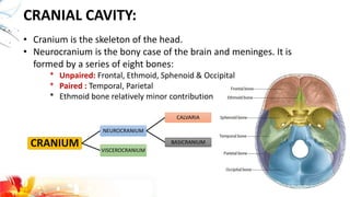
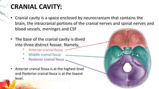
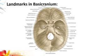
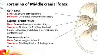
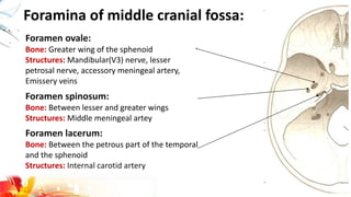
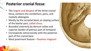
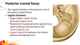
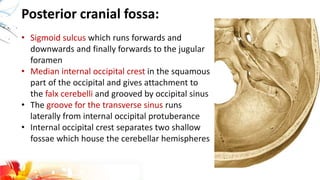
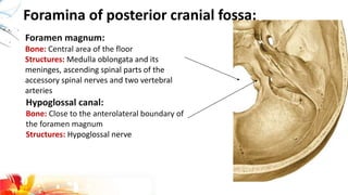
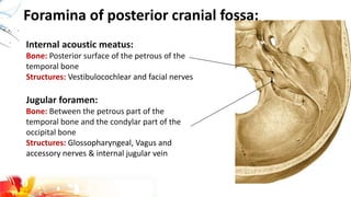
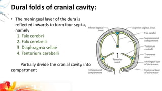
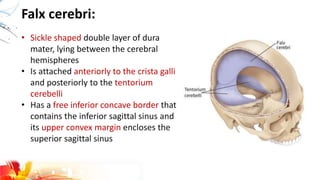
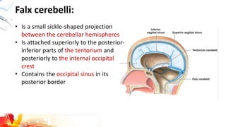
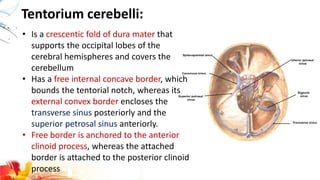
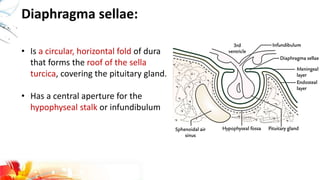
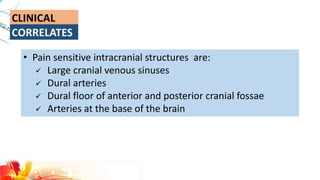
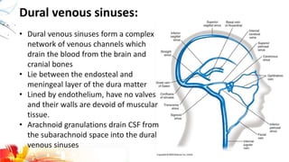
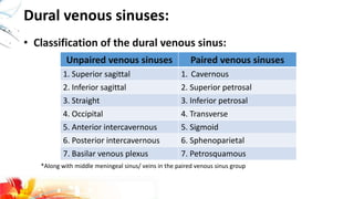
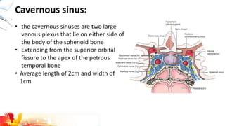
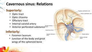
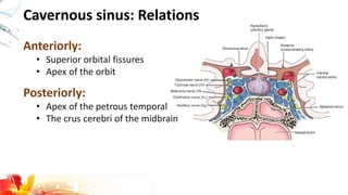
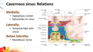
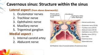
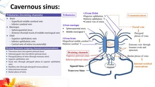
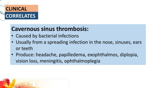
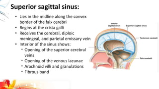
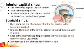
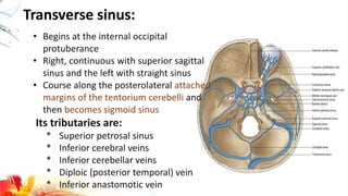
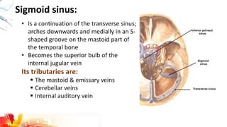
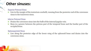
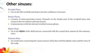
Cranium is the skeleton of the head.
Neurocranium is the bony case of the brain and meninges. It is formed by a series of eight bones:
Unpaired: Frontal, Ethmoid, Sphenoid & Occipital
Paired : Temporal, Parietal
Ethmoid bone relatively minor contribution
2. CRANIAL CAVITY:
• Cranium is the skeleton of the head.
• Neurocranium is the bony case of the brain and meninges. It is
formed by a series of eight bones:
* Unpaired: Frontal, Ethmoid, Sphenoid & Occipital
* Paired : Temporal, Parietal
* Ethmoid bone relatively minor contribution
CRANIUM
NEUROCRANIUM
CALVARIA
BASICRANIUM
VISCEROCRANIUM
3. CRANIAL CAVITY:
• Cranial cavity is a space enclosed by neurocranium that contains the
brain, the intracranial portions of the cranial nerves and spinal nerves and
blood vessels, meninges and CSF
• The base of the cranial cavity is dived
into three distinct fossae. Namely,
* Anterior cranial fossa
* Middle cranial fossa
* Posterior cranial fossa
• Anterior cranial fossa is at the highest level
and Posterior cranial fossa is at the lowest
level.
5. Anterior cranial fossa:
• Shallowest of the three cranial fossae
• Formed by the frontal bone anteriorly, the ethmoid bone is the
middle, and the lesser wings of the sphenoid posteriorly
• Greatest part of the fossa – orbital plate of the frontal bone
• Support the frontal lobes of the brain and form the roofs of the
orbit
• Crista galli- thick, median ridge of ethmoid, which project
superiorly and each of ridge is the sieve-like cribriform plate of
the ethmoid
• Olfactory bulbs of the brain, which lie on cribriform plate
6. Landmarks and Foramina/Apertures:
Foramen cecum:
Site: base of the frontal crest
Structures: emissary vein of frontal bone(1%)
Anterior and posterior ethmoidal foramina:
Structures: vessels and nerves with same name
Cribriform foramina:
Structures: Axons of olfactory cells in the olfactory
epithelium that form olfactory nerves
7. Middle cranial fossa:
• Butterfly shaped fossa
• It is bounded by:
* In front by sphenoid crest of lesser
wing and part of the body of the
sphenoid
* Behind superior border of the petrous
part of temporal bone dorsum sellae
* Laterally by the squamous parts of the
temporal bone
• Communicates with the orbits by the
superior orbital fissures
8. Middle cranial fossa:
• Sphenoid crests end medially in two sharp
bony projection, anterior clinoid processes
• Optic canal is lateral to the sulcus
chiasmatis which transmits the optic nerve
• Centrally the floor of the fossa is narrower
and formed by the sphenoid bone which
contains the sphenoidal sinuses
• Roof of the sphenoid sinus is deeply
concave and houses the pituitary fossa(aka
sella turcica) between turberculum and
dorsum sellae that gives attachment to the
tentorium cerebelli.
9. Foramina of Middle cranial fossa:
Optic canal:
Bone: Lesser wing of the sphenoid
Structures: Optic nerve and ophthalmic artery
Superior orbital fissure:
Bone: Between lesser and greater wings
Structures: Oculomotor, Trochlear, Lacrimal,
Frontal, Nasociliary and abducent nerve & Superior
ophthalmic vein
Foramen rotundum:
Bone: Greater wings of sphenoid
Structures: Maxillary division of the trigeminal
nerve
10. Foramina of middle cranial fossa:
Foramen ovale:
Bone: Greater wing of the sphenoid
Structures: Mandibular(V3) nerve, lesser
petrosal nerve, accessory meningeal artery,
Emissery veins
Foramen spinosum:
Bone: Between lesser and greater wings
Structures: Middle meningeal artey
Foramen lacerum:
Bone: Between the petrous part of the temporal
and the sphenoid
Structures: Internal carotid artery
11. Posterior cranial fossa:
• The largest and deepest of the three cranial
fossa, contains the cerebellum, pons, and
medulla oblongata
• Mostly by the occipital bone an sloping surface
of the basilar part, called clivus.
• Bounded anteriorly by dorsum sellae and
superior border of petrous part of temporal
• Corresponds extracranially with the posterior
part of the cranial base
• Most prominent feature – foramen magnum
12. Posterior cranial fossa:
• The jugular foramen at the posterior end of
the petro-occipital fissure
• Jugular foramen:
* Upper border- notch for the
glossopharyngeal nerve
* Posteriorly it is grooved by the sigmoid sinus
which continues into foramen as the
internal jugular vein
* Jugular tubercle lie between the inferior
petrosal and sigmoid sinus
13. Posterior cranial fossa:
• Sigmoid sulcus which runs forwards and
downwards and finally forwards to the jugular
foramen
• Median internal occipital crest in the squamous
part of the occipital and gives attachment to
the falx cerebelli and grooved by occipital sinus
• The groove for the transverse sinus runs
laterally from internal occipital protuberance
• Internal occipital crest separates two shallow
fossae which house the cerebellar hemispheres
14. Foramina of posterior cranial fossa:
Foramen magnum:
Bone: Central area of the floor
Structures: Medulla oblongata and its
meninges, ascending spinal parts of the
accessory spinal nerves and two vertebral
arteries
Hypoglossal canal:
Bone: Close to the anterolateral boundary of
the foramen magnum
Structures: Hypoglossal nerve
15. Foramina of posterior cranial fossa:
Jugular foramen:
Bone: Between the petrous part of the
temporal bone and the condylar part of the
occipital bone
Structures: Glossopharyngeal, Vagus and
accessory nerves & internal jugular vein
Internal acoustic meatus:
Bone: Posterior surface of the petrous of the
temporal bone
Structures: Vestibulocochlear and facial nerves
16. • Fracture of the anterior cranial fossa causes anosmia,
periorbital bruising, and CSF leakage from the nose
• Fracture of the anterior cranial fossa causes anosmia,
periorbital bruising, and CSF leakage from the nose
• A blow to the chin with the mouth closed, causing fracture
of the middle cranial fossa
18. Dural folds of cranial cavity:
• The meningeal layer of the dura is
reflected inwards to form four septa,
namely
1. Falx cerebri
2. Falx cerebelli
3. Diaphragma sellae
4. Tentorium cerebelli
Partially divide the cranial cavity into
compartment
19. Falx cerebri:
• Sickle shaped double layer of dura
mater, lying between the cerebral
hemispheres
• Is attached anteriorly to the crista galli
and posteriorly to the tentorium
cerebelli
• Has a free inferior concave border that
contains the inferior sagittal sinus and
its upper convex margin encloses the
superior sagittal sinus
20. Falx cerebelli:
• Is a small sickle-shaped projection
between the cerebellar hemispheres
• Is attached superiorly to the posterior-
inferior parts of the tentorium and
posteriorly to the internal occipital
crest
• Contains the occipital sinus in its
posterior border
21. Tentorium cerebelli:
• Is a crescentic fold of dura mater that
supports the occipital lobes of the
cerebral hemispheres and covers the
cerebellum
• Has a free internal concave border, which
bounds the tentorial notch, whereas its
external convex border encloses the
transverse sinus posteriorly and the
superior petrosal sinus anteriorly.
• Free border is anchored to the anterior
clinoid process, whereas the attached
border is attached to the posterior clinoid
process
22. Diaphragma sellae:
• Is a circular, horizontal fold of dura
that forms the roof of the sella
turcica, covering the pituitary gland.
• Has a central aperture for the
hypophyseal stalk or infundibulum
23. • Pain sensitive intracranial structures are:
Large cranial venous sinuses
Dural arteries
Dural floor of anterior and posterior cranial fossae
Arteries at the base of the brain
25. • Dural venous sinuses form a complex
network of venous channels which
drain the blood from the brain and
cranial bones
• Lie between the endosteal and
meningeal layer of the dura matter
• Lined by endothelium, have no valves
and their walls are devoid of muscular
tissue.
• Arachnoid granulations drain CSF from
the subarachnoid space into the dural
venous sinuses
Dural venous sinuses:
26. • Classification of the dural venous sinus:
Dural venous sinuses:
Unpaired venous sinuses Paired venous sinuses
1. Superior sagittal 1. Cavernous
2. Inferior sagittal 2. Superior petrosal
3. Straight 3. Inferior petrosal
4. Occipital 4. Transverse
5. Anterior intercavernous 5. Sigmoid
6. Posterior intercavernous 6. Sphenoparietal
7. Basilar venous plexus 7. Petrosquamous
*Along with middle meningeal sinus/ veins in the paired venous sinus group
27. Cavernous sinus:
• the cavernous sinuses are two large
venous plexus that lie on either side of
the body of the sphenoid bone
• Extending from the superior orbital
fissure to the apex of the petrous
temporal bone
• Average length of 2cm and width of
1cm
28. Cavernous sinus: Relations
Superiorly:
• Optic tract
• Optic chiasma
• Olfactory tract
• Internal carotid artery
• Anterior perforated substance
Inferiorly:
• Foramen lacerum
• Junction of the body and greater
wings of the sphenoid bone
29. Cavernous sinus: Relations
Anteriorly:
• Superior orbital fissures
• Apex of the orbit
Posteriorly:
• Apex of the petrous temporal
• The crus cerebri of the midbrain
33. Cavernous sinus thrombosis:
• Caused by bacterial infections
• Usually from a spreading infection in the nose, sinuses, ears
or teeth
• Produce: headache, papilledema, exophthalmos, diplopia,
vision loss, meningitis, ophthalmoplegia
34. Superior sagittal sinus:
• Lies in the midline along the convex
border of the falx cerebri
• Begins at the crista galli
• Receives the cerebral, diploic
meningeal, and parietal emissary vein
• Interior of the sinus shows:
* Opening of the superior cerebral
veins
* Opening of the venous lacunae
* Arachnoid villi and granulations
* Fibrous band
35. Inferior sagittal sinus:
• Lies in the free edge of the falx cerebri
• Ends in the strainght sinus
• Receives veins from the falx and medial
surface of the cerebral hemisphere
Straight sinus:
• Runs along the line of attachment of the falx cerebri to the
tentorium cerebelli
• Is formed by union of the inferior sagittal sinus and the great vein
of Galen
• Ends at the internal occipital protuberance by continuous as the
transverse sinus usually left
• Also receives a few of the superior cerebral vein
36. Transverse sinus:
• Begins at the internal occipital
protuberance
• Right, continuous with superior sagittal
sinus and the left with straight sinus
• Course along the posterolateral attached
margins of the tentorium cerebelli and
then becomes sigmoid sinus
Its tributaries are:
* Superior petrosal sinus
* Inferior cerebral veins
* Inferior cerebellar veins
* Diploic (posterior temporal) vein
* Inferior anastomotic vein
37. Sigmoid sinus:
• Is a continuation of the transverse sinus;
arches downwards and medially in an S-
shaped groove on the mastoid part of
the temporal bone
• Becomes the superior bulb of the
internal jugular vein
Its tributaries are:
The mastoid & emissary veins
Cerebellar veins
Internal auditory vein