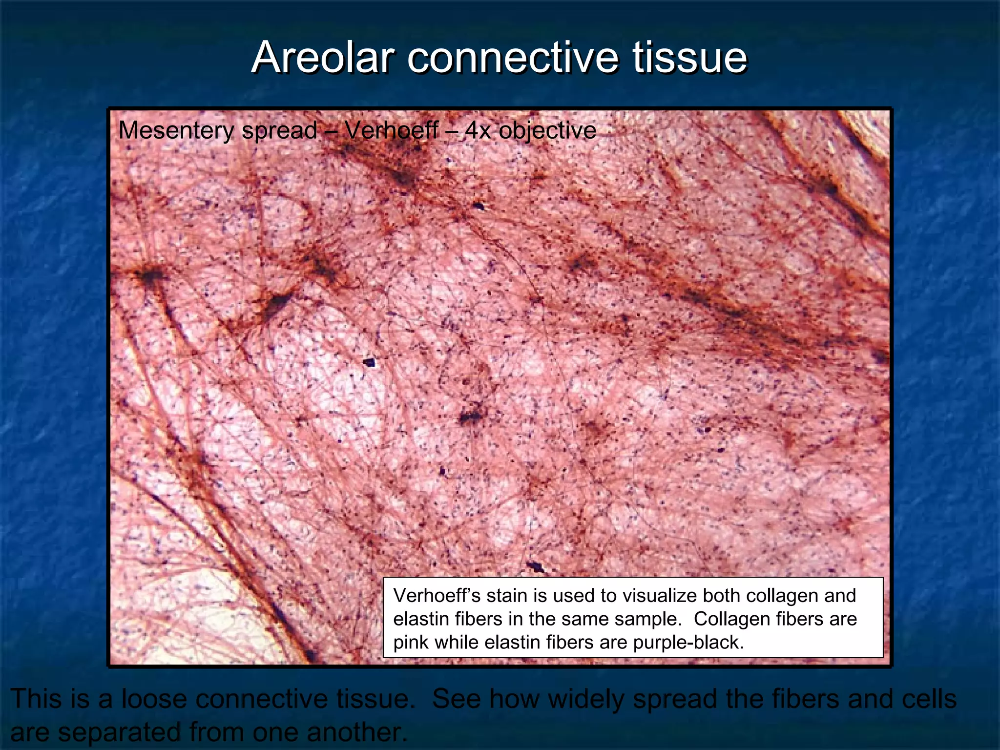
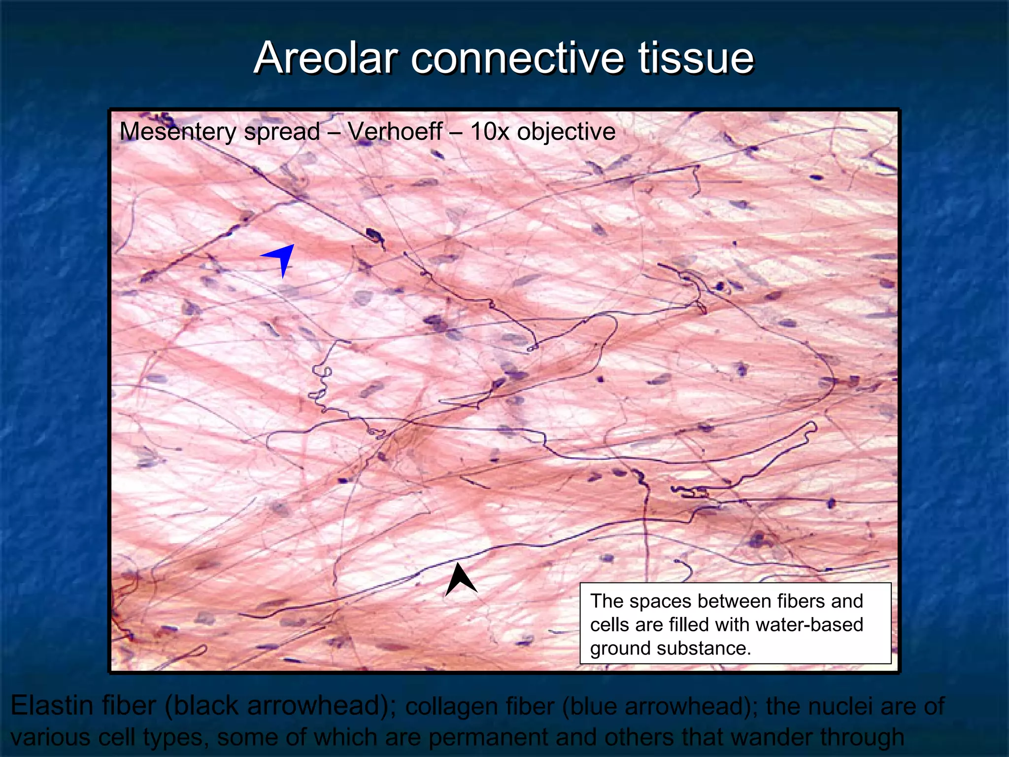
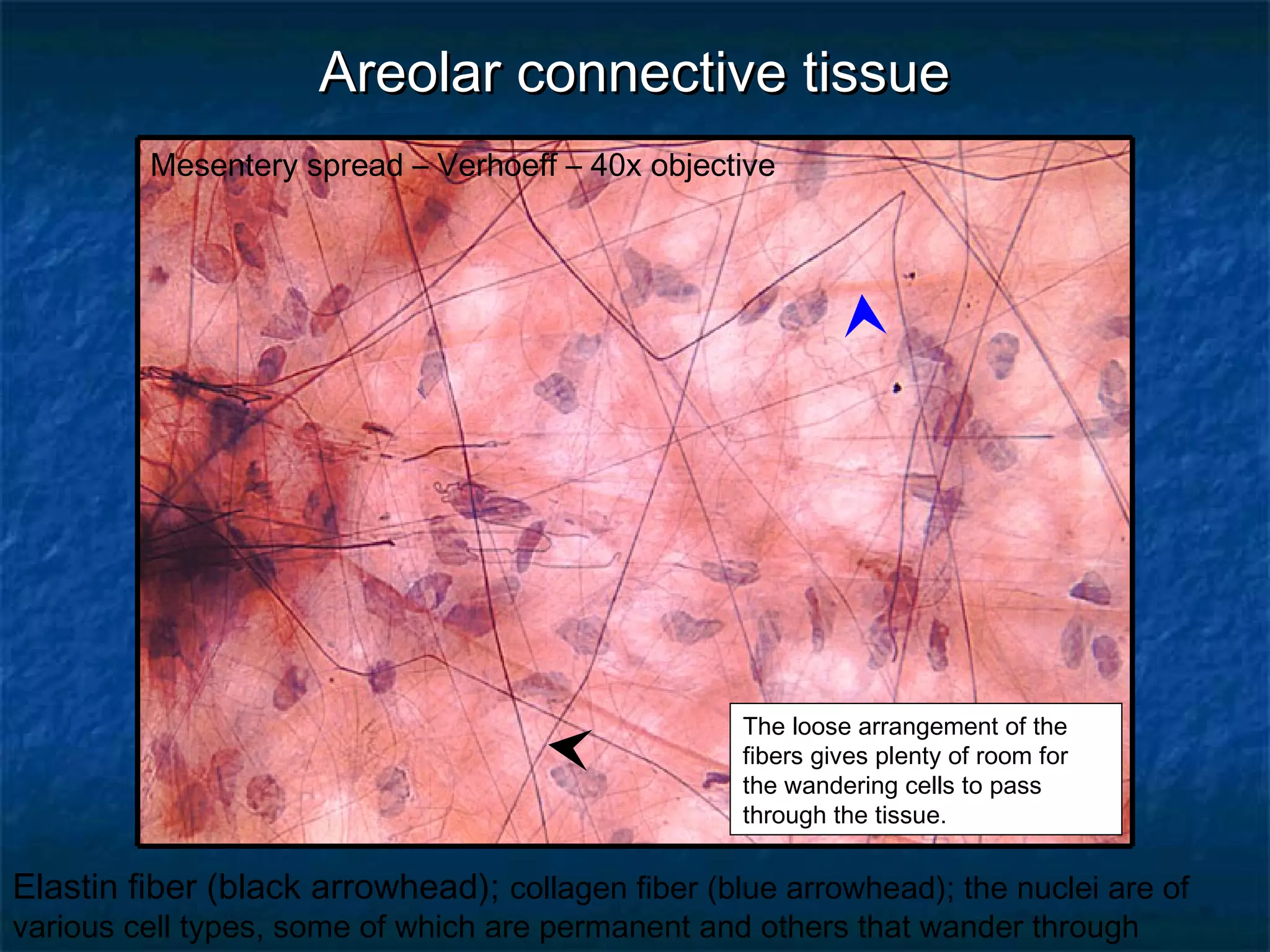
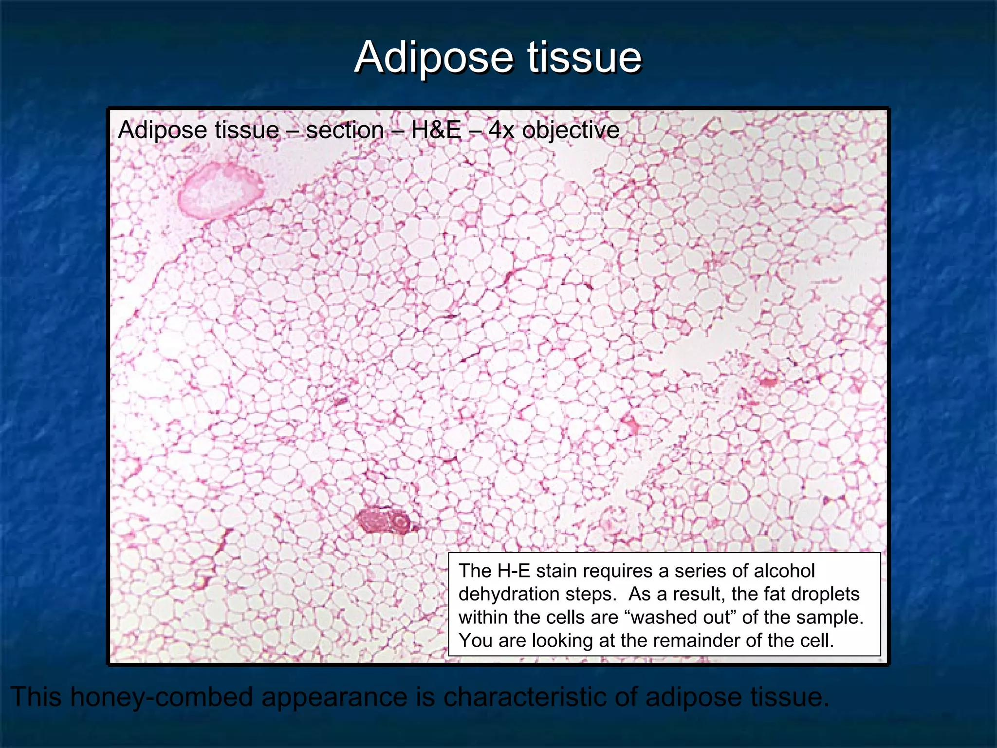
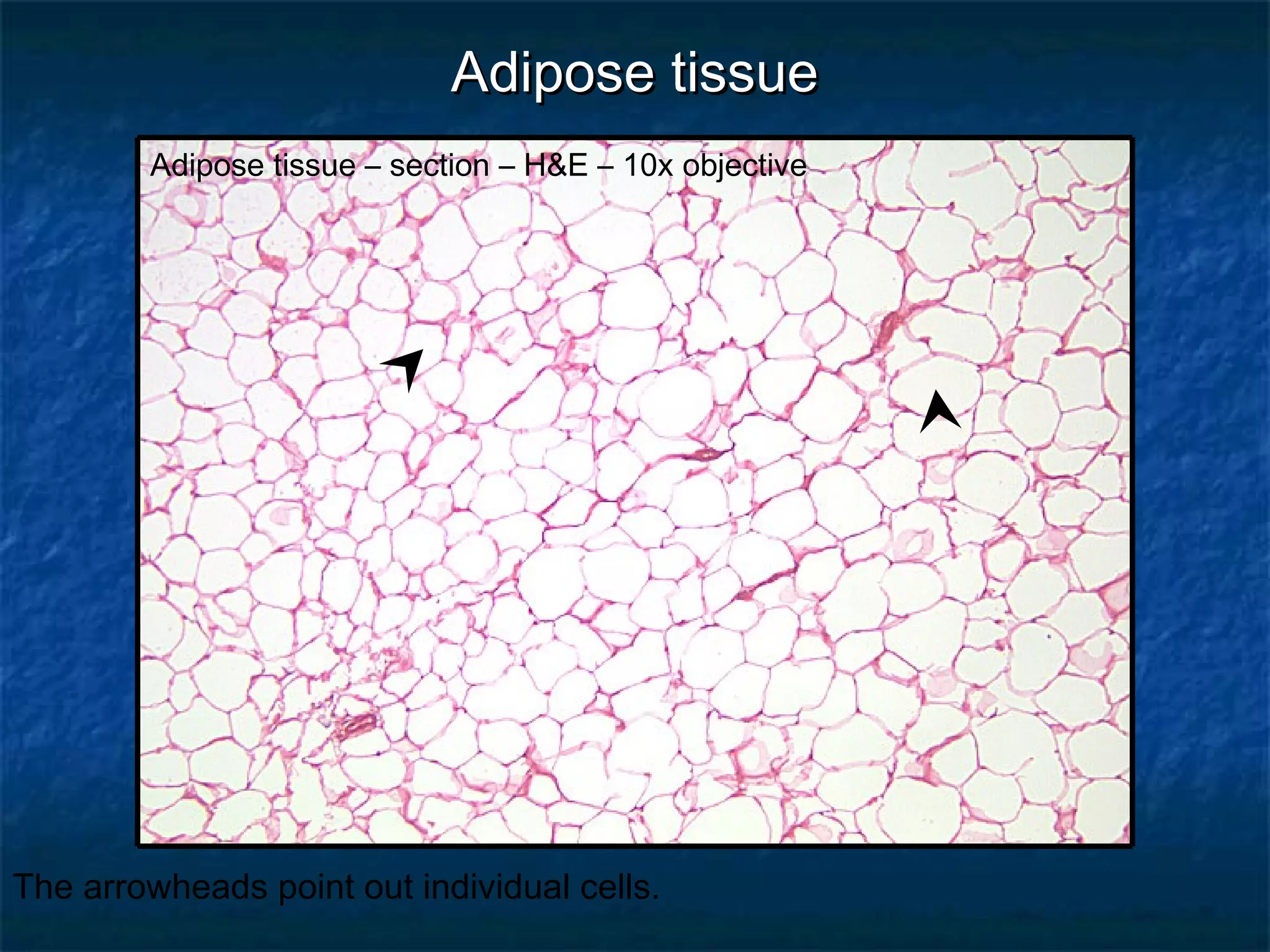
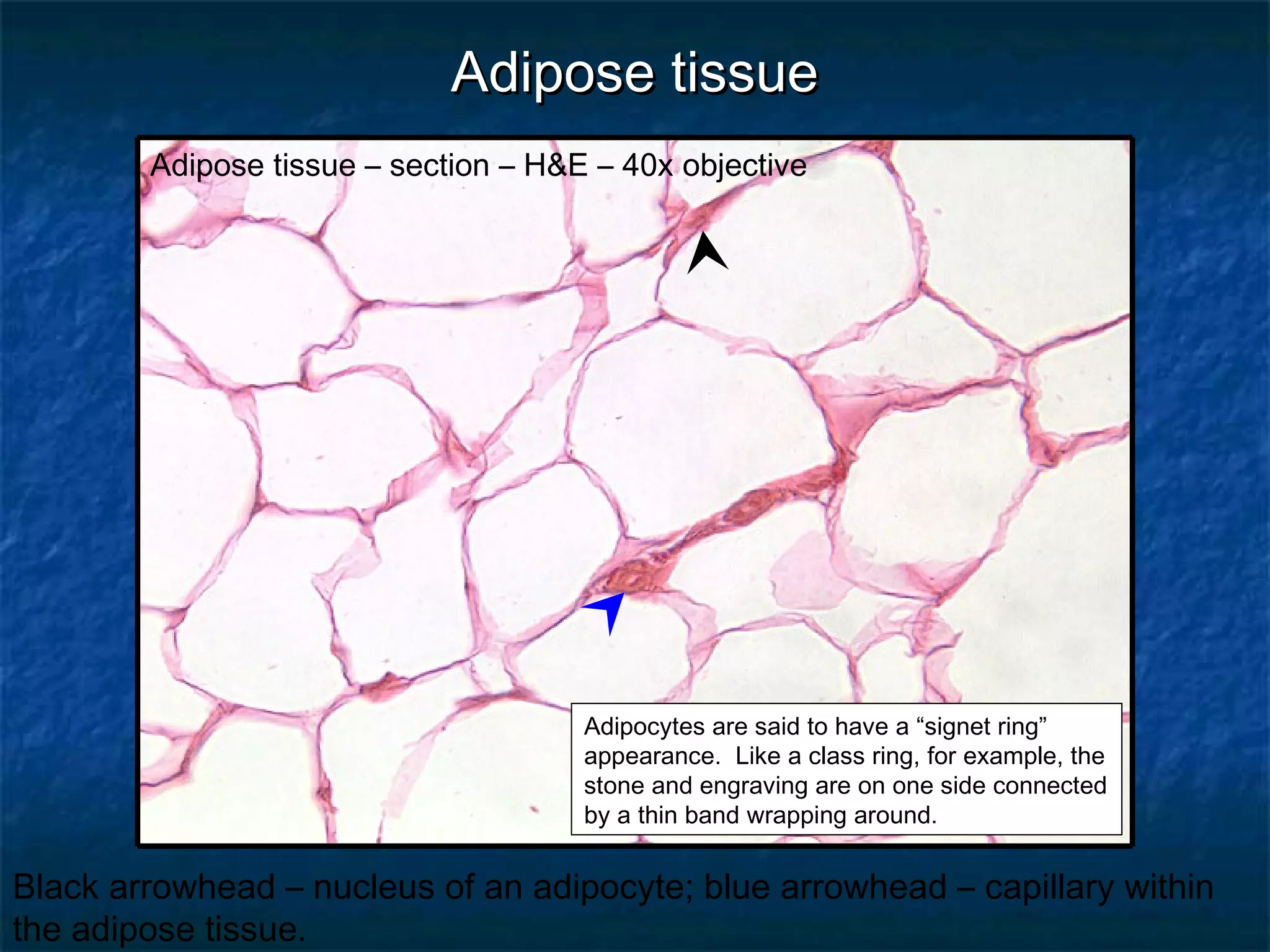
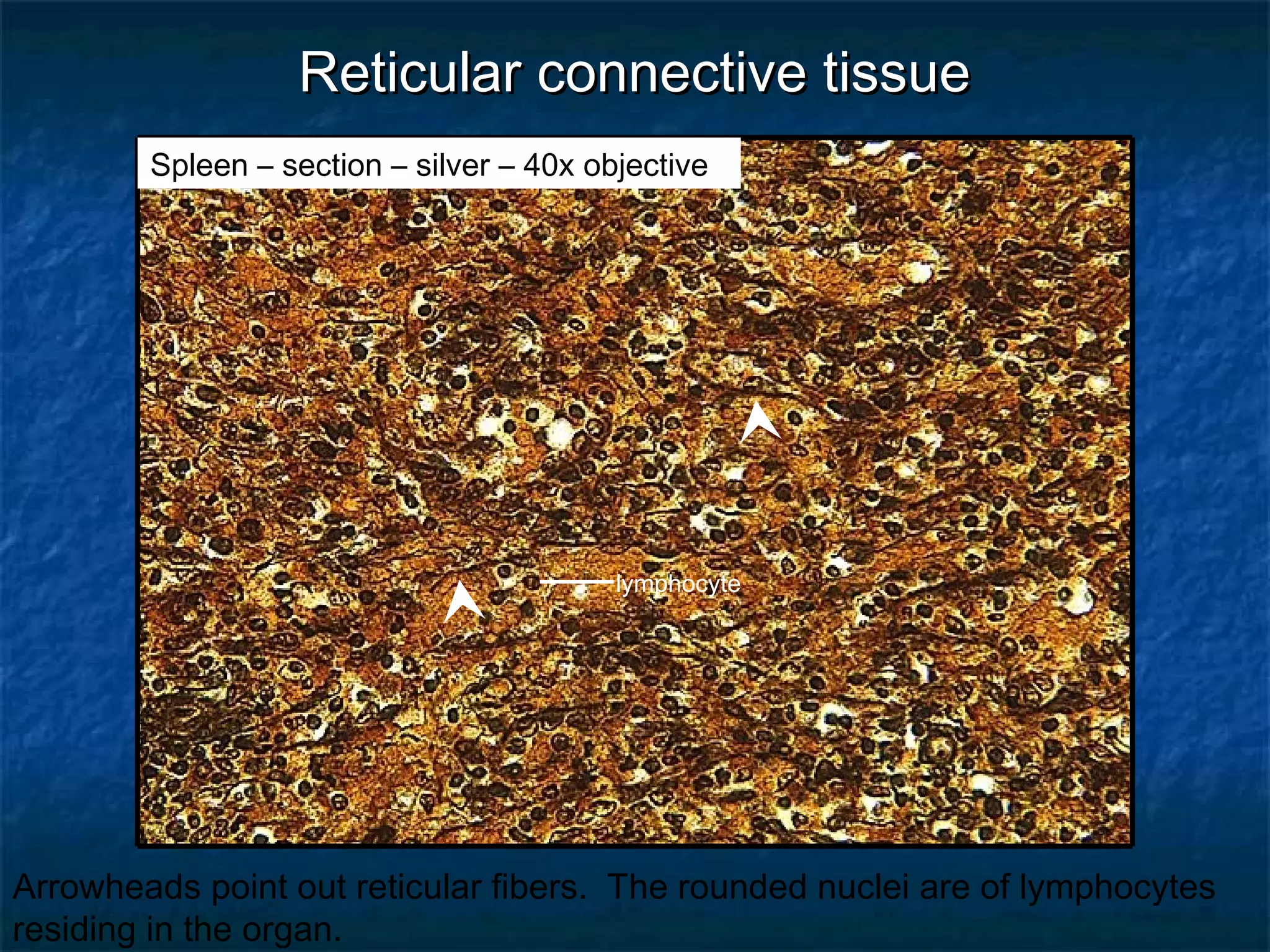
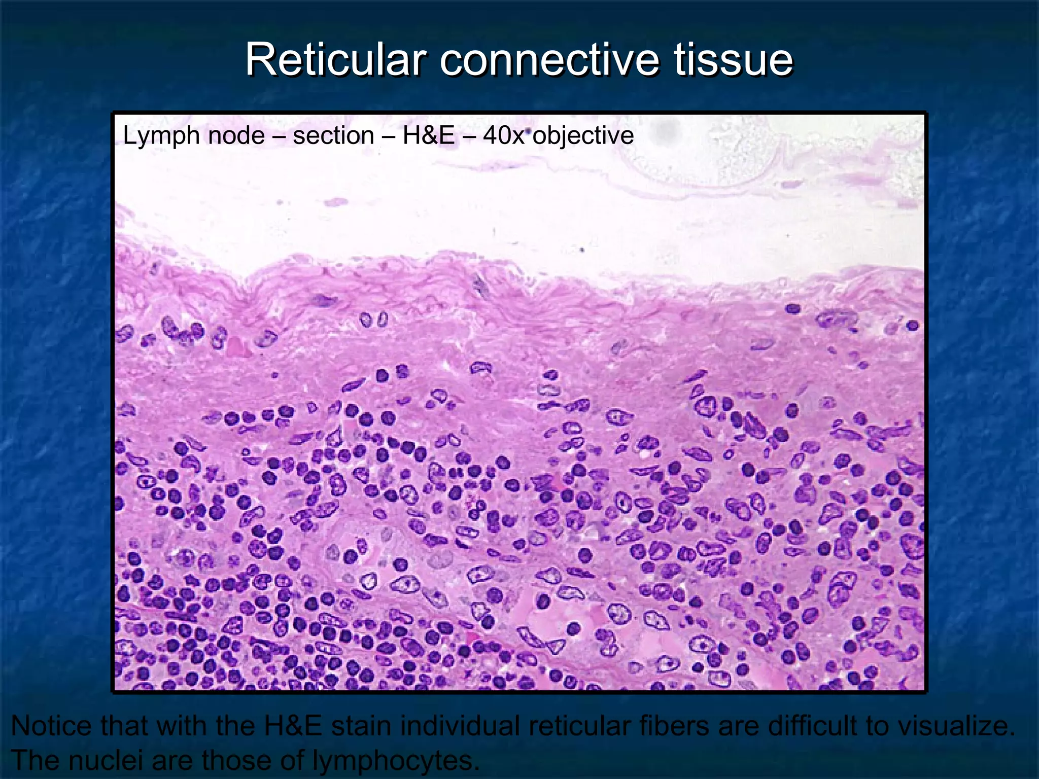
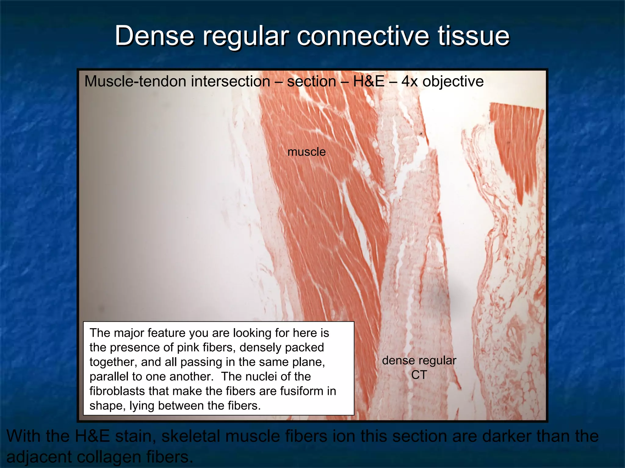
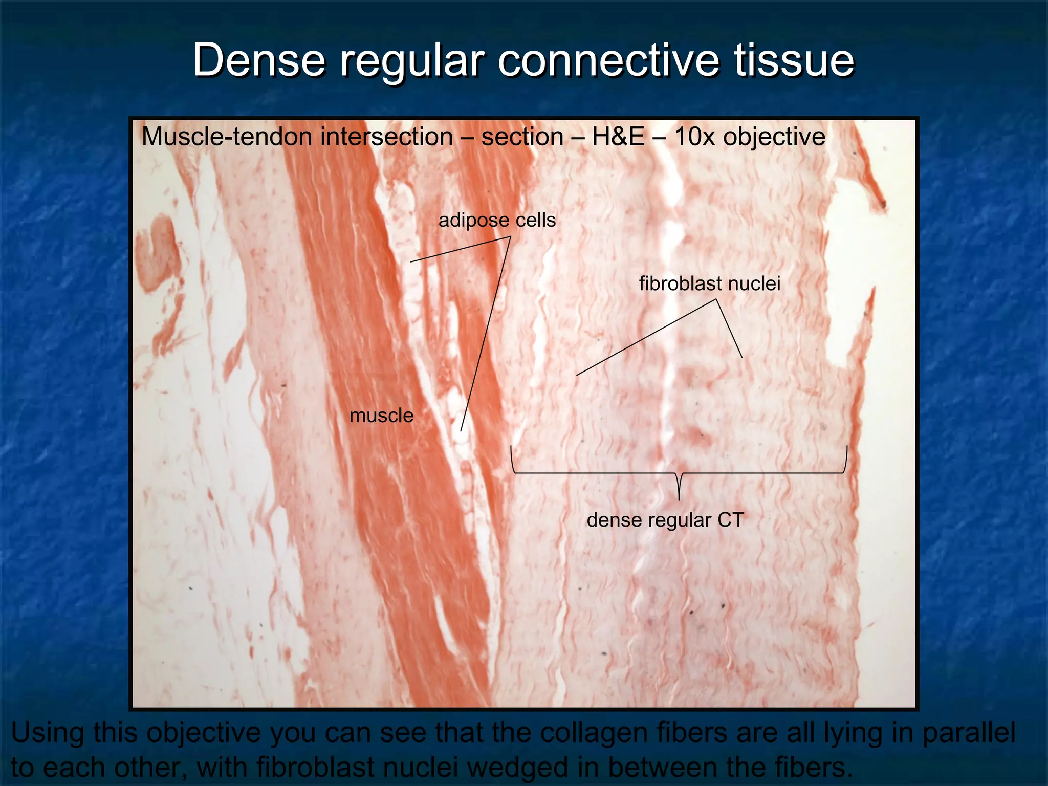
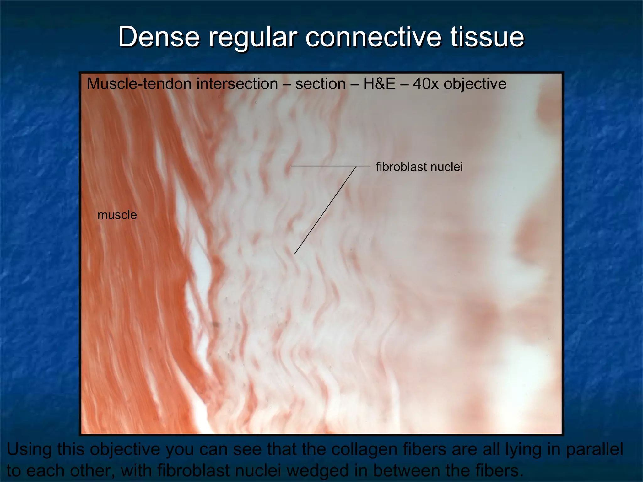
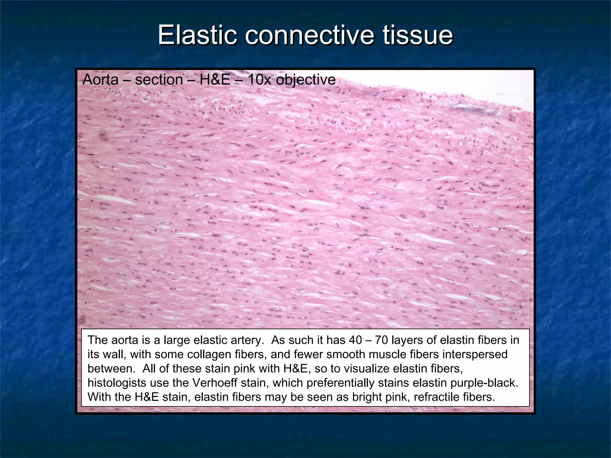
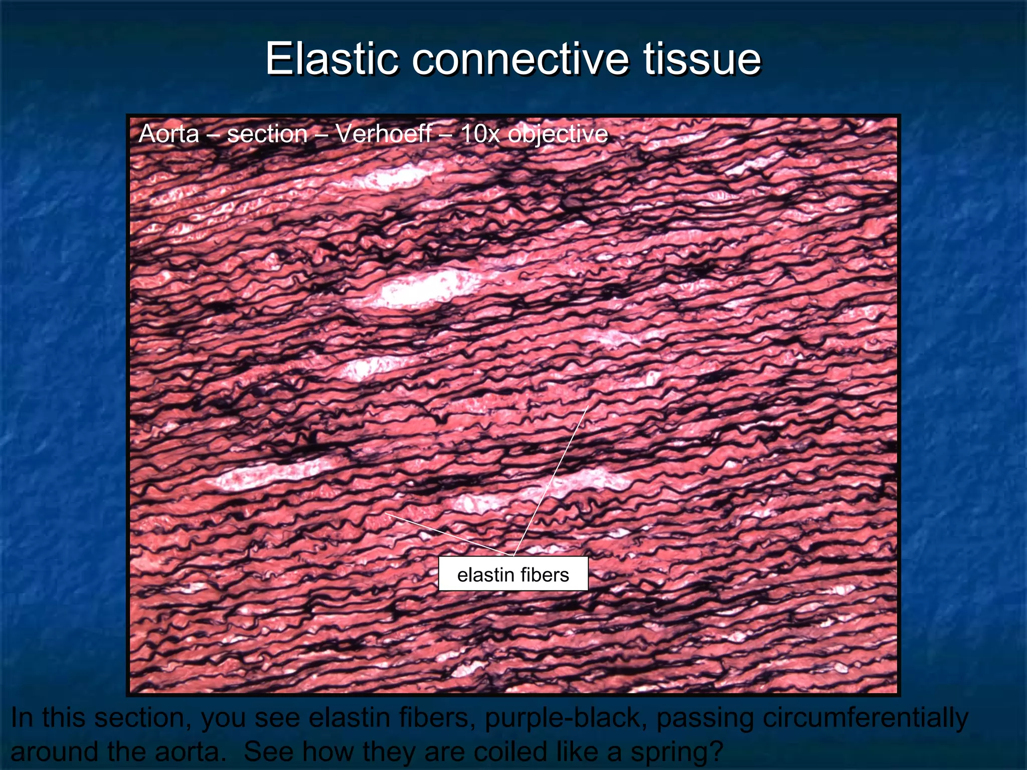
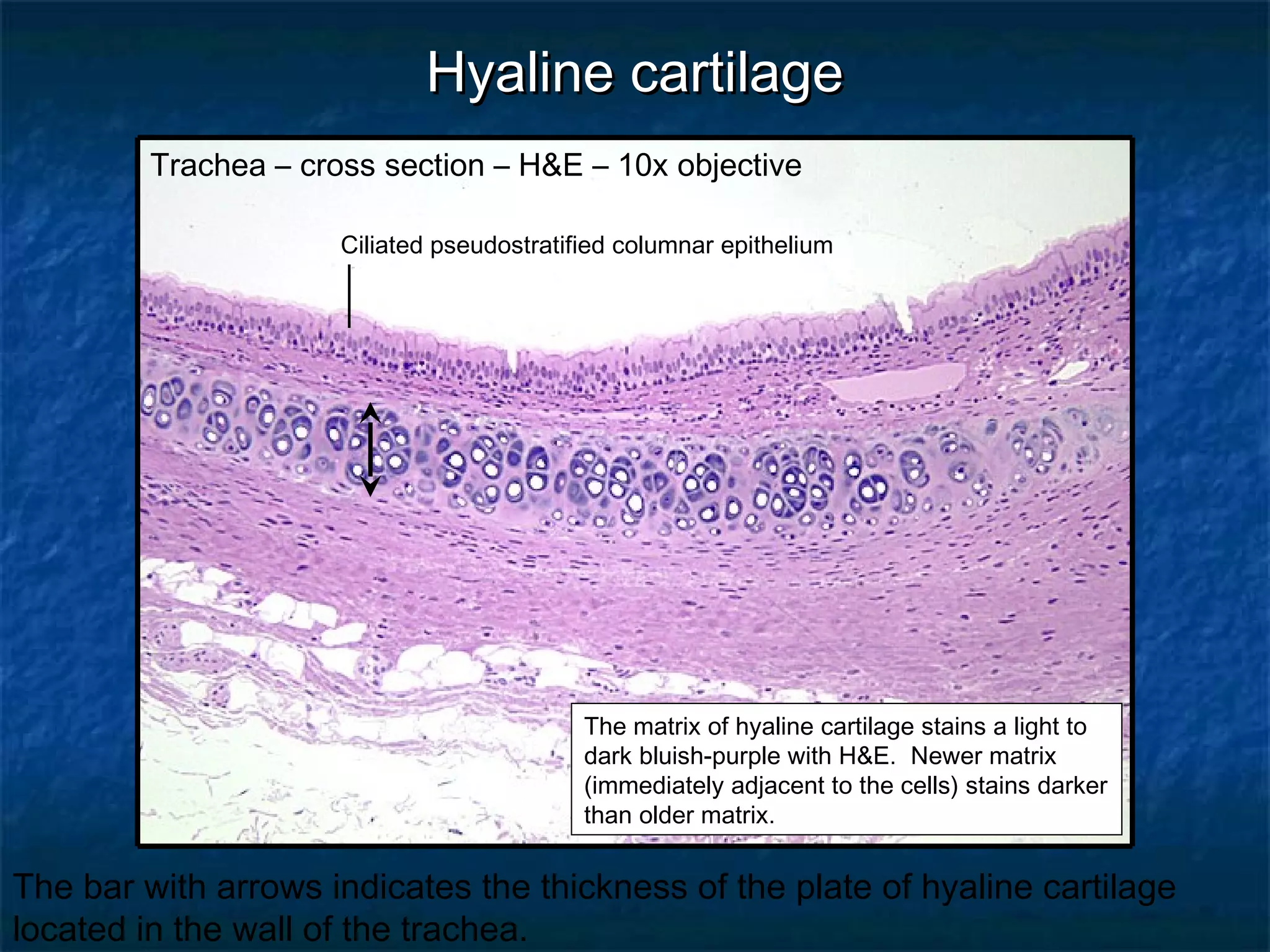
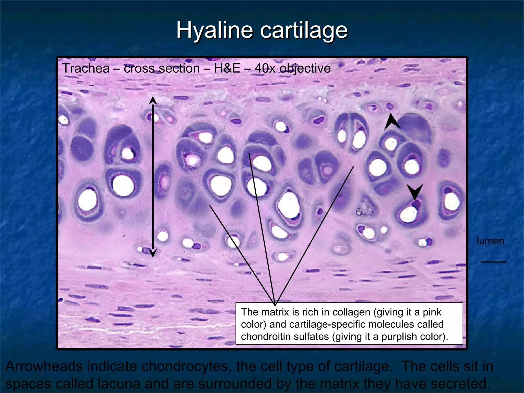
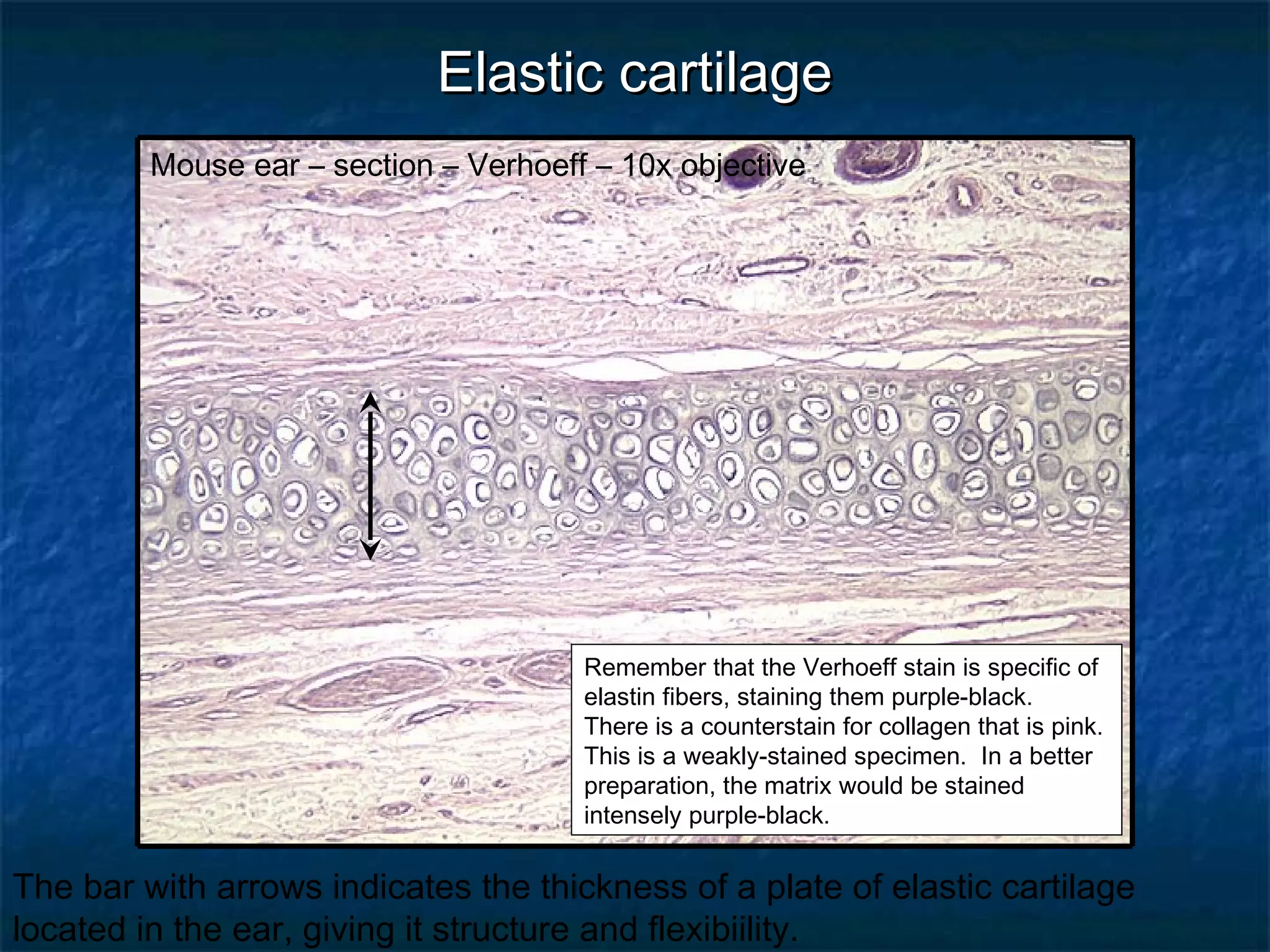
HHIISSTTOOLLOOGGYY RREEVVIIEEWW 
CCoonnnneeccttiivvee TTiissssuueess 
DDrr.. TTiimm BBaallllaarrdd 
DDeeppaarrttmmeenntt ooff BBiioollooggyy aanndd MMaarriinnee BBiioollooggyy
AArreeoollaarr ccoonnnneeccttiivvee ttiissssuuee 
Mesentery spread – Verhoeff – 4x objective 
Verhoeff’s stain is used to visualize both collagen and 
elastin fibers in the same sample. Collagen fibers are 
pink while elastin fibers are purple-black. 
This is a loose connective tissue. See how widely spread the fibers and cells 
are separated from one another.
AArreeoollaarr ccoonnnneeccttiivvee ttiissssuuee 
Mesentery spread – Verhoeff – 10x objective 
The spaces between fibers and 
cells are filled with water-based 
ground substance. 
Elastin fiber (black arrowhead); collagen fiber (blue arrowhead); the nuclei are of 
various cell types, some of which are permanent and others that wander through
AArreeoollaarr ccoonnnneeccttiivvee ttiissssuuee 
Mesentery spread – Verhoeff – 40x objective 
The loose arrangement of the 
fibers gives plenty of room for 
the wandering cells to pass 
through the tissue. 
Elastin fiber (black arrowhead); collagen fiber (blue arrowhead); the nuclei are of 
various cell types, some of which are permanent and others that wander through
AAddiippoossee ttiissssuuee 
Adipose tissue – section – H&E – 4x objective 
The H-E stain requires a series of alcohol 
dehydration steps. As a result, the fat droplets 
within the cells are “washed out” of the sample. 
You are looking at the remainder of the cell. 
This honey-combed appearance is characteristic of adipose tissue.
AAddiippoossee ttiissssuuee 
Adipose tissue – section – H&E – 10x objective 
The arrowheads point out individual cells.
AAddiippoossee ttiissssuuee 
Adipose tissue – section – H&E – 40x objective 
Adipocytes are said to have a “signet ring” 
appearance. Like a class ring, for example, the 
stone and engraving are on one side connected 
by a thin band wrapping around. 
Black arrowhead – nucleus of an adipocyte; blue arrowhead – capillary within 
the adipose tissue.
RReettiiccuullaarr ccoonnnneeccttiivvee ttiissssuuee 
Spleen – section – silver – 10x objective 
Reticular fibers are used to form the stroma or 
framework (think infrastructure) of some 
organs. For example, the spleen and lymph 
nodes are made by a meshwork of reticular 
fibers. Lymphocytes and macrophages then 
invade the organ and take up residence in the 
spaces. 
Reticular tissue consists of very small collagenous fibers. They are visualized 
as brownish-black fibers with the silver stain.
RReettiiccuullaarr ccoonnnneeccttiivvee ttiissssuuee 
Spleen – section – silver – 40x objective 
lymphocyte 
Arrowheads point out reticular fibers. The rounded nuclei are of lymphocytes 
residing in the organ.
RReettiiccuullaarr ccoonnnneeccttiivvee ttiissssuuee 
Lymph node – section – H&E – 40x objective 
Notice that with the H&E stain individual reticular fibers are difficult to visualize. 
The nuclei are those of lymphocytes.
DDeennssee rreegguullaarr ccoonnnneeccttiivvee ttiissssuuee 
Muscle-tendon intersection – section – H&E – 4x objective 
muscle 
dense regular 
CT 
The major feature you are looking for here is 
the presence of pink fibers, densely packed 
together, and all passing in the same plane, 
parallel to one another. The nuclei of the 
fibroblasts that make the fibers are fusiform in 
shape, lying between the fibers. 
With the H&E stain, skeletal muscle fibers ion this section are darker than the 
adjacent collagen fibers.
DDeennssee rreegguullaarr ccoonnnneeccttiivvee ttiissssuuee 
Muscle-tendon intersection – section – H&E – 10x objective 
muscle 
fibroblast nuclei 
dense regular CT 
adipose cells 
Using this objective you can see that the collagen fibers are all lying in parallel 
to each other, with fibroblast nuclei wedged in between the fibers.
DDeennssee rreegguullaarr ccoonnnneeccttiivvee ttiissssuuee 
Muscle-tendon intersection – section – H&E – 20x objective 
muscle 
fibroblast nuclei 
Using this objective you can see that the collagen fibers are all lying in parallel 
to each other, with fibroblast nuclei wedged in between the fibers.
DDeennssee rreegguullaarr ccoonnnneeccttiivvee ttiissssuuee 
Muscle-tendon intersection – section – H&E – 40x objective 
fibroblast nuclei 
bone 
collagen fibers 
Another example of dense regular connective tissue. In some of the following 
slides, look for it.
DDeennssee iirrrreegguullaarr ccoonnnneeccttiivvee ttiissssuuee 
Skin – section – H&E – 4x objective 
dermis 
epidermis 
The major feature you are looking for now is 
the presence of pink fibers, less densely 
packed together, and passing in multiple 
directions to resist tensile forces pulling from 
multiple directions. 
The dermis is where we go to see the features of dense irregular connective 
tissues. Look for collagen fibers passing in multiple directions.
DDeennssee iirrrreegguullaarr ccoonnnneeccttiivvee ttiissssuuee 
Skin – section – H&E – 10x objective 
epidermis 
Look at all the pink-stained collagen fibers. Can you see that they are passing 
in multiple directions?
DDeennssee iirrrreegguullaarr ccoonnnneeccttiivvee ttiissssuuee 
collagen fiber passing 
longitudinally 
Skin – section – H&E – 40x objective 
fibroblast nucleus 
collagen fiber cut obliquely 
collagen fiber passing out 
of the screen at you 
(in cross section) 
In this section, you see collagen fibers passing in multiple directions. Can you 
visualize how they appear in three-dimensions?
EEllaassttiicc ccoonnnneeccttiivvee ttiissssuuee 
Aorta – section – H&E – 10x objective 
The aorta is a large elastic artery. As such it has 40 – 70 layers of elastin fibers in 
its wall, with some collagen fibers, and fewer smooth muscle fibers interspersed 
between. All of these stain pink with H&E, so to visualize elastin fibers, 
histologists use the Verhoeff stain, which preferentially stains elastin purple-black. 
With the H&E stain, elastin fibers may be seen as bright pink, refractile fibers.
EEllaassttiicc ccoonnnneeccttiivvee ttiissssuuee 
Aorta – section – Verhoeff – 10x objective 
elastin fibers 
In this section, you see elastin fibers, purple-black, passing circumferentially 
around the aorta. See how they are coiled like a spring?
EEllaassttiicc ccoonnnneeccttiivvee ttiissssuuee 
Aorta – section – Verhoeff – 40x objective 
elastin fibers 
Since this section has been counterstained with H&E, you can also see 
collagen fibers and smooth muscle cells (pink), although you can’t differentiate 
them.
HHyyaalliinnee ccaarrttiillaaggee 
Trachea – cross section – H&E – 10x objective 
Ciliated pseudostratified columnar epithelium 
The matrix of hyaline cartilage stains a light to 
dark bluish-purple with H&E. Newer matrix 
(immediately adjacent to the cells) stains darker 
than older matrix. 
The bar with arrows indicates the thickness of the plate of hyaline cartilage 
located in the wall of the trachea.
lumen 
HHyyaalliinnee ccaarrttiillaaggee 
Trachea – cross section – H&E – 40x objective 
The matrix is rich in collagen (giving it a pink 
color) and cartilage-specific molecules called 
chondroitin sulfates (giving it a purplish color). 
Arrowheads indicate chondrocytes, the cell type of cartilage. The cells sit in 
spaces called lacuna and are surrounded by the matrix they have secreted.
EEllaassttiicc ccaarrttiillaaggee 
Mouse ear – section – Verhoeff – 10x objective 
Remember that the Verhoeff stain is specific of 
elastin fibers, staining them purple-black. 
There is a counterstain for collagen that is pink. 
This is a weakly-stained specimen. In a better 
preparation, the matrix would be stained 
intensely purple-black. 
The bar with arrows indicates the thickness of a plate of elastic cartilage 
located in the ear, giving it structure and flexibiility.
EEllaassttiicc ccaarrttiillaaggee 
Mouse ear – section – Verhoeff – 40x objective 
Chondrocytes in elastic cartilage retain a fairly 
high mitotic rate throughout life . As result, 
there are many more cells than you would see 
in hyaline cartilage. 
Chondrocyte within its lacuna (arrowhead). Although a weak stain, notice all of 
the dark-stained elastin fibers in the matrix.
FFiibbrrooccaarrttiillaaggee 
Pubic symphysis –section – Mallory’s – 4x objective 
All of the bluish fibers are collagen. Notice all of the rounded cells 
(chondrocytes) sitting inside lacunae.
FFiibbrrooccaarrttiillaaggee 
Pubic symphysis –section – Mallory’s – 10x objective 
All of the bluish fibers are collagen. Notice all of the rounded cells 
(chondrocytes) sitting inside lacunae (arrowheads).
FFiibbrrooccaarrttiillaaggee 
Pubic symphysis –section – Mallory’s – 40x objective 
lacuna 
Chondrocytes (arrowhead) within its lacuna; collagen fibers (arrow) 
end

Histology: Connective Tissues

  • 1.
    HHIISSTTOOLLOOGGYY RREEVVIIEEWW CCoonnnneeccttiivveeTTiissssuueess DDrr.. TTiimm BBaallllaarrdd DDeeppaarrttmmeenntt ooff BBiioollooggyy aanndd MMaarriinnee BBiioollooggyy
  • 2.
    AArreeoollaarr ccoonnnneeccttiivvee ttiissssuuee Mesentery spread – Verhoeff – 4x objective Verhoeff’s stain is used to visualize both collagen and elastin fibers in the same sample. Collagen fibers are pink while elastin fibers are purple-black. This is a loose connective tissue. See how widely spread the fibers and cells are separated from one another.
  • 3.
    AArreeoollaarr ccoonnnneeccttiivvee ttiissssuuee Mesentery spread – Verhoeff – 10x objective The spaces between fibers and cells are filled with water-based ground substance. Elastin fiber (black arrowhead); collagen fiber (blue arrowhead); the nuclei are of various cell types, some of which are permanent and others that wander through
  • 4.
    AArreeoollaarr ccoonnnneeccttiivvee ttiissssuuee Mesentery spread – Verhoeff – 40x objective The loose arrangement of the fibers gives plenty of room for the wandering cells to pass through the tissue. Elastin fiber (black arrowhead); collagen fiber (blue arrowhead); the nuclei are of various cell types, some of which are permanent and others that wander through
  • 5.
    AAddiippoossee ttiissssuuee Adiposetissue – section – H&E – 4x objective The H-E stain requires a series of alcohol dehydration steps. As a result, the fat droplets within the cells are “washed out” of the sample. You are looking at the remainder of the cell. This honey-combed appearance is characteristic of adipose tissue.
  • 6.
    AAddiippoossee ttiissssuuee Adiposetissue – section – H&E – 10x objective The arrowheads point out individual cells.
  • 7.
    AAddiippoossee ttiissssuuee Adiposetissue – section – H&E – 40x objective Adipocytes are said to have a “signet ring” appearance. Like a class ring, for example, the stone and engraving are on one side connected by a thin band wrapping around. Black arrowhead – nucleus of an adipocyte; blue arrowhead – capillary within the adipose tissue.
  • 8.
    RReettiiccuullaarr ccoonnnneeccttiivvee ttiissssuuee Spleen – section – silver – 10x objective Reticular fibers are used to form the stroma or framework (think infrastructure) of some organs. For example, the spleen and lymph nodes are made by a meshwork of reticular fibers. Lymphocytes and macrophages then invade the organ and take up residence in the spaces. Reticular tissue consists of very small collagenous fibers. They are visualized as brownish-black fibers with the silver stain.
  • 9.
    RReettiiccuullaarr ccoonnnneeccttiivvee ttiissssuuee Spleen – section – silver – 40x objective lymphocyte Arrowheads point out reticular fibers. The rounded nuclei are of lymphocytes residing in the organ.
  • 10.
    RReettiiccuullaarr ccoonnnneeccttiivvee ttiissssuuee Lymph node – section – H&E – 40x objective Notice that with the H&E stain individual reticular fibers are difficult to visualize. The nuclei are those of lymphocytes.
  • 11.
    DDeennssee rreegguullaarr ccoonnnneeccttiivveettiissssuuee Muscle-tendon intersection – section – H&E – 4x objective muscle dense regular CT The major feature you are looking for here is the presence of pink fibers, densely packed together, and all passing in the same plane, parallel to one another. The nuclei of the fibroblasts that make the fibers are fusiform in shape, lying between the fibers. With the H&E stain, skeletal muscle fibers ion this section are darker than the adjacent collagen fibers.
  • 12.
    DDeennssee rreegguullaarr ccoonnnneeccttiivveettiissssuuee Muscle-tendon intersection – section – H&E – 10x objective muscle fibroblast nuclei dense regular CT adipose cells Using this objective you can see that the collagen fibers are all lying in parallel to each other, with fibroblast nuclei wedged in between the fibers.
  • 13.
    DDeennssee rreegguullaarr ccoonnnneeccttiivveettiissssuuee Muscle-tendon intersection – section – H&E – 20x objective muscle fibroblast nuclei Using this objective you can see that the collagen fibers are all lying in parallel to each other, with fibroblast nuclei wedged in between the fibers.
  • 14.
    DDeennssee rreegguullaarr ccoonnnneeccttiivveettiissssuuee Muscle-tendon intersection – section – H&E – 40x objective muscle fibroblast nuclei Using this objective you can see that the collagen fibers are all lying in parallel to each other, with fibroblast nuclei wedged in between the fibers.
  • 15.
    DDeennssee rreegguullaarr ccoonnnneeccttiivveettiissssuuee Muscle-tendon intersection – section – H&E – 40x objective fibroblast nuclei bone collagen fibers Another example of dense regular connective tissue. In some of the following slides, look for it.
  • 16.
    DDeennssee iirrrreegguullaarr ccoonnnneeccttiivveettiissssuuee Skin – section – H&E – 4x objective dermis epidermis The major feature you are looking for now is the presence of pink fibers, less densely packed together, and passing in multiple directions to resist tensile forces pulling from multiple directions. The dermis is where we go to see the features of dense irregular connective tissues. Look for collagen fibers passing in multiple directions.
  • 17.
    DDeennssee iirrrreegguullaarr ccoonnnneeccttiivveettiissssuuee Skin – section – H&E – 10x objective epidermis Look at all the pink-stained collagen fibers. Can you see that they are passing in multiple directions?
  • 18.
    DDeennssee iirrrreegguullaarr ccoonnnneeccttiivveettiissssuuee collagen fiber passing longitudinally Skin – section – H&E – 40x objective fibroblast nucleus collagen fiber cut obliquely collagen fiber passing out of the screen at you (in cross section) In this section, you see collagen fibers passing in multiple directions. Can you visualize how they appear in three-dimensions?
  • 19.
    EEllaassttiicc ccoonnnneeccttiivvee ttiissssuuee Aorta – section – H&E – 10x objective The aorta is a large elastic artery. As such it has 40 – 70 layers of elastin fibers in its wall, with some collagen fibers, and fewer smooth muscle fibers interspersed between. All of these stain pink with H&E, so to visualize elastin fibers, histologists use the Verhoeff stain, which preferentially stains elastin purple-black. With the H&E stain, elastin fibers may be seen as bright pink, refractile fibers.
  • 20.
    EEllaassttiicc ccoonnnneeccttiivvee ttiissssuuee Aorta – section – Verhoeff – 10x objective elastin fibers In this section, you see elastin fibers, purple-black, passing circumferentially around the aorta. See how they are coiled like a spring?
  • 21.
    EEllaassttiicc ccoonnnneeccttiivvee ttiissssuuee Aorta – section – Verhoeff – 40x objective elastin fibers Since this section has been counterstained with H&E, you can also see collagen fibers and smooth muscle cells (pink), although you can’t differentiate them.
  • 22.
    HHyyaalliinnee ccaarrttiillaaggee Trachea– cross section – H&E – 10x objective Ciliated pseudostratified columnar epithelium The matrix of hyaline cartilage stains a light to dark bluish-purple with H&E. Newer matrix (immediately adjacent to the cells) stains darker than older matrix. The bar with arrows indicates the thickness of the plate of hyaline cartilage located in the wall of the trachea.
  • 23.
    lumen HHyyaalliinnee ccaarrttiillaaggee Trachea – cross section – H&E – 40x objective The matrix is rich in collagen (giving it a pink color) and cartilage-specific molecules called chondroitin sulfates (giving it a purplish color). Arrowheads indicate chondrocytes, the cell type of cartilage. The cells sit in spaces called lacuna and are surrounded by the matrix they have secreted.
  • 24.
    EEllaassttiicc ccaarrttiillaaggee Mouseear – section – Verhoeff – 10x objective Remember that the Verhoeff stain is specific of elastin fibers, staining them purple-black. There is a counterstain for collagen that is pink. This is a weakly-stained specimen. In a better preparation, the matrix would be stained intensely purple-black. The bar with arrows indicates the thickness of a plate of elastic cartilage located in the ear, giving it structure and flexibiility.
  • 25.
    EEllaassttiicc ccaarrttiillaaggee Mouseear – section – Verhoeff – 40x objective Chondrocytes in elastic cartilage retain a fairly high mitotic rate throughout life . As result, there are many more cells than you would see in hyaline cartilage. Chondrocyte within its lacuna (arrowhead). Although a weak stain, notice all of the dark-stained elastin fibers in the matrix.
  • 26.
    FFiibbrrooccaarrttiillaaggee Pubic symphysis–section – Mallory’s – 4x objective All of the bluish fibers are collagen. Notice all of the rounded cells (chondrocytes) sitting inside lacunae.
  • 27.
    FFiibbrrooccaarrttiillaaggee Pubic symphysis–section – Mallory’s – 10x objective All of the bluish fibers are collagen. Notice all of the rounded cells (chondrocytes) sitting inside lacunae (arrowheads).
  • 28.
    FFiibbrrooccaarrttiillaaggee Pubic symphysis–section – Mallory’s – 40x objective lacuna Chondrocytes (arrowhead) within its lacuna; collagen fibers (arrow) end