This document provides information on anatomy and physiology, including:
- Anatomy and physiology are closely integrated, with anatomical structures relating to physiological functions.
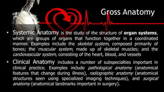
- Gross anatomy involves examining relatively large structures visible without a microscope, including surface, regional, systemic, clinical, developmental, and microscopic anatomy.
- Physiology is the study of the functions and workings of the body, including cell, organ, and systemic physiology as well as pathological physiology.
- The body is organized in a hierarchy from the chemical and cellular levels up through tissues, organs, organ systems, and the whole organism.
2. Anatomy and Physiology
• Anatomy and physiology are closely integrated,
both theoretically and practically. Anatomical information
provides clues about functions, and physiological
processes can be explained only in terms of the
underlying anatomy. This is a very important concept in
living systems: All specific functions are performed by
specific structures, and the form of a structure relates to
its function. This is known as the principle of
complementarity of structure and function.
3. Gross Anatomy
Gross anatomy, or macroscopic anatomy, involves
examining relatively large structures. Gross anatomy can be
seen without using a microscope and can involve the study
of anatomy by dissecting a cadaver.
• Surface Anatomy is the study of general form and superficial
(toward the body surface) markings.
• Regional Anatomy focuses on the anatomical organization of
specific areas of the body, such as the head, neck, or trunk. Many
advanced courses in anatomy stress a regional approach, because it
emphasizes the spatial relationships among structures already familiar
to students.
4. Gross Anatomy
• Systemic Anatomy is the study of the structure of organ systems,
which are groups of organs that function together in a coordinated
manner. Examples include the skeletal system, composed primarily of
bones; the muscular system, made up of skeletal muscles; and the
cardiovascular system, consisting of the heart, blood, and vessels
• Clinical Anatomy includes a number of subspecialties important in
clinical practice. Examples include pathological anatomy (anatomical
features that change during illness), radiographic anatomy (anatomical
structures seen using specialized imaging techniques), and surgical
anatomy (anatomical landmarks important in surgery).
5. Gross Anatomy
Developmental Anatomy describes the changes in form that
take place between conception and adulthood. The techniques of
developmental anatomists are similar to those used in gross anatomy
and in microscopic anatomy because developmental anatomy considers
anatomical structures over a broad range of sizes—from a single cell to
an adult human. The most extensive structural changes take place
during the first two months of development. The study of these early
developmental processes is called embryology.
6. Microscopic Anatomy
Microscopic anatomy deals with structures that we
cannot see without magnification. The boundaries of
microscopic anatomy are set by the limits of the equipment
we use. With a dissecting microscope you can see tissue
structure. With a light microscope, you can see basic
details of cell structure. And with an electron microscope,
you can see individual molecules that are only a few
nanometers (billionths of a meter) across.
7. Microscopic Anatomy
• Cytology is the study of the internal structure of individual
cells, the simplest units of life. Cells are made up of
chemical substances in various combinations, and our lives
depend on the chemical processes taking place in the
trillions of cells in the body.
• Histology is the examination of tissues—groups of
specialized cells and cell products that work together to
perform specific functions. Tissues combine to form organs,
such as the heart, kidney, liver, or brain, each with specific
functions.
8. Physiology
Human physiology is the study of the functions, or workings, of the
human body. These functions are complex processes and much more difficult to
examine than most anatomical structures.
• Cell Physiology the study of the functions of cells, is the
cornerstone of human physiology. Cell physiology looks at events
involving the atoms and molecules important to life. It includes both
chemical processes within cells and chemical interactions among cells.
• Organ Physiology is the study of the function of specific
organs. An example is cardiac physiology, the study of heart
function—how the heart works.
9. Physiology
• Systemic Physiology includes all aspects of the
functioning of specific organ systems. Cardiovascular physiology,
respiratory physiology, and reproductive physiology are examples.
• Pathological Physiology the study of the effects of
diseases on organ functions or system functions. Modern medicine
depends on an understanding of both normal physiology and
pathological physiology.
13. Levels of Organization
• The Chemical Level. Atoms are the smallest stable units of
matter. They can combine to form molecules with complex
shapes. The atomic components and unique three dimensional
shape of a particular molecule determine its function. For
example, complex protein molecules form filaments that produce
the contractions of muscle cells in the heart.
• The Cellular Level. Cells are the smallest living units in the body.
Complex molecules can form various types of larger structures
called organelles. Each organelle has a specific function in a cell.
Energy-producing organelles provide the energy needed for heart
muscle cell contractions.
14. Levels of Organization
• The Tissue Level. A tissue is a group of cells working
together to perform one or more specific functions.
Heart muscle cells, also called cardiac muscle cells
(cardium, heart), interact with other types of cells and
with materials outside the cell to form cardiac muscle
tissue.
• The Organ Level. Organs are made of two or more
tissues working together to perform specific functions.
Layers of cardiac muscle tissue, in combination with
another type of tissue called connective tissue, form
the bulk of the wall of the heart, which is a hollow,
15. • The Organ System Level. A group of organs interacting to
perform a particular function forms an organ system. Each
time it contracts, the heart pushes blood into a network of
blood vessels. Together, the heart, blood, and blood vessels
make up the cardiovascular system, one of 11 organ systems
in the body. This system functions to distribute oxygen and
nutrients throughout the body.
• The Organism Level. An organism—in this case, an
individual human—is the highest level of organization that
we consider. All of the body’s organ systems must work
together to maintain the life and health of the organism.
Levels of
Organization
17. Main Subdivisions of the Human
Body
The Head
The
Thorax
The
Abdomen
The Pelvis
The Trunk
Limbs/
Extremities
Arm Forearm
Thigh Leg Great Toe
18. The Head
Head, in human anatomy, the upper
portion of the body, consisting of
the skull with its coverings and contents,
including the lower jaw. It is attached to
the spinal column by way of the first
cervical vertebra, the atlas, and connected
with the trunk of the body by the
muscles, blood vessels, and nerves
that constitute the neck. The term also is
used to describe the anterior or fore part of
animals other than humans
20. The Abdomen
• The abdomen refers to the region
between the pelvis (pelvic brim) and the
thorax (thoracic diaphragm) in
vertebrates, including humans.
• The abdomen functions to house the
digestive system and provides muscles
essential for posture, balance, and
breathing.
21. The Pelvis
• Pelvis, also called bony
pelvis or pelvic girdle, in
human anatomy, basin-shaped
complex of bones that connects the
trunk and the legs, supports and
balances the trunk, and contains
and supports the intestines,
the urinary bladder, and the internal
sex organs.
22. The Trunk
• In our body, the Trunk Region (Torso), an
anatomical term for the central part of the body, is a
combination of both the thoracic region (chest),
including the mammary region (breasts), and
the abdomen region (belly region) including
the naval (umbilicus region), coxal region, and pubic
region.
In our body, the Trunk Region (Torso) as an
anatomical term for the central part of the body,
including the thoracic region (chest) and abdomen
region (belly region), from which extends (but are
not included) the neck, the upper limbs and
the lower limbs. *
23. Limbs/
Extremities
• ‘The limbs’ describes the
anatomy of the upper limb—
divided into the arm between
shoulder and elbow; the
forearm between elbow and
wrist; and the hand below the
wrist—and the lower limb,
which is divided into the thigh
between hip and knee; the leg
between knee and ankle; and
the foot below the ankle.
25. Superficial Anatomy
• Superficial anatomy involves
locating structures on or near the
body surface. A familiarity with
anatomical landmarks (structures
that can be felt or palpated),
anatomical regions (specific areas
used for reference purposes), and
terms for anatomical directions will
make the material in subsequent
chapters easier to understand.
26. Sectional Anatomy
• Sometimes the only way to understand the relationships among the
parts of a three-dimensional object is to slice through it and look at the
internal organization. A slice through a threedimensional object is called
a section.
• An understanding of sectional views is particularly important now that
imaging techniques enable us to see inside the living body. These views
are sometimes difficult to interpret, but it is worth spending the time
required to understand what they show. Once you are able to interpret
sectional views, you will have a good mental model for studying the
anatomy and physiology of a particular region or system. Radiologists
and other medical professionals responsible for interpreting medical
scans spend much of their time analyzing sectional views of the body
27. Basic Anatomical Terms
• Anterior and posterior, which describe structures at the front (anterior) and back
(posterior) of the body. For example, the toes are anterior to the heel, and the
popliteus is posterior to the patella.
• Superior and inferior, which describe a position above (superior) or below
(inferior) another part of the body. For example, the orbits are superior to the oris,
and the pelvis is inferior to the abdomen.
• Proximal and distal, which describe a position that is closer (proximal) or further
(distal) from the trunk of the body. For example, the shoulder is proximal to the
arm, and the foot is distal to the knee.
• Superficial and deep, which describe structures that are closer to (superficial) or
further from (deep) the surface of the body. For example, the skin is superficial to
the bones, and the brain is deep to the skull. Sometimes profound is used
synonymously with deep.
28. Basic Anatomical Terms
• Medial and lateral, which describe a position that is closer to
(medial) or further from (lateral) the midline of the body. For example,
the nose is medial to the eyes, and the thumb is lateral to the other
fingers.
• Ventral and Dorsal, which describe structures derived from the front
(ventral) and back (dorsal) of the embryo, before limb rotation.
• Cranial and caudal, which describe structures close to the top of the
skull (cranial), and towards the bottom of the body (caudal).
• Occasionally, sinister for left, and dexter for right are used.
• Paired, referring to a structure that is present on both sides of the
body. For example, the hands are paired structures.
30. Anatomical Landmarks
• The standard anatomical reference for the human form is the
anatomical position. When the body is in this position, the
hands are at the sides with the palms facing forward, and
the feet are together.
• Anatomical position is very important because the relations
of all structures are described as presumed to be in
anatomical position. In standard anatomical position, the
human body is standing erect and at rest. Unlike the
situation in other vertebrates, the limbs are placed in
positions reminiscent of the supine position imposed on
cadavers during autopsy.
31.
32.
33.
34. • Supine position: In this position, the body is lying down with face
pointing upwards. All the remaining positions are similar to anatomical
position with the only difference of being in a horizontal plane rather
than a vertical plane.
35. • Prone position: This is the position in which the back of the
body is directed upwards. The body lies in a horizontal plane
with face directed downwards.
36. • Lithotomy position: In
this position the body is
lying in a supine with hips
and knees fully extended.
The feet are strapped in
position to support the
flexed knees and hips.
38. Anatomy is often described in planes, referring to two-
dimensional sections of the body. A section is a two-dimensional
surface of a three-dimensional structure that has been cut. A
plane is an imaginary two-dimensional surface that passes
through the body. Three planes are commonly referred to in
anatomy and medicine:
1. The sagittal plane is the plane that divides the body or an
organ vertically into right and left sides. If this vertical plane
runs directly down the middle of the body, it is called the
midsagittal or median plane. If it divides the body into
unequal right and left sides, it is called a parasagittal plane, or
less commonly a longitudinal section.
Planes and Sections
39. 2. The frontal plane is the plane that divides the body
or an organ into an anterior (front) portion and a
posterior (rear) portion. The frontal plane is often
referred to as a coronal plane, following Latin corona,
which means "crown".
3. The transverse plane is the plane that divides the
body or organ horizontally into upper and lower
portions. Transverse planes produce images referred to
as cross sections.
40.
41. Anatomical Directions
Sectional Anatomy introduces the main directional terms and
some examples of their use. There are many different terms,
and some can be used interchangeably. For example,
anterior refers to the front of the body when viewed in the
anatomical position. In humans, this term is equivalent to
ventral, which refers to the belly. Posterior refers to the back
of the body; this term is equivalent to dorsal. When reading
anatomical descriptions, remember that the terms left and
right always refer to the left and right sides of the subject,
not of the observer.
44. Joints, especially synovial joints allow the
body a tremendous range of movements.
Each movement at a synovial joint results
from the contraction or relaxation of the
muscles that are attached to the bones on
either side of the articulation. The type of
movement that can be produced at a
synovial joint is determined by its structural
type.
Movement types are generally paired, with
one being the opposite of the other. Body
movements are always described in relation
to the anatomical position of the body:
upright stance, with upper limbs to the side
of body and palms facing forward.
45. 1. Flexion and Extension, which refer to a movement that decreases
(flexion) or increases (extension) the angle between body parts. For example,
when standing up, the knees are extended.
2. Abduction and adduction refers to a motion that pulls a structure away
from (abduction) or towards (adduction) the midline of the body or limb. For
example, a star jump requires the legs to be abducted.
3. Internal rotation (or medial rotation) and External rotation (or lateral
rotation) refers to rotation towards (internal) or away from (external) the center
of the body. For example, the asana posture in yoga requires the legs to be
externally rotated.[citation needed]
4. Elevation and Depression refer to movement in a superior (elevation) or
inferior (depression) direction. Primarily refers to movements involving the
scapula and mandible.
General Motions
46.
47.
48. Elevation refers to movement of a body part
in a superior direction, or moving upward
Depression refers to movement of a body
part in an inferior direction or moving
downward.
In anatomy, elevation and depression most
commonly describe movements of the
mandible (lower jaw) or scapulae (shoulder
blades) within the frontal plane. When you
move your lower jaw (mandible) in a
downward direction, depression occurs. When
you move your mandible upward, elevation
occurs.
Elevation and
Depression
49. • Protraction moves the scapula
forward (anteriorly) and toward
the side of the body (laterally) in
an anterolateral direction.
• Retraction is the opposite
movement. It causes the shoulder
blades to move back (posteriorly)
and toward the body’s midline
(medially). This movement is
known as
a posteromedial movement.
Protraction and
Refraction
50. Special Motions of the Hands And Feet
1. Dorsiflexion and Plantarflexion refers to flexion (dorsiflexion) or
extension of the foot at the ankle. For example, plantarflexion occurs
when pressing the brake pedal of a car.
2. Palmarflexion and dorsiflexion refer to movement of the flexion
(palmarflexion) or extension (dorsiflexion) of the hand at the wrist. For
example, prayer is often conducted with the hands dorsiflexed.
3. Pronation and Supination refer to rotation of the forearm or foot so
that in the anatomical position the palm or sole is facing anteriorly
(supination) or posteriorly (pronation) rotation of the forearm. For
example, a person skiing must pronate their arms in order to grasp the
skis.
4. Eversion and Inversion refer to movements that tilt the sole of the
foot away from (eversion) or towards (inversion) the midline of the body.
51.
52. Opposition and
Reposition
• Opposition of the thumb occurs
when the tip of the thumb comes to
meet (and oppose) the tip of another
finger from the same hand. A super
easy way to remember this is that
you’ve probably heard someone say
that humans have opposable
thumbs. Opposition is the special
movement of our opposable thumbs.
• Reposition is the opposite action of
opposition. During reposition, the
thumb and finger return to their
original position.
53. Anatomical Terms For Describing
Muscles
• Muscle action that moves the axial skeleton work over a
joint with an origin and insertion of the muscle on
respective side. The insertion is on the bone deemed to
move towards the origin during muscle contraction.
Muscles are often present that engage in several actions
of the joint; able to perform for example both flexion and
extension of the forearm as in the biceps and triceps
respectively. This is not only to be able to revert actions
of muscles, but also brings on stability of the actions
though muscle co-activation.
54. Anatomical Terms For Describing
Muscles
1. Origin: The relatively fixed end of muscle during natural
movements of the muscle
2. Insertion: The relatively mobile end of the muscle during
natural movements of the muscle
3. Belly: The fat fleshy part of the muscle which is contractile
in function
4. Tendon: The fibrous and non-contractile part of the
muscle which attaches muscle to the bone.
5. Aponeurosis: It is a flattened tendon arising from the
connective tissues around the muscle.
57. • The muscle performing an action is the agonist, while the
muscle which contraction brings about an opposite action
is the antagonist. For example, an extension of the lower
arm is performed by the triceps as the agonist and the
biceps as the antagonist (which contraction will perform
flexion over the same joint). Muscles that work together
to perform the same action are called synergists.
58.
59.
60.
61. Anatomical Regions
• Clinicians refer to four abdominopelvic quadrants formed by a
pair of imaginary perpendicular lines that intersect at the umbilicus
(navel). This simple method provides useful references for the
description of aches, pains, and injuries. The location can help the
clinician determine the possible cause. For example, tenderness in
the right lower quadrant (RLQ) is a symptom of appendicitis.
Tenderness in the right upper quadrant (RUQ), however, may
indicate gallbladder or liver problems.
• Anatomists prefer more precise terms to describe the location and
orientation of internal organs. They recognize nine
abdominopelvic regions shows the relationships among
quadrants, regions, and internal organs.
66. Cavities of the Body
The cavities of the body house the internal organs, which
commonly referred to as the viscera. The two main body
cavities are the larger ventral (anterior) and the smaller, dorsal
(posterior) body cavity.
1. The ventral body cavity constitutes the thoracic cavity and
the abdomino-pelvic body cavity.
2. The Thoracic cavity houses lung and heart. It is protected
by the rib cage & associated musculature and the sternum
anteriorly. It consists of the right and left pleural cavities and
mediastinum (the portion of tissues and organs that separates
the left and right lung).
67. 3. Abdomino-pelvic Cavity extends from the diaphragm
inferior to the floor of the pelvis. It is divided into superior
abdominal and inferior pelvic cavity by imaginary line passing at
upper pelvis.
4. Abdominal cavity contains the stomach, intestine, liver,
spleen and gallbladder.
5. The pelvic cavity contains urinary bladder, rectum, and
portions of the reproductive organs
6. The dorsal body cavity constitutes the cephalic cavity
containing brain and the vertebral canal containing the spinal
cord.
Editor's Notes
human body, the physical substance of the human organism, composed of living cells and extracellular materials and organized into tissues, organs, and systems
These terms refer to movements that are regarded as unique to the hands and feet: