Downloaded 109 times
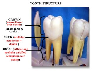
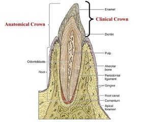
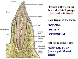
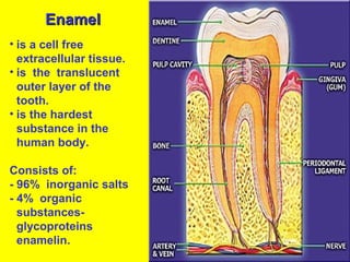
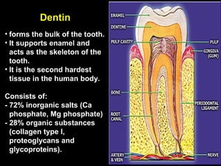
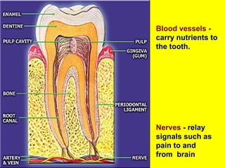
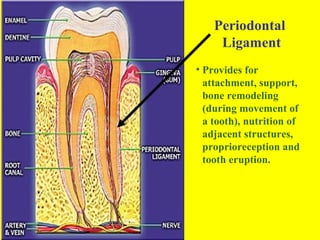
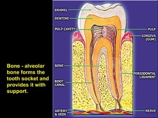
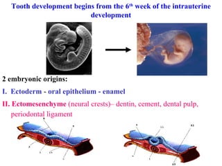
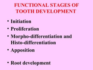
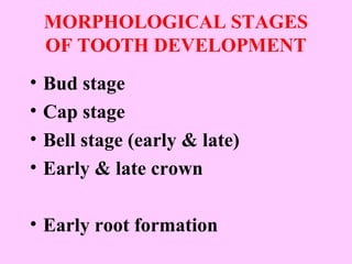
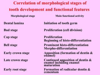
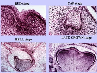
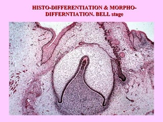

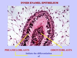
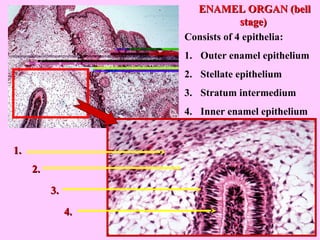
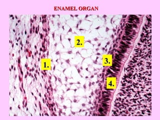
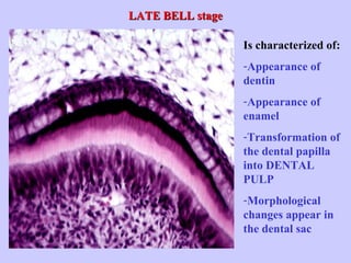
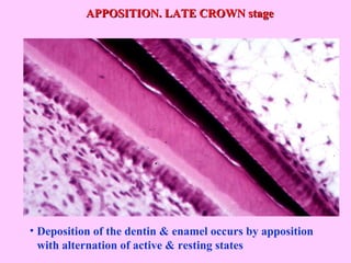
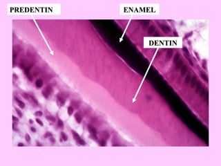
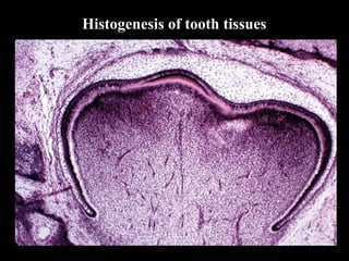
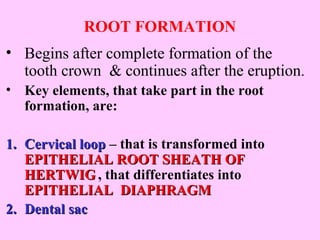


This document provides information about tooth structure and development. It discusses the hard tissues that make up teeth (enamel, dentin, cementum) and the soft dental pulp tissue. It describes the stages of tooth development from the bud stage to root formation. Key events include the differentiation of enamel-forming ameloblasts and dentin-forming odontoblasts during the bell stage, and the deposition of enamel and dentin layers during the late crown stage. The root develops after crown formation is complete.


