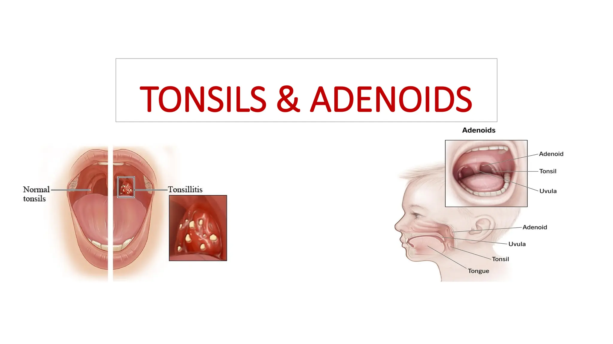
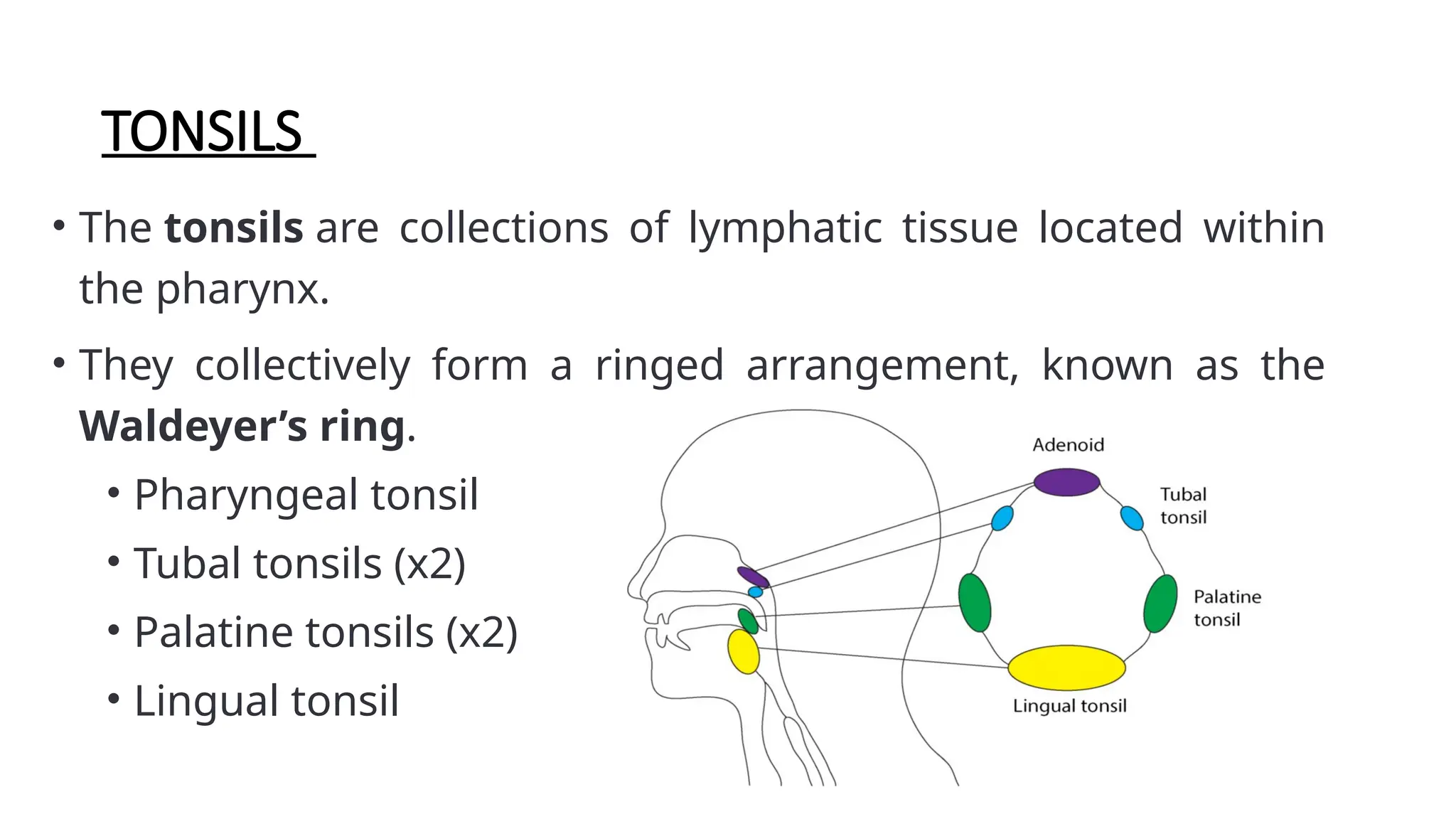
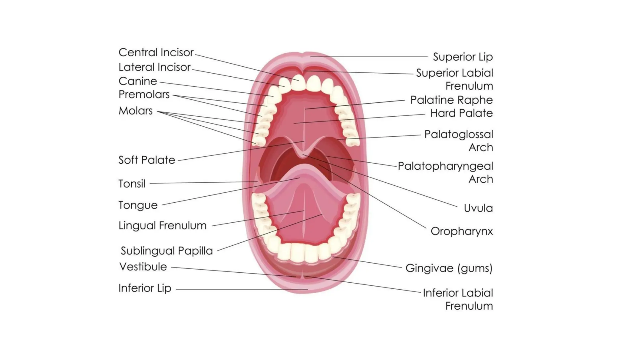
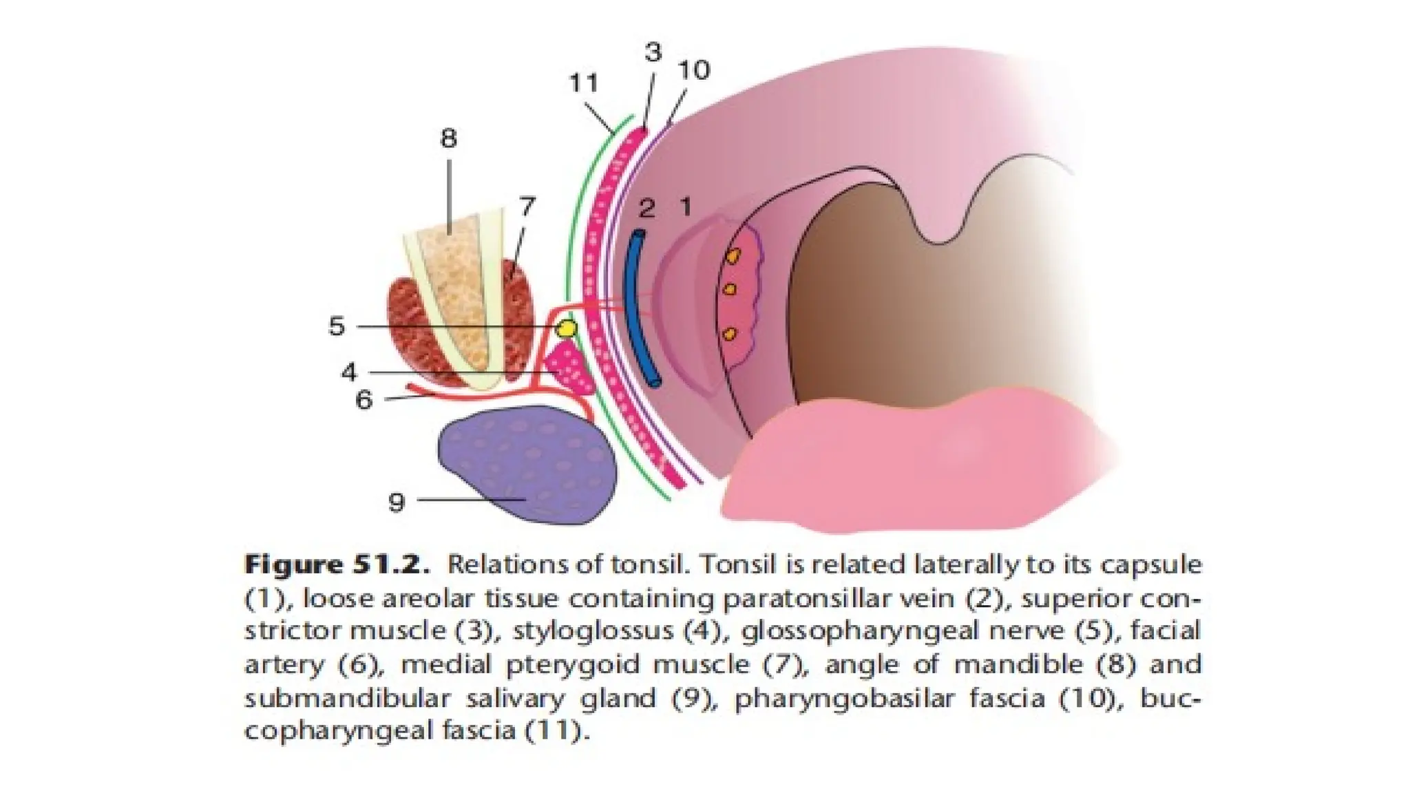
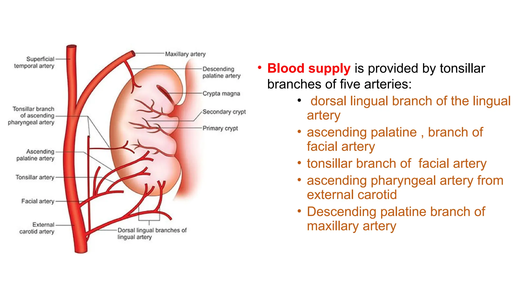
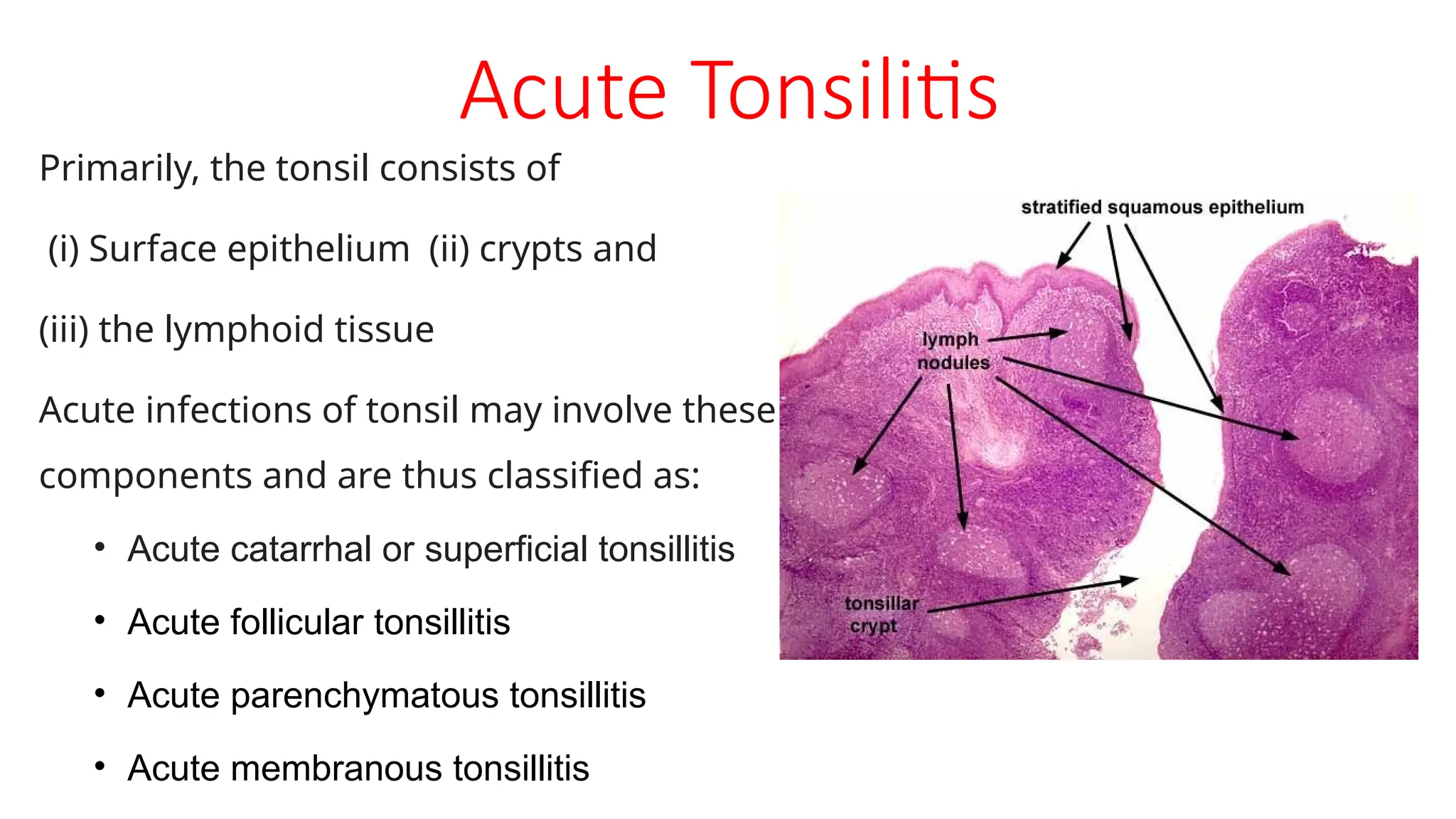
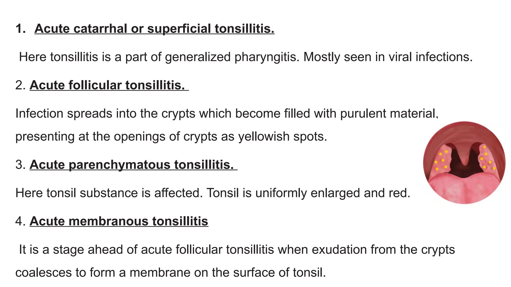
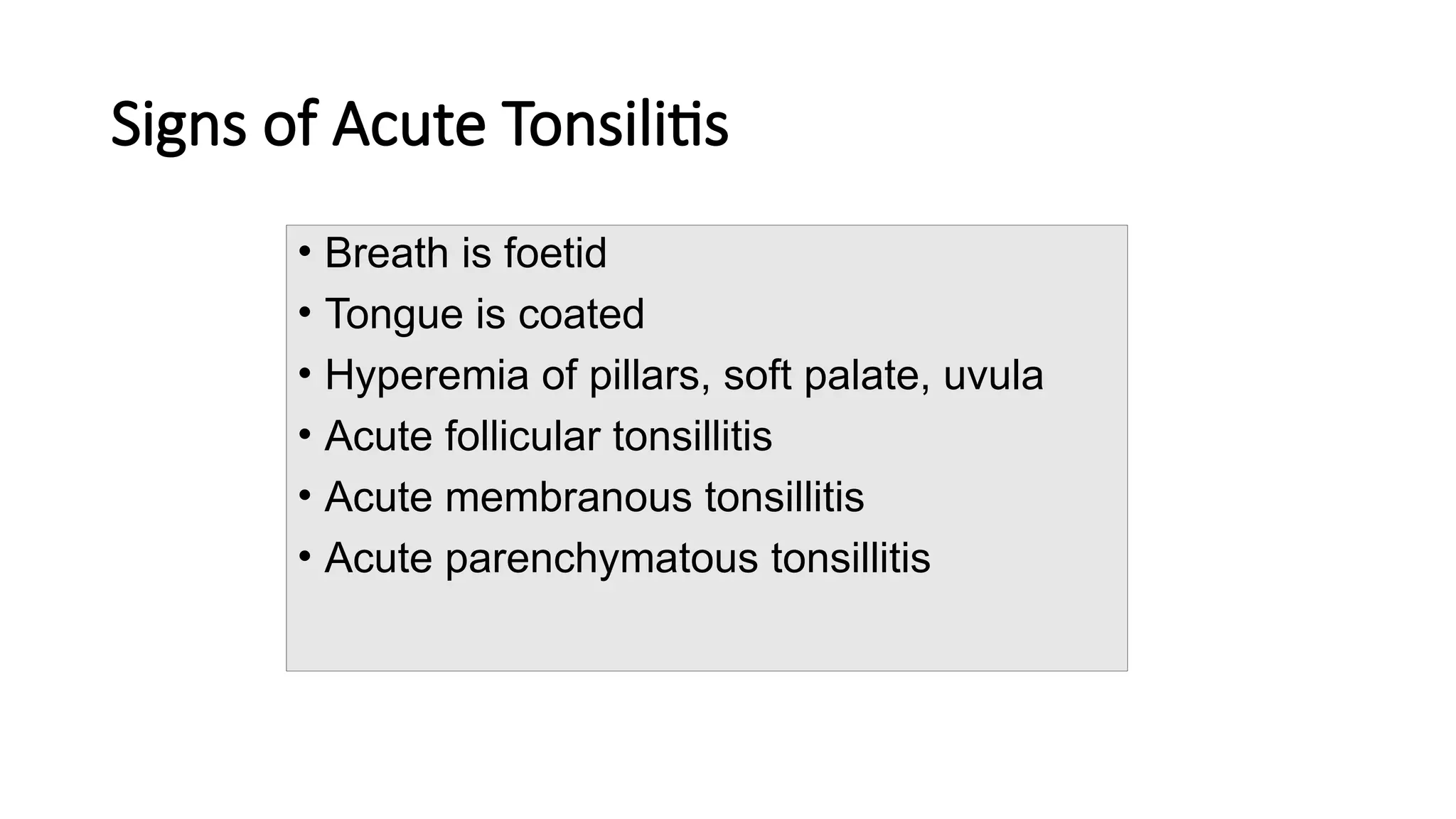
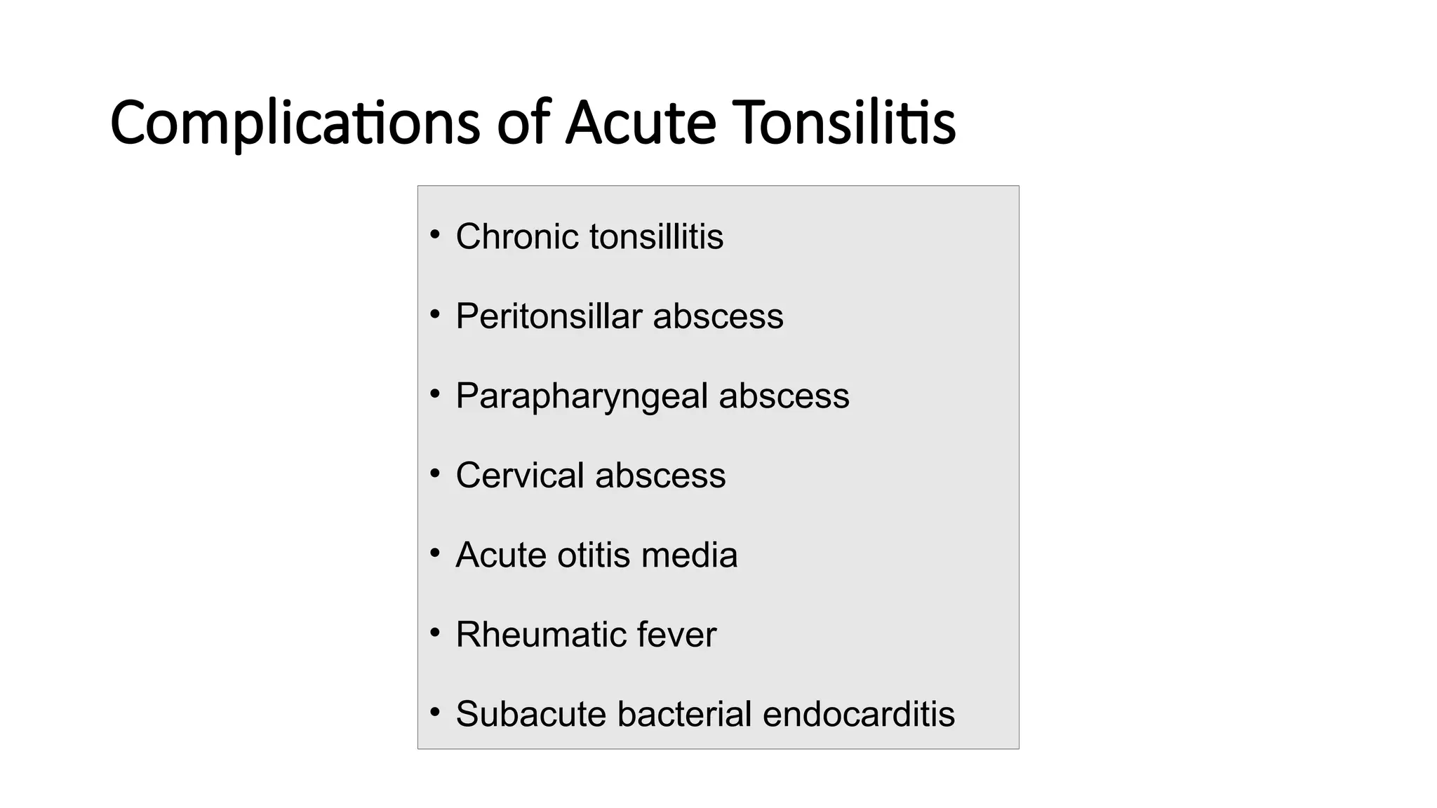
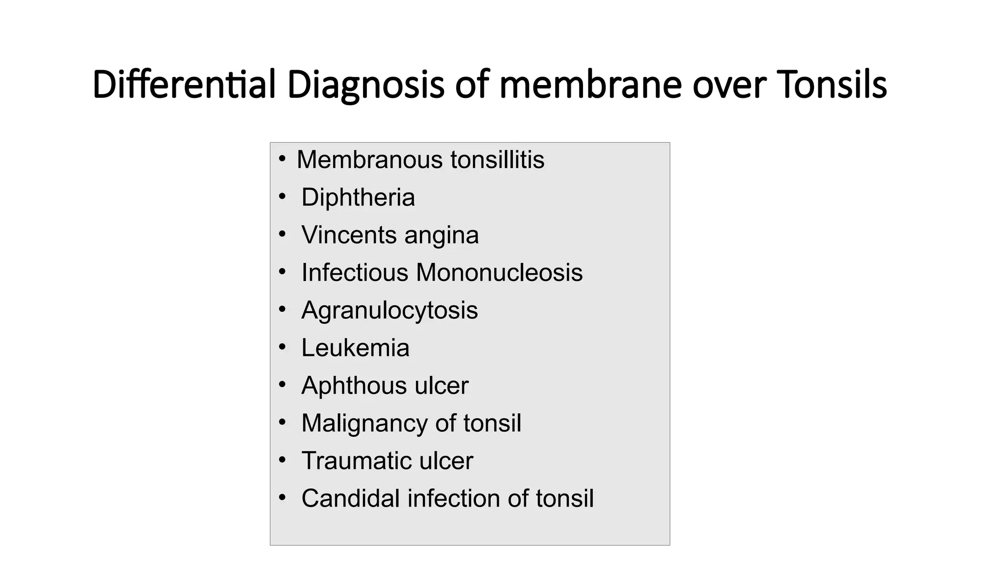
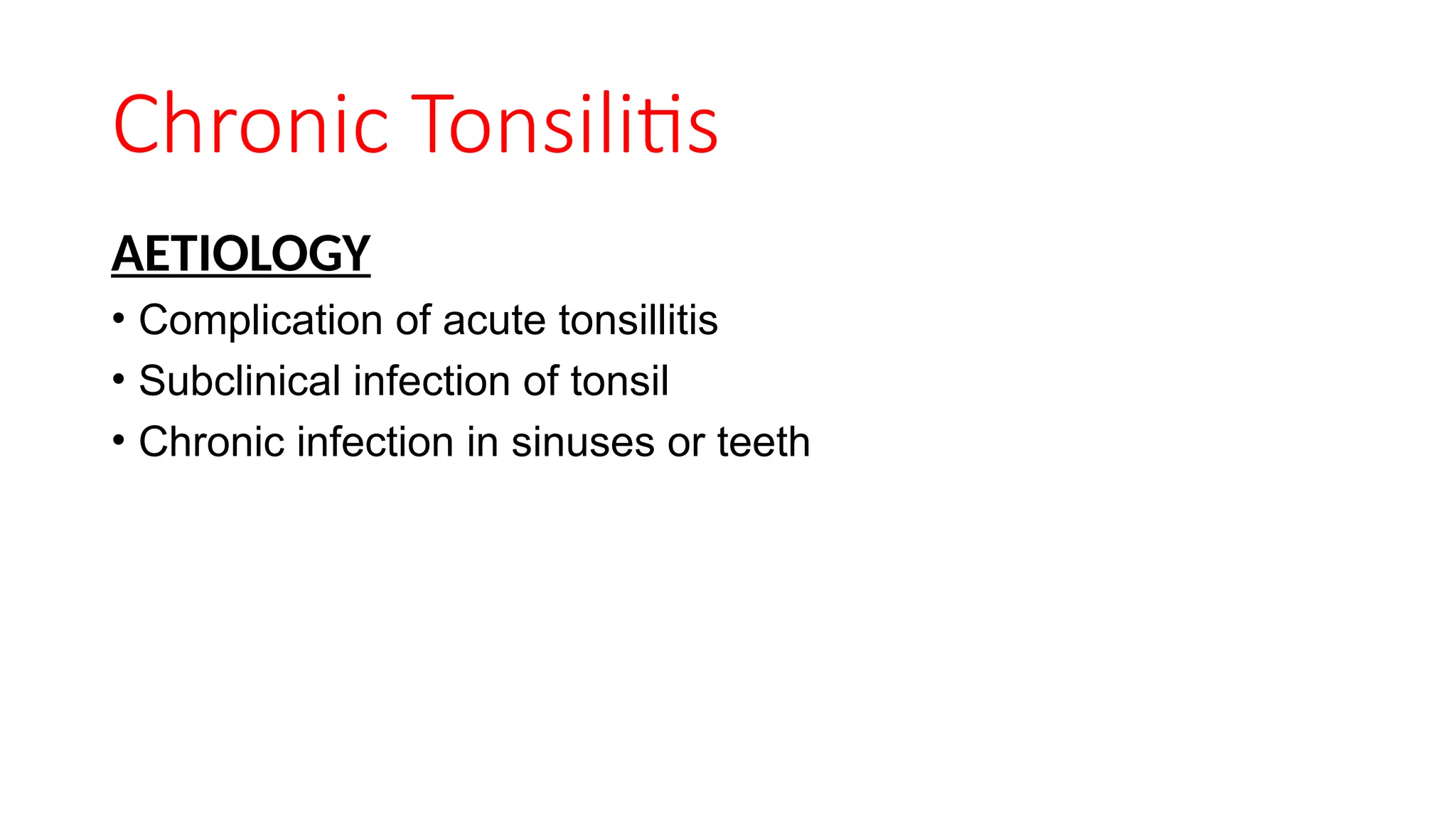
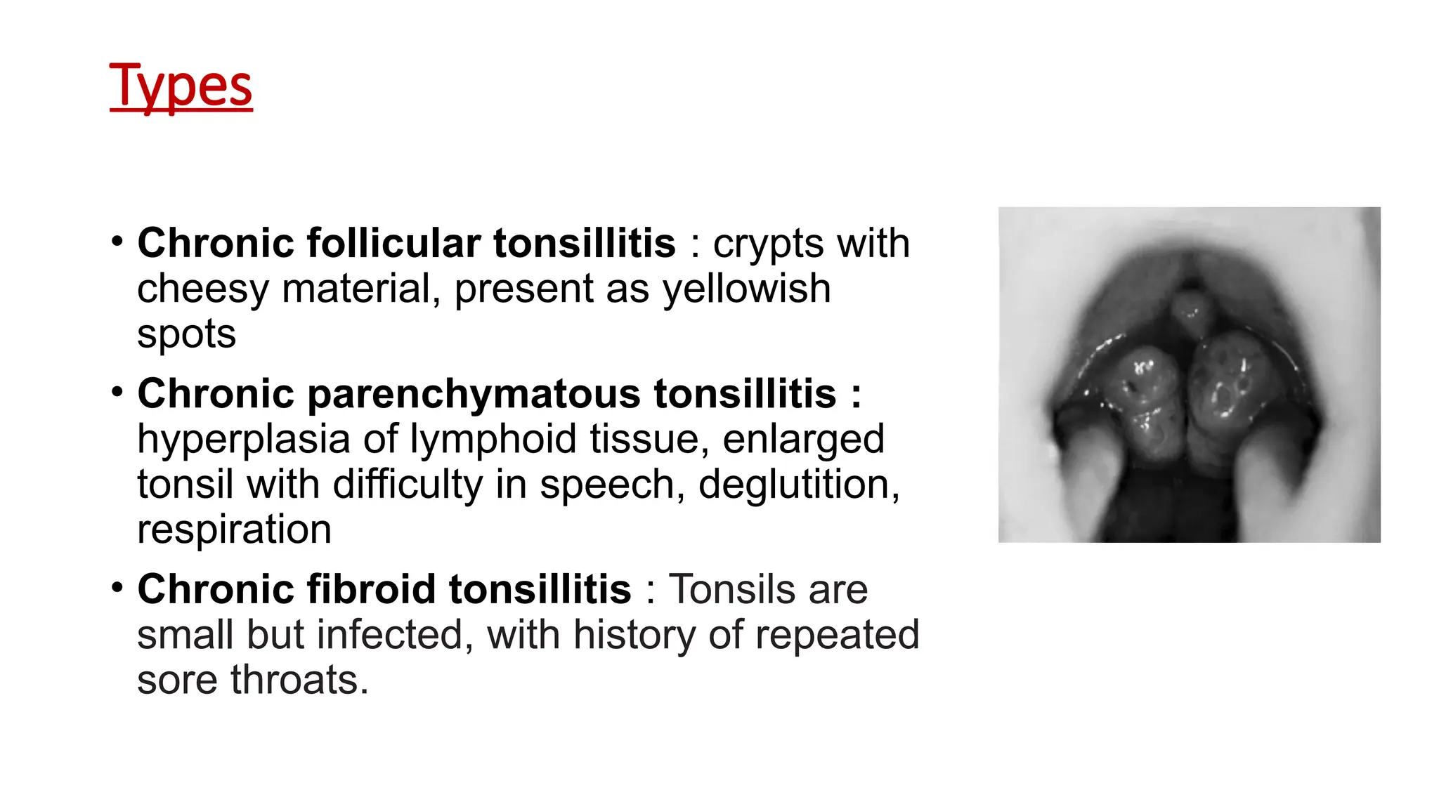
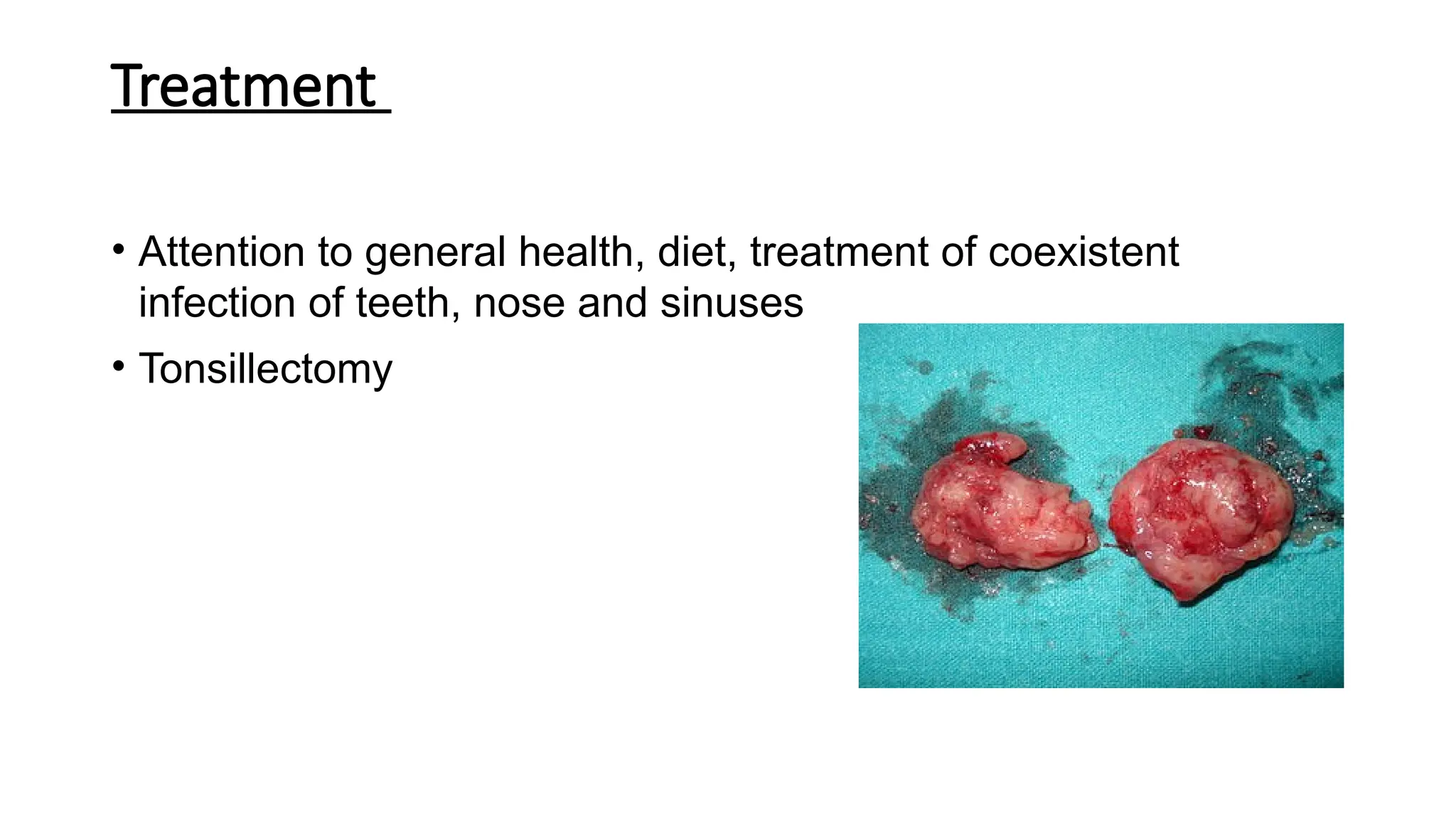
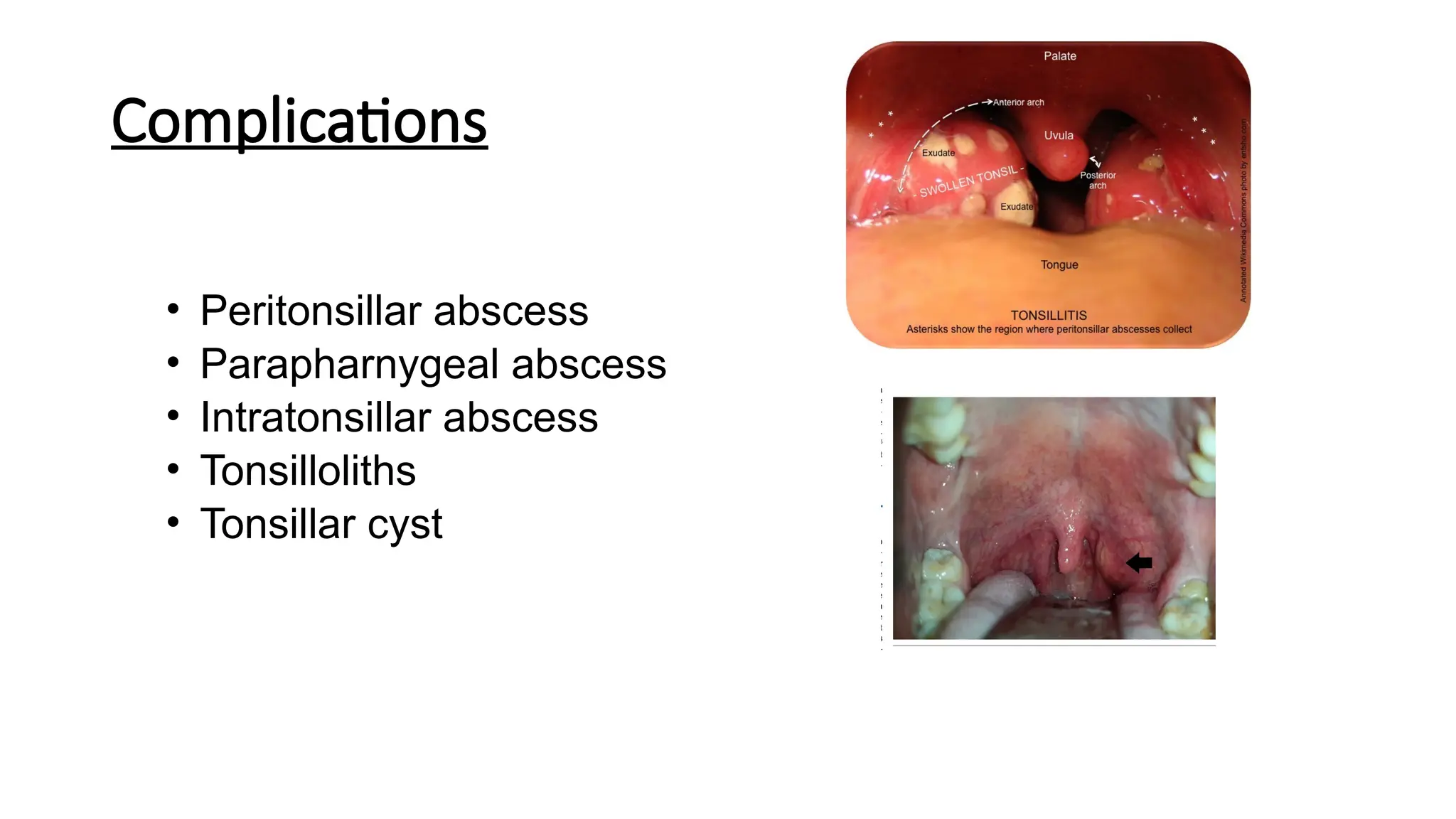
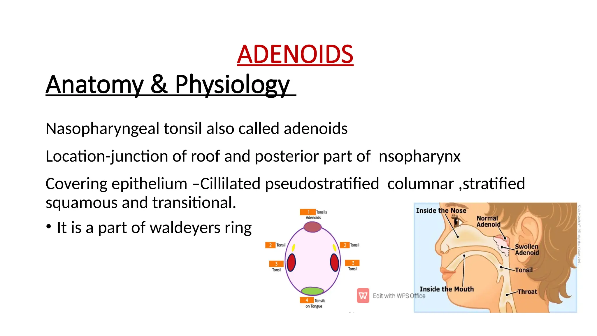
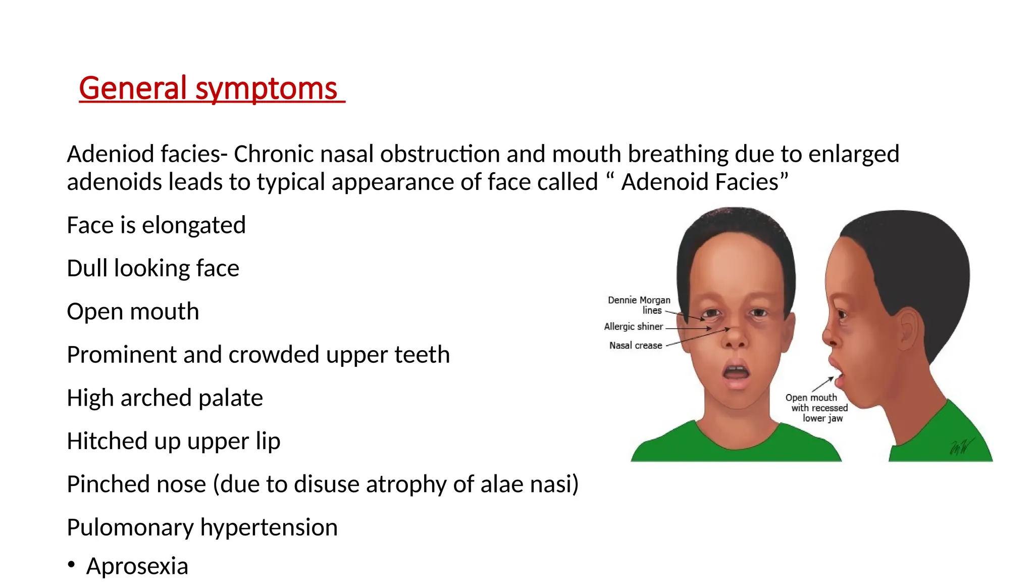
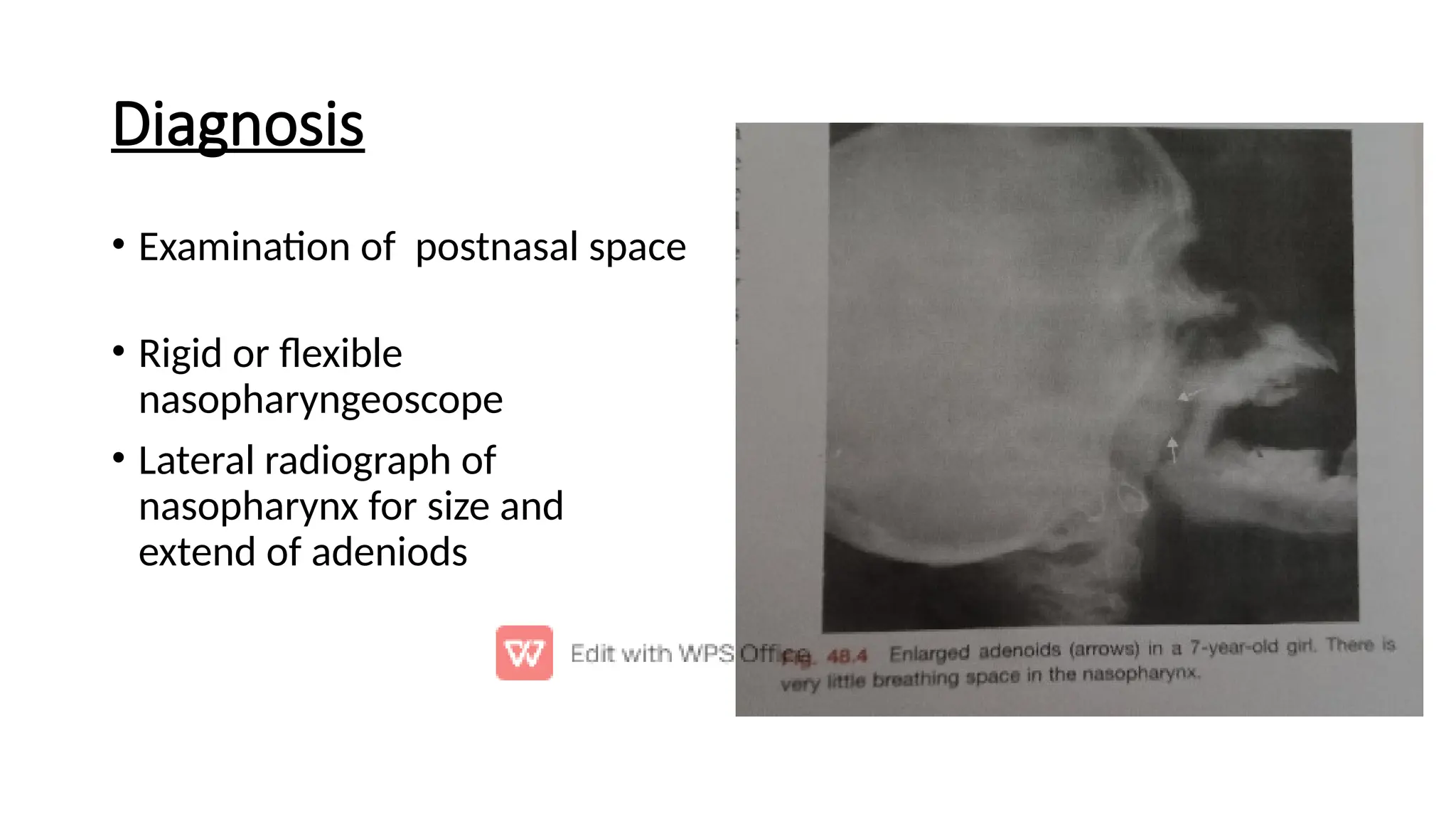
TONSILS & ADENOIDS
TONSILS
• The tonsils are collections of lymphatic tissue located within
the pharynx.
• They collectively form a ringed arrangement, known as the
Waldeyer’s ring.
• Pharyngeal tonsil
• Tubal tonsils (x2)
• Palatine tonsils (x2)
• Lingual tonsil
Palatine Tonsils
• These are commonly referred to as ‘the tonsils’.
• They are located within the tonsillar bed of the lateral oropharynx wall –
between the palatoglossal arch (anteriorly) and palatopharyngeal arch
(posteriorly). They form the lateral part of the Waldeyer’s ring.
• Each tonsil has free medial surface which projects into the pharynx. The lateral
surface is covered by a fibrous capsule, and is separated from the superior
constrictor of the tonsillar bed by loose areolar connective tissue.
• They are covered by a stratified non-keratinised squamous epithelium.
• Blood supply is provided by tonsillar
branches of five arteries:
• dorsal lingual branch of the lingual
artery
• ascending palatine , branch of
facial artery
• tonsillar branch of facial artery
• ascending pharyngeal artery from
external carotid
• Descending palatine branch of
maxillary artery
• The Nerves supplying the palatine tonsils come from the
maxillary division of the trigeminal nerve via the lesser palatine
nerves, and from the tonsillar branches of the glossopharyngeal
nerve.
• Venous drainage is by the peritonsillar plexus, which drain into
the lingual and pharyngeal veins, which in turn drain into the
internal jugular vein.
Tonsilitis
• Inflammation of the tonsils in the upper part of the throat.
• Tonsils play an important role in the immune system.
• It is a type of pharyngitis.
• It is usually caused by viral infection, with bacterial causes accounting for
approximately 1/3 of cases.
• Typically the infection is spread between people through the air.
• Major Complication is Peritonsillar Abscess
Acute Tonsilitis
Primarily, the tonsil consists of
(i) Surface epithelium (ii) crypts and
(iii) the lymphoid tissue
Acute infections of tonsil may involve these
components and are thus classified as:
• Acute catarrhal or superficial tonsillitis
• Acute follicular tonsillitis
• Acute parenchymatous tonsillitis
• Acute membranous tonsillitis
1. Acute catarrhal or superficial tonsillitis.
Here tonsillitis is a part of generalized pharyngitis. Mostly seen in viral infections.
2. Acute follicular tonsillitis.
Infection spreads into the crypts which become filled with purulent material,
presenting at the openings of crypts as yellowish spots.
3. Acute parenchymatous tonsillitis.
Here tonsil substance is affected. Tonsil is uniformly enlarged and red.
4. Acute membranous tonsillitis
It is a stage ahead of acute follicular tonsillitis when exudation from the crypts
coalesces to form a membrane on the surface of tonsil.
Aetiology of Acute Tonsilitis
• Haemolytic streptococcus is the most commonly infecting
organism.
• Other causes of infection may be staphylococci,
pneumococci or H. influenzae.
• These bacteria may primarily infect the tonsil or may be
secondary to a viral infection.
Symptoms of Acute Tonsilitis
• Sore throat
• Difficulty in swallowing
• Fever with chills and rigors
• Earache
• Other constitutional symptoms like
headache, body ache, malaise, constipation
Signs of Acute Tonsilitis
• Breath is foetid
• Tongue is coated
• Hyperemia of pillars, soft palate, uvula
• Acute follicular tonsillitis
• Acute membranous tonsillitis
• Acute parenchymatous tonsillitis
Treatment of Acute Tonsilitis
• Patient is put to bed
• Give plenty of oral fluids
• Analgesics like aspirin or paracetamol to relieve local pain and
bring down the fever.
• Antimicrobial therapy like penicillin, ampicillin, amoxicillin and to
penicillin sensitive individuals,Erythromycin is administered.
Complications of Acute Tonsilitis
• Chronic tonsillitis
• Peritonsillar abscess
• Parapharyngeal abscess
• Cervical abscess
• Acute otitis media
• Rheumatic fever
• Subacute bacterial endocarditis
Differential Diagnosis of membrane over Tonsils
• Membranous tonsillitis
• Diphtheria
• Vincents angina
• Infectious Mononucleosis
• Agranulocytosis
• Leukemia
• Aphthous ulcer
• Malignancy of tonsil
• Traumatic ulcer
• Candidal infection of tonsil
Chronic Tonsilitis
AETIOLOGY
• Complication of acute tonsillitis
• Subclinical infection of tonsil
• Chronic infection in sinuses or teeth
Types
• Chronic follicular tonsillitis : crypts with
cheesy material, present as yellowish
spots
• Chronic parenchymatous tonsillitis :
hyperplasia of lymphoid tissue, enlarged
tonsil with difficulty in speech, deglutition,
respiration
• Chronic fibroid tonsillitis : Tonsils are
small but infected, with history of repeated
sore throats.
Clinical Features
• Recurrent attacks of sore throat or acute tonsillitis.
• Chronic irritation in throat with cough.
• Bad taste in mouth, foul breath (halitosis).
• Thick speech, difficulty in swallowing, choking spells at night.
Treatment
• Attention to general health, diet, treatment of coexistent
infection of teeth, nose and sinuses
• Tonsillectomy
Complications
• Peritonsillar abscess
• Parapharnygeal abscess
• Intratonsillar abscess
• Tonsilloliths
• Tonsillar cyst
ADENOIDS
Nasopharyngeal tonsil also called adenoids
Location-junction of roof and posterior part of nsopharynx
Covering epithelium –Cillilated pseudostratified columnar ,stratified
squamous and transitional.
• It is a part of waldeyers ring
Anatomy & Physiology
Blood supply
Ascending palatine bbranch of facial
Ascending pharyngeal branch of external carotid artery
Pharyngeal branch of third part of maxillary artery
• Ascending cervical branch of inf thyroid artey of thyrocervical trunk
Lymphatics – upper jugular or through retropharyngeal and
parapharyngeal nodes
• Nerve Supply – CN 9& 10
Aetiology
Mainly seen in children
Certain children have tendancy to generalize lymphoid hyperplasia in
which adenoids also take part
Recurrent attack of rhinitis, sinusitis,or chronic tonsilitis
• Allergy of URT
Clinical features
• Nasal symptoms
• Nasal obstruction - commenest symptom
• mouth breathing, interfers with feeding
• Nasal discharge -wet blubby nose , partly due to choanal obstruction
and partly due to assosciated c/c rhinosinusitis
• Sinusitis
• Epistaxis
• Voice change
• Aural symptoms
• Tubal obstruction – retracted TM and conductive hearing loss
• Recurrent attacks of acute otitis media
• CSOM
• Otitis media with effusion
General symptoms
Adeniod facies- Chronic nasal obstruction and mouth breathing due to enlarged
adenoids leads to typical appearance of face called “ Adenoid Facies”
Face is elongated
Dull looking face
Open mouth
Prominent and crowded upper teeth
High arched palate
Hitched up upper lip
Pinched nose (due to disuse atrophy of alae nasi)
Pulomonary hypertension
• Aprosexia
Diagnosis
• Examination of postnasal space
• Rigid or flexible
nasopharyngeoscope
• Lateral radiograph of
nasopharynx for size and
extend of adeniods
Treatment
When there is no marked symptoms-
Breathing exercises
Nasal decongestant
Antihistaminics
When marked symptoms present
Adeniodectomy
Currette
• Endoscopically by using a debrider or coblation
Tonsillitis new.pptx adenoids - ENT , adenoids hypertrophy and tonsillitis

Tonsillitis new.pptx adenoids - ENT , adenoids hypertrophy and tonsillitis

  • 1.
  • 2.
    TONSILS • The tonsilsare collections of lymphatic tissue located within the pharynx. • They collectively form a ringed arrangement, known as the Waldeyer’s ring. • Pharyngeal tonsil • Tubal tonsils (x2) • Palatine tonsils (x2) • Lingual tonsil
  • 3.
    Palatine Tonsils • Theseare commonly referred to as ‘the tonsils’. • They are located within the tonsillar bed of the lateral oropharynx wall – between the palatoglossal arch (anteriorly) and palatopharyngeal arch (posteriorly). They form the lateral part of the Waldeyer’s ring. • Each tonsil has free medial surface which projects into the pharynx. The lateral surface is covered by a fibrous capsule, and is separated from the superior constrictor of the tonsillar bed by loose areolar connective tissue. • They are covered by a stratified non-keratinised squamous epithelium.
  • 6.
    • Blood supplyis provided by tonsillar branches of five arteries: • dorsal lingual branch of the lingual artery • ascending palatine , branch of facial artery • tonsillar branch of facial artery • ascending pharyngeal artery from external carotid • Descending palatine branch of maxillary artery
  • 7.
    • The Nervessupplying the palatine tonsils come from the maxillary division of the trigeminal nerve via the lesser palatine nerves, and from the tonsillar branches of the glossopharyngeal nerve. • Venous drainage is by the peritonsillar plexus, which drain into the lingual and pharyngeal veins, which in turn drain into the internal jugular vein.
  • 8.
    Tonsilitis • Inflammation ofthe tonsils in the upper part of the throat. • Tonsils play an important role in the immune system. • It is a type of pharyngitis. • It is usually caused by viral infection, with bacterial causes accounting for approximately 1/3 of cases. • Typically the infection is spread between people through the air. • Major Complication is Peritonsillar Abscess
  • 9.
    Acute Tonsilitis Primarily, thetonsil consists of (i) Surface epithelium (ii) crypts and (iii) the lymphoid tissue Acute infections of tonsil may involve these components and are thus classified as: • Acute catarrhal or superficial tonsillitis • Acute follicular tonsillitis • Acute parenchymatous tonsillitis • Acute membranous tonsillitis
  • 10.
    1. Acute catarrhalor superficial tonsillitis. Here tonsillitis is a part of generalized pharyngitis. Mostly seen in viral infections. 2. Acute follicular tonsillitis. Infection spreads into the crypts which become filled with purulent material, presenting at the openings of crypts as yellowish spots. 3. Acute parenchymatous tonsillitis. Here tonsil substance is affected. Tonsil is uniformly enlarged and red. 4. Acute membranous tonsillitis It is a stage ahead of acute follicular tonsillitis when exudation from the crypts coalesces to form a membrane on the surface of tonsil.
  • 11.
    Aetiology of AcuteTonsilitis • Haemolytic streptococcus is the most commonly infecting organism. • Other causes of infection may be staphylococci, pneumococci or H. influenzae. • These bacteria may primarily infect the tonsil or may be secondary to a viral infection.
  • 12.
    Symptoms of AcuteTonsilitis • Sore throat • Difficulty in swallowing • Fever with chills and rigors • Earache • Other constitutional symptoms like headache, body ache, malaise, constipation
  • 13.
    Signs of AcuteTonsilitis • Breath is foetid • Tongue is coated • Hyperemia of pillars, soft palate, uvula • Acute follicular tonsillitis • Acute membranous tonsillitis • Acute parenchymatous tonsillitis
  • 14.
    Treatment of AcuteTonsilitis • Patient is put to bed • Give plenty of oral fluids • Analgesics like aspirin or paracetamol to relieve local pain and bring down the fever. • Antimicrobial therapy like penicillin, ampicillin, amoxicillin and to penicillin sensitive individuals,Erythromycin is administered.
  • 15.
    Complications of AcuteTonsilitis • Chronic tonsillitis • Peritonsillar abscess • Parapharyngeal abscess • Cervical abscess • Acute otitis media • Rheumatic fever • Subacute bacterial endocarditis
  • 16.
    Differential Diagnosis ofmembrane over Tonsils • Membranous tonsillitis • Diphtheria • Vincents angina • Infectious Mononucleosis • Agranulocytosis • Leukemia • Aphthous ulcer • Malignancy of tonsil • Traumatic ulcer • Candidal infection of tonsil
  • 17.
    Chronic Tonsilitis AETIOLOGY • Complicationof acute tonsillitis • Subclinical infection of tonsil • Chronic infection in sinuses or teeth
  • 18.
    Types • Chronic folliculartonsillitis : crypts with cheesy material, present as yellowish spots • Chronic parenchymatous tonsillitis : hyperplasia of lymphoid tissue, enlarged tonsil with difficulty in speech, deglutition, respiration • Chronic fibroid tonsillitis : Tonsils are small but infected, with history of repeated sore throats.
  • 19.
    Clinical Features • Recurrentattacks of sore throat or acute tonsillitis. • Chronic irritation in throat with cough. • Bad taste in mouth, foul breath (halitosis). • Thick speech, difficulty in swallowing, choking spells at night.
  • 20.
    Treatment • Attention togeneral health, diet, treatment of coexistent infection of teeth, nose and sinuses • Tonsillectomy
  • 21.
    Complications • Peritonsillar abscess •Parapharnygeal abscess • Intratonsillar abscess • Tonsilloliths • Tonsillar cyst
  • 22.
    ADENOIDS Nasopharyngeal tonsil alsocalled adenoids Location-junction of roof and posterior part of nsopharynx Covering epithelium –Cillilated pseudostratified columnar ,stratified squamous and transitional. • It is a part of waldeyers ring Anatomy & Physiology
  • 23.
    Blood supply Ascending palatinebbranch of facial Ascending pharyngeal branch of external carotid artery Pharyngeal branch of third part of maxillary artery • Ascending cervical branch of inf thyroid artey of thyrocervical trunk Lymphatics – upper jugular or through retropharyngeal and parapharyngeal nodes • Nerve Supply – CN 9& 10
  • 24.
    Aetiology Mainly seen inchildren Certain children have tendancy to generalize lymphoid hyperplasia in which adenoids also take part Recurrent attack of rhinitis, sinusitis,or chronic tonsilitis • Allergy of URT
  • 25.
    Clinical features • Nasalsymptoms • Nasal obstruction - commenest symptom • mouth breathing, interfers with feeding • Nasal discharge -wet blubby nose , partly due to choanal obstruction and partly due to assosciated c/c rhinosinusitis • Sinusitis • Epistaxis • Voice change
  • 26.
    • Aural symptoms •Tubal obstruction – retracted TM and conductive hearing loss • Recurrent attacks of acute otitis media • CSOM • Otitis media with effusion
  • 27.
    General symptoms Adeniod facies-Chronic nasal obstruction and mouth breathing due to enlarged adenoids leads to typical appearance of face called “ Adenoid Facies” Face is elongated Dull looking face Open mouth Prominent and crowded upper teeth High arched palate Hitched up upper lip Pinched nose (due to disuse atrophy of alae nasi) Pulomonary hypertension • Aprosexia
  • 28.
    Diagnosis • Examination ofpostnasal space • Rigid or flexible nasopharyngeoscope • Lateral radiograph of nasopharynx for size and extend of adeniods
  • 29.
    Treatment When there isno marked symptoms- Breathing exercises Nasal decongestant Antihistaminics When marked symptoms present Adeniodectomy Currette • Endoscopically by using a debrider or coblation