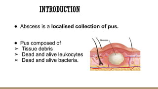
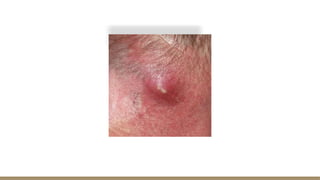
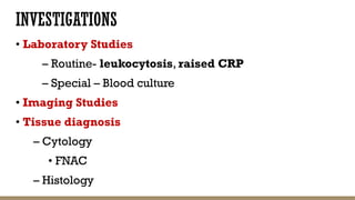
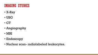
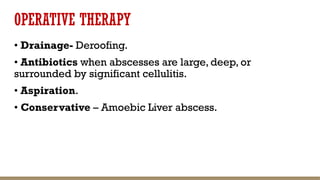
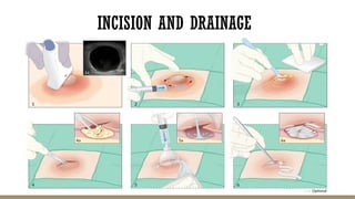
ABSCESS
Dr. Murshida Jabin
Surgery Intern
INTRODUCTION
● Abscess is a localised collection of pus.
● Pus composed of
➢ Tissue debris
➢ Dead and alive leukocytes
➢ Dead and alive bacteria.
ETIOLOGY
1. Idiopathic
2. Congenital/Genetic
3. Traumatic
4. Infections/Infestation
5. Autoimmune
6. Neoplastic (Benign/Malignant)
7. Degenerative
8. Iatrogenic
ETIOLOGY: PREDISPOSING FACTORS
• Impaired host defense mechanisms eg: HIV
• The presence of foreign bodies
• Obstruction to normal drainage (urinary, biliary, or resp tract)
• Tissue ischemia or necrosis
• Hematoma or excessive fluid accumulation in tissue
• Trauma
• Numerous organisms can cause abscesses,(MC: Staph aureus)
PATHOPHYSIOLOGY
• Abscesses may begin in an area of cellulitis or in
compromised tissue where leukocytes accumulate.
• Progressive dissection by pus or necrosis of surrounding
cells expands the abscess.
• Highly vascularized connective tissue may then surround
the necrotic tissue, leukocytes, and debris to wall off the
abscess and limit further spread.
• Organisms may enter the tissue by-
– Direct implantation eg:IM injection.
– Spread from an established, contiguous infection
– Dissemination via lymphatic or hematogenous routes
from a distant site
– Migration from a location where there are resident flora
into an adjacent, normally sterile area because natural
barriers are disrupted (eg, by perforation of an abdominal
viscus causing an intra-abdominal abscess)
CLINICAL FEATURES
Symptoms
• Fever with spikes and chills.
• Local pain and tenderness
• Anorexia
• Weight loss
• Fatigue
Signs
• Signs of cutaneous and subcutaneous abscesses are pain,
heat, swelling, tenderness, and redness.
• If superficial abscesses are ready to spontaneously
rupture, the skin over the center of the abscess may thin,
sometimes appearing white or yellow because of the
underlying pus (termed pointing).
• The predominant manifestation of some abscesses is
abnormal organ function (eg, hemiplegia due to a brain
abscess).
COMPLICATIONS
• Bacteremic spread
• Rupture into adjacent tissue
• Bleeding from vessels eroded by inflammation
• Impaired function of a vital organ
• Inanition due to anorexia and increased metabolic needs
• Antibioma
• Sterile abscess
• Chronic draining sinuses
INVESTIGATIONS
• Laboratory Studies
– Routine- leukocytosis, raised CRP
– Special – Blood culture
• Imaging Studies
• Tissue diagnosis
– Cytology
• FNAC
– Histology
IMAGING STUDIES
• X-Ray
• USG
• CT
• Angiography
• MRI
• Endoscopy
• Nuclear scan- radiolabeled leukocytes.
OPERATIVE THERAPY
• Drainage- Deroofing.
• Antibiotics when abscesses are large, deep, or
surrounded by significant cellulitis.
• Aspiration.
• Conservative – Amoebic Liver abscess.
INCISION AND DRAINAGE
ASPIRATION
SOME COMMON ABSCESS
• Brain Abscess
• Gluteal Injection abscess.
• Breast Abscess
• Lung abscess
• Liver Abscess
• Subphrenic Abscess
• Pelvic abscess.
• Psoas abscess.
THANK YOU

abscess....pptx surgery ppt....types and management

  • 1.
  • 2.
    INTRODUCTION ● Abscess isa localised collection of pus. ● Pus composed of ➢ Tissue debris ➢ Dead and alive leukocytes ➢ Dead and alive bacteria.
  • 3.
    ETIOLOGY 1. Idiopathic 2. Congenital/Genetic 3.Traumatic 4. Infections/Infestation 5. Autoimmune 6. Neoplastic (Benign/Malignant) 7. Degenerative 8. Iatrogenic
  • 4.
    ETIOLOGY: PREDISPOSING FACTORS •Impaired host defense mechanisms eg: HIV • The presence of foreign bodies • Obstruction to normal drainage (urinary, biliary, or resp tract) • Tissue ischemia or necrosis • Hematoma or excessive fluid accumulation in tissue • Trauma • Numerous organisms can cause abscesses,(MC: Staph aureus)
  • 5.
    PATHOPHYSIOLOGY • Abscesses maybegin in an area of cellulitis or in compromised tissue where leukocytes accumulate. • Progressive dissection by pus or necrosis of surrounding cells expands the abscess. • Highly vascularized connective tissue may then surround the necrotic tissue, leukocytes, and debris to wall off the abscess and limit further spread.
  • 6.
    • Organisms mayenter the tissue by- – Direct implantation eg:IM injection. – Spread from an established, contiguous infection – Dissemination via lymphatic or hematogenous routes from a distant site – Migration from a location where there are resident flora into an adjacent, normally sterile area because natural barriers are disrupted (eg, by perforation of an abdominal viscus causing an intra-abdominal abscess)
  • 7.
    CLINICAL FEATURES Symptoms • Feverwith spikes and chills. • Local pain and tenderness • Anorexia • Weight loss • Fatigue
  • 8.
    Signs • Signs ofcutaneous and subcutaneous abscesses are pain, heat, swelling, tenderness, and redness. • If superficial abscesses are ready to spontaneously rupture, the skin over the center of the abscess may thin, sometimes appearing white or yellow because of the underlying pus (termed pointing). • The predominant manifestation of some abscesses is abnormal organ function (eg, hemiplegia due to a brain abscess).
  • 10.
    COMPLICATIONS • Bacteremic spread •Rupture into adjacent tissue • Bleeding from vessels eroded by inflammation • Impaired function of a vital organ • Inanition due to anorexia and increased metabolic needs • Antibioma • Sterile abscess • Chronic draining sinuses
  • 11.
    INVESTIGATIONS • Laboratory Studies –Routine- leukocytosis, raised CRP – Special – Blood culture • Imaging Studies • Tissue diagnosis – Cytology • FNAC – Histology
  • 12.
    IMAGING STUDIES • X-Ray •USG • CT • Angiography • MRI • Endoscopy • Nuclear scan- radiolabeled leukocytes.
  • 13.
    OPERATIVE THERAPY • Drainage-Deroofing. • Antibiotics when abscesses are large, deep, or surrounded by significant cellulitis. • Aspiration. • Conservative – Amoebic Liver abscess.
  • 14.
  • 15.
  • 16.
    SOME COMMON ABSCESS •Brain Abscess • Gluteal Injection abscess. • Breast Abscess • Lung abscess • Liver Abscess • Subphrenic Abscess • Pelvic abscess. • Psoas abscess.
  • 17.