INTRAOCULAR PRESSURE
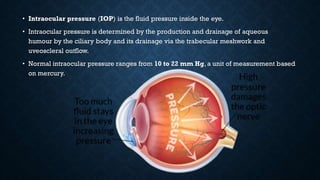
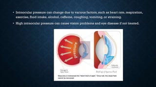
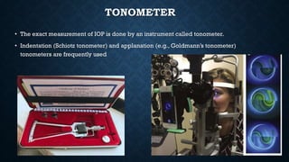
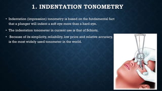
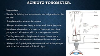
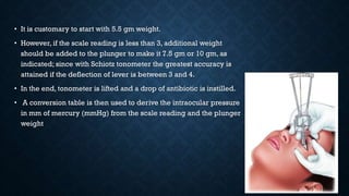
• Intraocular pressure (IOP) is the fluid pressure inside the eye.
• Intraocular pressure is determined by the production and drainage of aqueous
humour by the ciliary body and its drainage via the trabecular meshwork and
uveoscleral outflow.
• Normal intraocular pressure ranges from 10 to 22 mm Hg, a unit of measurement based
on mercury.
• Intraocular pressure can change due to various factors, such as heart rate, respiration,
exercise, fluid intake, alcohol, caffeine, coughing, vomiting, or straining.
• High intraocular pressure can cause vision problems and eye disease if not treated.
• Normal IOP range is 10-21 mm of Hg with an average tension of 16 ± 2.5 mm of Hg.
• When IOP is less than 10 mm of Hg, it is called hypotony.
• An IOP of more than 21 mm of Hg should always arouse suspicion of glaucoma and
such patients should be thoroughly investigated.
MEASUREMENT OF IOP
• The measurement of IOP (ocular tension) should be made in all suspected cases of
glaucoma and in routine after the age of 40 years.
DIGITAL TONOMETRY
• A rough estimate of IOP can be made by digital tonometry.
• For this procedure patient is asked to look down and the eyeball is palpated by index
fingers of both the hands, through the upper lid, beyond the tarsal plate.
• One finger is kept stationary which feels the fluctuation produced by indentation of globe
by the other finger.
• It is a subjective method and needs experience.
• When IOP is raised, fluctuation produced is feeble or absent and the eyeball feels firm to
hard.
• When IOP is very low eye feels soft like a partially filled water bag
TONOMETER
• The exact measurement of IOP is done by an instrument called tonometer.
• Indentation (Schiotz tonometer) and applanation (e.g., Goldmann’s tonometer)
tonometers are frequently used
1. INDENTATION TONOMETRY
• Indentation (impression) tonometry is based on the fundamental fact
that a plunger will indent a soft eye more than a hard eye.
• The indentation tonometer in current use is that of Schiotz.
• Because of its simplicity, reliability, low price and relative accuracy, it
is the most widely used tonometer in the world.
SCHIOTZ TONOMETER.
• It consists of :
- Handle for holding the instrument in vertical position on the
cornea;
- Footplate which rests on the cornea;
- Plunger which moves freely within a shaft in the footplate;
- Bent lever whose short arm rests on the upper end of the
plunger and a long arm which acts as a pointer needle.
- The degree to which the plunger indents the cornea is
indicated by the movement of this needle on a scale; and
- Weights: a 5.5 g weight is permanently fixed to the plunger,
which can be increased to 7.5 and 10 gm.
TECHNIQUE OF SCHIOTZ TONOMETRY.
• Before tonometry, the footplate and lower end of plunger should be
sterilized.
• For repeated use in multiple patients it can be sterilized by dipping
the footplate in ether, absolute alcohol, acetone or by heating the
footplate in the flame of spirit.
• After anaesthetising the cornea with 2-4 per cent topical xylocaine,
patient is made to lie supine on a couch and instructed to fix at a target
on the ceiling.
• Then the examiner separates the lids with left hand and gently rests
the footplate of the tonometer vertically on the centre of cornea.
• The reading on scale is recorded as soon as the needle becomes
steady
• It is customary to start with 5.5 gm weight.
• However, if the scale reading is less than 3, additional weight
should be added to the plunger to make it 7.5 gm or 10 gm, as
indicated; since with Schiotz tonometer the greatest accuracy is
attained if the deflection of lever is between 3 and 4.
• In the end, tonometer is lifted and a drop of antibiotic is instilled.
• A conversion table is then used to derive the intraocular pressure
in mm of mercury (mmHg) from the scale reading and the plunger
weight
ADVANTAGE AND DISADVANTAGE
• The main advantages of Schiotz tonometer are that it is cheap, handy and easy to use.
• Its main disadvantage is that it gives a false reading when used in eyes with abnormal
scleral rigidity.
• False low levels of IOP are obtained in eyes with low scleral rigidity seen in high
myopes and following ocular surgery.
2. APPLANATION TONOMETRY
• The concept of applanation tonometry was introduced by Goldmann in 1954.
• It is based on ImbertFick law which states that the pressure inside a sphere (P) is
equal to the force (W) required to flatten its surface divided by the area of flattening
(A); i.e., P = W/A.
THE COMMONLY USED APPLANATION
TONOMETERS ARE:
• 1. goldmann tonometer
• 2. Perkin’s applanation tonometer
• 3. Pneumatic tonometer
• 4. Air- puff tonometer
• 5. Pulse air tonometer
• 6.Tono pen
1. GOLDMANN TONOMETER
• Currently, it is the most popular and accurate tonometer.
- It consists of a double prism mounted on a standard slit-lamp.
- The prism applanates the cornea in an area of 3.06 mm diameter.
TECHNIQUE
• After anaesthetising the cornea with a drop of 2 per cent xylocaine and staining the
tear film with fluorescein patient is made to sit in front of slit-lamp.
• The cornea and biprisms are illuminated with cobalt blue light from the slit-lamp.
• Biprism is then advanced until it just touches the apex of cornea.
• At this point two fluorescent semicircles are viewed through the prism.
• Then, the applanation force against cornea is adjusted until the inner edges of the two
semicircles just touch .
• This is the end point.
• The intraocular pressure is determined by multiplying the dial reading with ten
2. PERKIN’S APPLANATION TONOMETER
• This is a hand-held tonometer utilizing the same biprism as in the Goldmann
applanation tonometer.
• It is small, easy to carry and does not require slit lamp.
• However, it requires considerable practice before, reliable readings can be obtained.
3. PNEUMATIC TONOMETER.
• In this, the cornea is applanated by touching its apex by a silastic diaphragm
covering the sensing nozzle (which is connected to a central chamber containing
pressurised air).
• In this tonometer, there is a pneumatic-to-electronic transducer, which converts the
air pressure to a recording on a paper-strip, from where IOP is read
• 4. Pulse air tonometer is a hand-held, non-contact tonometer that can be used with the
patient in any position.
• 5.Tono-Pen is a computerised pocket tonometer. It employs a microscopic
transducer which applanates the cornea and converts IOP into electric waves.
• 6. air puff tonometer: non contact tonometer based on the principle of goldmann
tonometry. In this, the central part of cornea is flattened by a jet of air.
TONOGRAPHY
• Tonography is a non-invasive technique for determining the facility of aqueous
outflow (C-value).
• The C-value is expressed as aqueous outflow in microlitres per minute per millimetre
of mercury.
• It is estimated by placing Schiotz tonometer on the eye for 4 minutes.
• For a graphic record the electronic Schiotz tonometer is used.
• C-value is calculated from special tonographic tables taking into consideration the
initial IOP (P0) and the change in scale reading over the 4 minutes.
• Clinically, C-value does not play much role in the management of a glaucoma patient.
• Although, in general, C-values more than 0.20 are considered normal, between 0.2
and 0.11 border line, and those below 0.11 abnormal

INTRAOCULAR PRESSURE.pdf

  • 1.
  • 2.
    • Intraocular pressure(IOP) is the fluid pressure inside the eye. • Intraocular pressure is determined by the production and drainage of aqueous humour by the ciliary body and its drainage via the trabecular meshwork and uveoscleral outflow. • Normal intraocular pressure ranges from 10 to 22 mm Hg, a unit of measurement based on mercury.
  • 3.
    • Intraocular pressurecan change due to various factors, such as heart rate, respiration, exercise, fluid intake, alcohol, caffeine, coughing, vomiting, or straining. • High intraocular pressure can cause vision problems and eye disease if not treated.
  • 4.
    • Normal IOPrange is 10-21 mm of Hg with an average tension of 16 ± 2.5 mm of Hg. • When IOP is less than 10 mm of Hg, it is called hypotony. • An IOP of more than 21 mm of Hg should always arouse suspicion of glaucoma and such patients should be thoroughly investigated.
  • 5.
    MEASUREMENT OF IOP •The measurement of IOP (ocular tension) should be made in all suspected cases of glaucoma and in routine after the age of 40 years.
  • 6.
    DIGITAL TONOMETRY • Arough estimate of IOP can be made by digital tonometry. • For this procedure patient is asked to look down and the eyeball is palpated by index fingers of both the hands, through the upper lid, beyond the tarsal plate. • One finger is kept stationary which feels the fluctuation produced by indentation of globe by the other finger. • It is a subjective method and needs experience. • When IOP is raised, fluctuation produced is feeble or absent and the eyeball feels firm to hard. • When IOP is very low eye feels soft like a partially filled water bag
  • 8.
    TONOMETER • The exactmeasurement of IOP is done by an instrument called tonometer. • Indentation (Schiotz tonometer) and applanation (e.g., Goldmann’s tonometer) tonometers are frequently used
  • 9.
    1. INDENTATION TONOMETRY •Indentation (impression) tonometry is based on the fundamental fact that a plunger will indent a soft eye more than a hard eye. • The indentation tonometer in current use is that of Schiotz. • Because of its simplicity, reliability, low price and relative accuracy, it is the most widely used tonometer in the world.
  • 10.
    SCHIOTZ TONOMETER. • Itconsists of : - Handle for holding the instrument in vertical position on the cornea; - Footplate which rests on the cornea; - Plunger which moves freely within a shaft in the footplate; - Bent lever whose short arm rests on the upper end of the plunger and a long arm which acts as a pointer needle. - The degree to which the plunger indents the cornea is indicated by the movement of this needle on a scale; and - Weights: a 5.5 g weight is permanently fixed to the plunger, which can be increased to 7.5 and 10 gm.
  • 11.
    TECHNIQUE OF SCHIOTZTONOMETRY. • Before tonometry, the footplate and lower end of plunger should be sterilized. • For repeated use in multiple patients it can be sterilized by dipping the footplate in ether, absolute alcohol, acetone or by heating the footplate in the flame of spirit. • After anaesthetising the cornea with 2-4 per cent topical xylocaine, patient is made to lie supine on a couch and instructed to fix at a target on the ceiling. • Then the examiner separates the lids with left hand and gently rests the footplate of the tonometer vertically on the centre of cornea. • The reading on scale is recorded as soon as the needle becomes steady
  • 13.
    • It iscustomary to start with 5.5 gm weight. • However, if the scale reading is less than 3, additional weight should be added to the plunger to make it 7.5 gm or 10 gm, as indicated; since with Schiotz tonometer the greatest accuracy is attained if the deflection of lever is between 3 and 4. • In the end, tonometer is lifted and a drop of antibiotic is instilled. • A conversion table is then used to derive the intraocular pressure in mm of mercury (mmHg) from the scale reading and the plunger weight
  • 14.
    ADVANTAGE AND DISADVANTAGE •The main advantages of Schiotz tonometer are that it is cheap, handy and easy to use. • Its main disadvantage is that it gives a false reading when used in eyes with abnormal scleral rigidity. • False low levels of IOP are obtained in eyes with low scleral rigidity seen in high myopes and following ocular surgery.
  • 15.
    2. APPLANATION TONOMETRY •The concept of applanation tonometry was introduced by Goldmann in 1954. • It is based on ImbertFick law which states that the pressure inside a sphere (P) is equal to the force (W) required to flatten its surface divided by the area of flattening (A); i.e., P = W/A.
  • 16.
    THE COMMONLY USEDAPPLANATION TONOMETERS ARE: • 1. goldmann tonometer • 2. Perkin’s applanation tonometer • 3. Pneumatic tonometer • 4. Air- puff tonometer • 5. Pulse air tonometer • 6.Tono pen
  • 17.
    1. GOLDMANN TONOMETER •Currently, it is the most popular and accurate tonometer. - It consists of a double prism mounted on a standard slit-lamp. - The prism applanates the cornea in an area of 3.06 mm diameter.
  • 18.
    TECHNIQUE • After anaesthetisingthe cornea with a drop of 2 per cent xylocaine and staining the tear film with fluorescein patient is made to sit in front of slit-lamp. • The cornea and biprisms are illuminated with cobalt blue light from the slit-lamp. • Biprism is then advanced until it just touches the apex of cornea. • At this point two fluorescent semicircles are viewed through the prism. • Then, the applanation force against cornea is adjusted until the inner edges of the two semicircles just touch . • This is the end point. • The intraocular pressure is determined by multiplying the dial reading with ten
  • 19.
    2. PERKIN’S APPLANATIONTONOMETER • This is a hand-held tonometer utilizing the same biprism as in the Goldmann applanation tonometer. • It is small, easy to carry and does not require slit lamp. • However, it requires considerable practice before, reliable readings can be obtained.
  • 20.
    3. PNEUMATIC TONOMETER. •In this, the cornea is applanated by touching its apex by a silastic diaphragm covering the sensing nozzle (which is connected to a central chamber containing pressurised air). • In this tonometer, there is a pneumatic-to-electronic transducer, which converts the air pressure to a recording on a paper-strip, from where IOP is read
  • 21.
    • 4. Pulseair tonometer is a hand-held, non-contact tonometer that can be used with the patient in any position.
  • 22.
    • 5.Tono-Pen isa computerised pocket tonometer. It employs a microscopic transducer which applanates the cornea and converts IOP into electric waves. • 6. air puff tonometer: non contact tonometer based on the principle of goldmann tonometry. In this, the central part of cornea is flattened by a jet of air.
  • 23.
    TONOGRAPHY • Tonography isa non-invasive technique for determining the facility of aqueous outflow (C-value). • The C-value is expressed as aqueous outflow in microlitres per minute per millimetre of mercury. • It is estimated by placing Schiotz tonometer on the eye for 4 minutes. • For a graphic record the electronic Schiotz tonometer is used. • C-value is calculated from special tonographic tables taking into consideration the initial IOP (P0) and the change in scale reading over the 4 minutes.
  • 24.
    • Clinically, C-valuedoes not play much role in the management of a glaucoma patient. • Although, in general, C-values more than 0.20 are considered normal, between 0.2 and 0.11 border line, and those below 0.11 abnormal