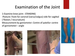
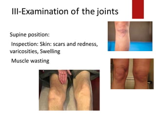
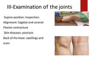
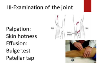
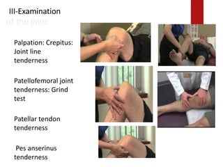
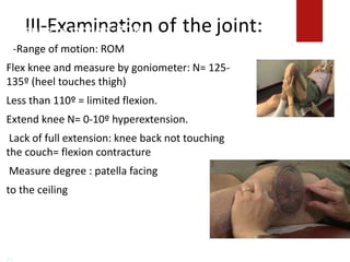
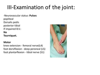
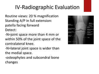
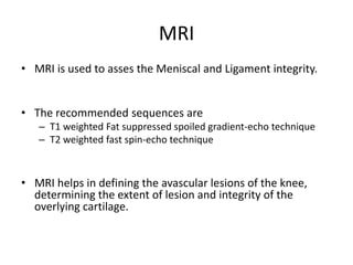
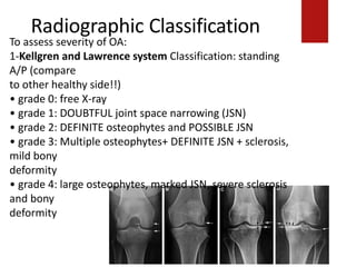
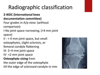
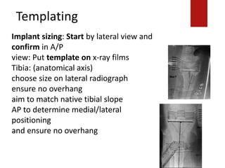
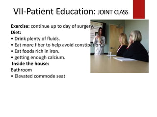
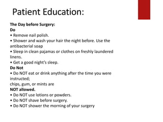
Download to read offline


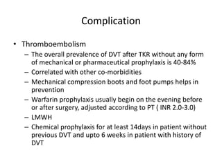
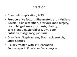


This document discusses the pre-operative evaluation and planning for total knee replacement (TKR) surgery. It covers indications and contraindications for the surgery, patient evaluation including history, physical exam, and imaging, optimization of co-morbidities, surgical planning including templating and sizing, education of the patient, and potential complications of the surgery such as thromboembolism. The goal of the pre-operative process is to adequately prepare the patient and surgical team so that the TKR journey begins with the initial patient meeting and ends safely at the operating room door.


![CTEV [ clubfoot] DR ARUN LAL ,DR MOHAMED ASHRAF travancore medical college k...](https://cdn.slidesharecdn.com/ss_thumbnails/ctevclubfootdrarunlaldrmohamedashraftravancoremedicalcollegekollamkeralaindia-260208063247-18fc466c-thumbnail.jpg?width=640&height=640&fit=bounds)


![PERI-PROSTHETIC FRACTURE NAIL-PLATE CONSTRUCT [NPC].pptx](https://cdn.slidesharecdn.com/ss_thumbnails/drarunkumardrmohamedashrafperiprostheticfrasturenail-plateconstructnpc-260209164459-7e9d15a1-thumbnail.jpg?width=640&height=640&fit=bounds)
![ONFH[AVN HIP] -TRIPLE REGIME -A NOVAL SURGICAL CONCEPT .pptx](https://cdn.slidesharecdn.com/ss_thumbnails/onfhavnhip2026koaconcalicutdrgokuldevdrmashraf-260210064517-213ec005-thumbnail.jpg?width=640&height=640&fit=bounds)


