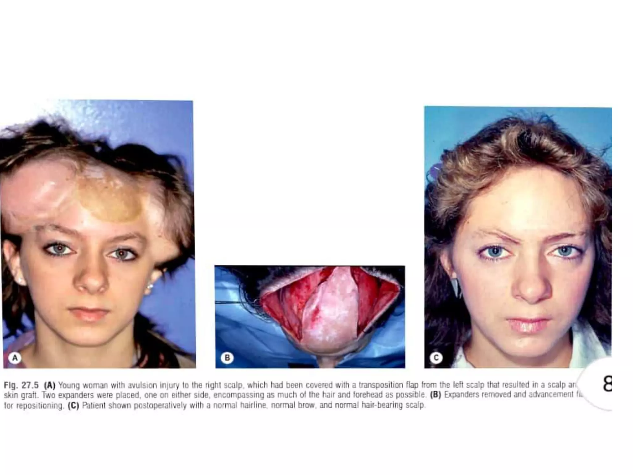
Tissue expansion is a technique used to generate additional soft tissue for reconstruction. It involves surgically placing an implant under the skin that is gradually inflated over time using saline injections, causing the overlying tissue to stretch and grow. The document discusses the history, biology, types of implants, principles and applications of tissue expansion. It can be used to reconstruct defects on the scalp, forehead, face, neck and nose by expanding the surrounding tissue.
'Surgical Incisions on Abdominal Wall', a Surgical Anatomy Seminar by 1st yr MBBS students of Venkateswara Institute of Medical Science, Galraula, UP. India
Knee anatomy and clinical tests 2024.pdfvimalpl1234
This includes all relevant anatomy and clinical tests compiled from standard textbooks, Campbell,netter etc..It is comprehensive and best suited for orthopaedicians and orthopaedic residents.
Explore natural remedies for syphilis treatment in Singapore. Discover alternative therapies, herbal remedies, and lifestyle changes that may complement conventional treatments. Learn about holistic approaches to managing syphilis symptoms and supporting overall health.
Recomendações da OMS sobre cuidados maternos e neonatais para uma experiência pós-natal positiva.
Em consonância com os ODS – Objetivos do Desenvolvimento Sustentável e a Estratégia Global para a Saúde das Mulheres, Crianças e Adolescentes, e aplicando uma abordagem baseada nos direitos humanos, os esforços de cuidados pós-natais devem expandir-se para além da cobertura e da simples sobrevivência, de modo a incluir cuidados de qualidade.
Estas diretrizes visam melhorar a qualidade dos cuidados pós-natais essenciais e de rotina prestados às mulheres e aos recém-nascidos, com o objetivo final de melhorar a saúde e o bem-estar materno e neonatal.
Uma “experiência pós-natal positiva” é um resultado importante para todas as mulheres que dão à luz e para os seus recém-nascidos, estabelecendo as bases para a melhoria da saúde e do bem-estar a curto e longo prazo. Uma experiência pós-natal positiva é definida como aquela em que as mulheres, pessoas que gestam, os recém-nascidos, os casais, os pais, os cuidadores e as famílias recebem informação consistente, garantia e apoio de profissionais de saúde motivados; e onde um sistema de saúde flexível e com recursos reconheça as necessidades das mulheres e dos bebês e respeite o seu contexto cultural.
Estas diretrizes consolidadas apresentam algumas recomendações novas e já bem fundamentadas sobre cuidados pós-natais de rotina para mulheres e neonatos que recebem cuidados no pós-parto em unidades de saúde ou na comunidade, independentemente dos recursos disponíveis.
É fornecido um conjunto abrangente de recomendações para cuidados durante o período puerperal, com ênfase nos cuidados essenciais que todas as mulheres e recém-nascidos devem receber, e com a devida atenção à qualidade dos cuidados; isto é, a entrega e a experiência do cuidado recebido. Estas diretrizes atualizam e ampliam as recomendações da OMS de 2014 sobre cuidados pós-natais da mãe e do recém-nascido e complementam as atuais diretrizes da OMS sobre a gestão de complicações pós-natais.
O estabelecimento da amamentação e o manejo das principais intercorrências é contemplada.
Recomendamos muito.
Vamos discutir essas recomendações no nosso curso de pós-graduação em Aleitamento no Instituto Ciclos.
Esta publicação só está disponível em inglês até o momento.
Prof. Marcus Renato de Carvalho
www.agostodourado.com
ARTIFICIAL INTELLIGENCE IN HEALTHCARE.pdfAnujkumaranit
Artificial intelligence (AI) refers to the simulation of human intelligence processes by machines, especially computer systems. It encompasses tasks such as learning, reasoning, problem-solving, perception, and language understanding. AI technologies are revolutionizing various fields, from healthcare to finance, by enabling machines to perform tasks that typically require human intelligence.
These simplified slides by Dr. Sidra Arshad present an overview of the non-respiratory functions of the respiratory tract.
Learning objectives:
1. Enlist the non-respiratory functions of the respiratory tract
2. Briefly explain how these functions are carried out
3. Discuss the significance of dead space
4. Differentiate between minute ventilation and alveolar ventilation
5. Describe the cough and sneeze reflexes
Study Resources:
1. Chapter 39, Guyton and Hall Textbook of Medical Physiology, 14th edition
2. Chapter 34, Ganong’s Review of Medical Physiology, 26th edition
3. Chapter 17, Human Physiology by Lauralee Sherwood, 9th edition
4. Non-respiratory functions of the lungs https://academic.oup.com/bjaed/article/13/3/98/278874
Title: Sense of Smell
Presenter: Dr. Faiza, Assistant Professor of Physiology
Qualifications:
MBBS (Best Graduate, AIMC Lahore)
FCPS Physiology
ICMT, CHPE, DHPE (STMU)
MPH (GC University, Faisalabad)
MBA (Virtual University of Pakistan)
Learning Objectives:
Describe the primary categories of smells and the concept of odor blindness.
Explain the structure and location of the olfactory membrane and mucosa, including the types and roles of cells involved in olfaction.
Describe the pathway and mechanisms of olfactory signal transmission from the olfactory receptors to the brain.
Illustrate the biochemical cascade triggered by odorant binding to olfactory receptors, including the role of G-proteins and second messengers in generating an action potential.
Identify different types of olfactory disorders such as anosmia, hyposmia, hyperosmia, and dysosmia, including their potential causes.
Key Topics:
Olfactory Genes:
3% of the human genome accounts for olfactory genes.
400 genes for odorant receptors.
Olfactory Membrane:
Located in the superior part of the nasal cavity.
Medially: Folds downward along the superior septum.
Laterally: Folds over the superior turbinate and upper surface of the middle turbinate.
Total surface area: 5-10 square centimeters.
Olfactory Mucosa:
Olfactory Cells: Bipolar nerve cells derived from the CNS (100 million), with 4-25 olfactory cilia per cell.
Sustentacular Cells: Produce mucus and maintain ionic and molecular environment.
Basal Cells: Replace worn-out olfactory cells with an average lifespan of 1-2 months.
Bowman’s Gland: Secretes mucus.
Stimulation of Olfactory Cells:
Odorant dissolves in mucus and attaches to receptors on olfactory cilia.
Involves a cascade effect through G-proteins and second messengers, leading to depolarization and action potential generation in the olfactory nerve.
Quality of a Good Odorant:
Small (3-20 Carbon atoms), volatile, water-soluble, and lipid-soluble.
Facilitated by odorant-binding proteins in mucus.
Membrane Potential and Action Potential:
Resting membrane potential: -55mV.
Action potential frequency in the olfactory nerve increases with odorant strength.
Adaptation Towards the Sense of Smell:
Rapid adaptation within the first second, with further slow adaptation.
Psychological adaptation greater than receptor adaptation, involving feedback inhibition from the central nervous system.
Primary Sensations of Smell:
Camphoraceous, Musky, Floral, Pepperminty, Ethereal, Pungent, Putrid.
Odor Detection Threshold:
Examples: Hydrogen sulfide (0.0005 ppm), Methyl-mercaptan (0.002 ppm).
Some toxic substances are odorless at lethal concentrations.
Characteristics of Smell:
Odor blindness for single substances due to lack of appropriate receptor protein.
Behavioral and emotional influences of smell.
Transmission of Olfactory Signals:
From olfactory cells to glomeruli in the olfactory bulb, involving lateral inhibition.
Primitive, less old, and new olfactory systems with different path
Tom Selleck Health: A Comprehensive Look at the Iconic Actor’s Wellness Journeygreendigital
Tom Selleck, an enduring figure in Hollywood. has captivated audiences for decades with his rugged charm, iconic moustache. and memorable roles in television and film. From his breakout role as Thomas Magnum in Magnum P.I. to his current portrayal of Frank Reagan in Blue Bloods. Selleck's career has spanned over 50 years. But beyond his professional achievements. fans have often been curious about Tom Selleck Health. especially as he has aged in the public eye.
Follow us on: Pinterest
Introduction
Many have been interested in Tom Selleck health. not only because of his enduring presence on screen but also because of the challenges. and lifestyle choices he has faced and made over the years. This article delves into the various aspects of Tom Selleck health. exploring his fitness regimen, diet, mental health. and the challenges he has encountered as he ages. We'll look at how he maintains his well-being. the health issues he has faced, and his approach to ageing .
Early Life and Career
Childhood and Athletic Beginnings
Tom Selleck was born on January 29, 1945, in Detroit, Michigan, and grew up in Sherman Oaks, California. From an early age, he was involved in sports, particularly basketball. which played a significant role in his physical development. His athletic pursuits continued into college. where he attended the University of Southern California (USC) on a basketball scholarship. This early involvement in sports laid a strong foundation for his physical health and disciplined lifestyle.
Transition to Acting
Selleck's transition from an athlete to an actor came with its physical demands. His first significant role in "Magnum P.I." required him to perform various stunts and maintain a fit appearance. This role, which he played from 1980 to 1988. necessitated a rigorous fitness routine to meet the show's demands. setting the stage for his long-term commitment to health and wellness.
Fitness Regimen
Workout Routine
Tom Selleck health and fitness regimen has evolved. adapting to his changing roles and age. During his "Magnum, P.I." days. Selleck's workouts were intense and focused on building and maintaining muscle mass. His routine included weightlifting, cardiovascular exercises. and specific training for the stunts he performed on the show.
Selleck adjusted his fitness routine as he aged to suit his body's needs. Today, his workouts focus on maintaining flexibility, strength, and cardiovascular health. He incorporates low-impact exercises such as swimming, walking, and light weightlifting. This balanced approach helps him stay fit without putting undue strain on his joints and muscles.
Importance of Flexibility and Mobility
In recent years, Selleck has emphasized the importance of flexibility and mobility in his fitness regimen. Understanding the natural decline in muscle mass and joint flexibility with age. he includes stretching and yoga in his routine. These practices help prevent injuries, improve posture, and maintain mobilit
Flu Vaccine Alert in Bangalore Karnatakaaddon Scans
As flu season approaches, health officials in Bangalore, Karnataka, are urging residents to get their flu vaccinations. The seasonal flu, while common, can lead to severe health complications, particularly for vulnerable populations such as young children, the elderly, and those with underlying health conditions.
Dr. Vidisha Kumari, a leading epidemiologist in Bangalore, emphasizes the importance of getting vaccinated. "The flu vaccine is our best defense against the influenza virus. It not only protects individuals but also helps prevent the spread of the virus in our communities," he says.
This year, the flu season is expected to coincide with a potential increase in other respiratory illnesses. The Karnataka Health Department has launched an awareness campaign highlighting the significance of flu vaccinations. They have set up multiple vaccination centers across Bangalore, making it convenient for residents to receive their shots.
To encourage widespread vaccination, the government is also collaborating with local schools, workplaces, and community centers to facilitate vaccination drives. Special attention is being given to ensuring that the vaccine is accessible to all, including marginalized communities who may have limited access to healthcare.
Residents are reminded that the flu vaccine is safe and effective. Common side effects are mild and may include soreness at the injection site, mild fever, or muscle aches. These side effects are generally short-lived and far less severe than the flu itself.
Healthcare providers are also stressing the importance of continuing COVID-19 precautions. Wearing masks, practicing good hand hygiene, and maintaining social distancing are still crucial, especially in crowded places.
Protect yourself and your loved ones by getting vaccinated. Together, we can help keep Bangalore healthy and safe this flu season. For more information on vaccination centers and schedules, residents can visit the Karnataka Health Department’s official website or follow their social media pages.
Stay informed, stay safe, and get your flu shot today!
1. TISSUE EXPANSION PRINCIPLE
AND APPLICATION
Dr. Amit Kumar Choudhary
SR Plastic and Reconstructive Surgery
RIMS,Imphal
Moderator-Dr. AK. Ibohal Singh
2. HISTORY OF TISSUE EXPANSION
• Neumann induced soft tissue growth with a subcutaneously implanted balloon in an attempt to
reconstruct an external ear deformity.
• Radovan and Austad simultaneously evolved the concept of purposeful soft tissue expansion with
use of an implanted silicone balloon.
Radovan's device contained a self-sealing valve through which saline was periodically injected to
increase size of the prosthesis.
• Austad's prosthesis was devised as a self-inflating device using osmotic gradients driven by salt
placed within the expander.
4. Epidermis
• Early after placement of the prosthesis, significant thickening of the epidermis is evident. Within 4 to
6 weeks, epidermal thickness generally returns to initial levels, but some increase in thickness
persists for many months.
• Hair follicles and accessory skin structures are compressed but no evidence of degeneration.
• Animal studies demonstrate that there may be an increase in number of hairs and density
proportional with expansion.
• Melanocytic activity is increased during expansion but returns to normal within several months after
completion of reconstruction
5.
6. Dermis
• The dermis decreases rapidly in thickness over the entire implant during expansion.
• Dermal thinning persists at least 36 weeks after expansion is completed . A dense fibrous capsule is
formed around the implant, which becomes less cellular over time.
• The capsule is thickest at 2 months of expansion. Progressive collagenization with well-organized
bundles develops during 3 months.
• Dystrophic calcification may occur when a hematoma resolves or when the prosthesis is repeatedly
traumatized.
• Expanded tissue demonstrates a quantitative increase in collagen content of the dermis. After
expansion, the relative proportions of type I and type III collagen are not significantly changed in
the dermalepidermal or subcutaneous-capsular interface.
• Mitotic activity in the capsule fibroblast is maximum about 96 hours after expansion.
• The application of a constant pressure beyond 96 hours results in progressive decrease in mitotic
activity
7. Muscle
• Muscle atrophies significantly during the process of
expansion, whether the prosthesis is placed above or below a
specific muscle .
• The effects on human muscle after expansion have demonstrated
occasional histologic ulceration.
• Focal muscle fiber degeneration with glycogen deposits and mild
interstitial fibrosis have been noted. Some muscle fibers show
disorganization of the myofibrils in the sarcomeres.
• Expansion of skeletal muscle is not a stretching process but rather a
growth of the muscle cell accompanied by an increase in the
number of sarcomeres per fiber.
8.
9. Cranial Bone
• There is a decrease in bone thickness and volume in cranial bone beneath the expander, but bone
density is unaffected.
• An increase in bone volume and thickness occurs predominantly at the periphery of the expander.
• Osteoplastic bone resorption occurs beneath expanders, and a periosteal inflammatory reaction is
seen at the periphery of the expander.
• Cranial bone appears to be significantly more affected than long bone is. Long bone remodeling
begins within 5 days after removal of the expander, and the long bone is completely normal within
2 months.
10. Vascularity of Expanded Tissue
• It has been clinically and histologically demonstrated that a large number of new vessels are
formed adjacent to the capsule.
• The content of collagen fibers in existing vessels initially decreases after expansion.
• Elastic fibers in existing blood vessels initially increase, probably as a response to mechanical stress.
• Angiogenesis probably occurs secondary to ischemia of the expanded tissues.
• Cells expressing vascular growth factor is significantly increase.
11. SURVIVAL
• Flaps elevated in expanded tissue have
significantly greater survival areas compared
with acutely raised and delayed flaps.
12. Ultrastructure of Expanded Tissue
• The epidermis demonstrates a reduction of intercellular distance and a significant
decrease in the undulation of the basal lamina .
• The dermis displays large, compact bundles of collagen fibers oriented in parallel
fashion over the implant surface.
• Active fibroblasts are found in the expanded dermis.
• Myofibroblasts develop in the deep dermis adjacent to the capsule.
• Skeletal muscle demonstrates pressure atrophy with increased mitochondria and
abnormal rearrangement of sarcomeres.
13. Molecular Basis for Tissue Expansion
• Intracellular tension and cell structure are maintained by a system of microfilaments within
the cytoplasm.
• These microfilaments act to transduce signals to adjacent cells and play a critical role in
initiating transduction cascades within the cell.
• Protein kinase C plays a pivotal role in signal transduction.
• Mechanical strain on cell walls activates inositol phosphatase, phospholipase A2,
phospholipase D, and other messengers.
14.
15.
16. • Activation of these components results in protein kinase C activation. Protein kinase C is
associated with nuclear proteins, intracellular signals can be transmitted to the nucleus.
• Many growth factors, including platelet-derived growth factor and angiotensin II, play a role
in strain induced cell growth.
• Platelet-derived growth factor has a effect of stimulating cutaneous cell proliferation.
• Transforming growth factor-β production has also been demonstrated in stretch in vitro
models and has been implicated in extracellular matrix products.
17. The Source of Increased Tissue from Expansion
• The increase in skin surface area over the expander includes normal skin brought
in from adjacent areas as well as new skin generated by increased mitosis.
• Increased mitotic activity in the epidermis directly overlying the expansion.Serial
inflations of the prosthesis result in serial increases in tritiated thymidine uptake.
• With deflation of the implant, a significant decrease in the rate of the epidermal
mitosis below normal baseline occurs.
19. Radovan's expander
• Radovan's expander consisted of a silicone prosthesis with two valves, each connected to the
main reservoir by silicone tubing.
• One valve was used for injection; the other was used as a means to withdraw fluid.
Technologic improvements resulted in a single valve for both purposes.
• The filling reservoir may be incorporated directly into the prosthesis. Such devices have the
advantage of avoiding the remote port.
• The valve in the integrated prosthesis can be difficult to palpate.
• Breast reconstruction with these prostheses has become popular..
20. Expanders with distal ports
• Remote filling ports.
• Advantage of minimizing the risk of implant
puncture during inflation.
• The distal , self-sealing injection port and inflation
reservoir are connected to the prosthesis by a
length of tubing.
• This allows the injection port to be placed away
from the expander pocket.
• It is also possible to move the inflation port of a
distant reservoir to the exterior of the body; this
location facilitates inflation, particularly when the
expansion is accomplished by family member.
22. Expanders with integrated ports
• The inflation reservoir may be incorporated directly in the prosthesis.
• Advantage of avoiding the remote port and its associated mechanical problems.
• Risk of inadvertent perforation of the prosthesis during inflation is higher .
• Magnetic and ultrasonic devices can be useful when the valve is difficult to locate and
metal finding devices have been designed.
• Expander prostheses with integrated valves are particularly popular for breast
reconstruction where adequate soft tissue and pocket can accommodate the added
projection of the injection port.
23. Self-inflating expanders
• Self-inflating expanders have become available largely through
Europe.
• These contain osmotic hydrocolloids that cause migration of
extracellular water through the silicone membrane of the device.
• The first such expander was devised by Dr. Austad and was used
experimentally.
• It was not approved by the Food and Drug Administration
24. BASIC PRINCIPLES
• Tissue expansion is a protracted procedure that may involve temporary, but very
obvious, cosmetic deformity.
• Emotionally stable patients of all ages tolerate tissue expansion well.
Noncompliant or mentally impaired patients are poor candidates.
• Smokers have a higher risk of complications.
• Tissue expansion is generally best performed as a secondary reconstructive
procedure rather than in the acute trauma period.
• Expansion can be performed adjacent to an area of an open wound before
definitive closure, but such a procedure carries the risks of infection, extrusion,
and less than-optimal results.
• Tissue expansion is best suited to those patients who require definitive, optimal
coverage ,when time is not of the essence.
25. Incision planning and implant
selection
• The proposed type of flap
– advancement
- rotation
- Interpolation
The simpler the flap, the less the potential for complication.
• planning
(1) incisions are incorporated into tissue that will become one margin of the flap
(2) aesthetic units are reconstructed
(3) scars are in minimally conspicuous locations
(4) tension on suture lines is reduced.
26. • Incisions should be planned to minimize tension on the suture line and risk of
extrusion.
• Tension from the initial inflation on the suture line will be greater when incisions
are parallel to the direction of expansion than when they are perpendicular to it.
• Undermining of the prosthesis should be sufficient enough that the prosthesis can
be easily accommodated and the wound can be closed in multiple layers.
• The inflation valve and tubing should be maintained at a site away from the
incision.
27. The size of the implant
• The size of the implant selected should closely relate to the size and shape of the donor
surface.
• An implant equal to or slightly smaller than the donor area is selected.
• In general, the use of multiple small expanders is better than the use of one large expander.
Inflation of multiple prostheses proceeds more rapidly and complications are fewer.
• Multiple expanders also allow the surgeon to vary the plan for reconstruction after expansion
• An integrated valve and a distal inflation port should be considered on a case-by-case basis.
28. Implant and distal port positioning
• If a remote filling port and reservoir is chosen,it must be
placed superficially in subcutaneous tissue,where even an
extremely small port is easily palpable under stable skin.
• To minimize discomfort, it is occasionally possible to
position a filling port in an area that is relatively less sensitive.
• The port should be placed in a location that will not be
subjected to pressure .
• Bony prominences are avoided
29. Implant inflation strategy and
technique
• Implants should be partially inflated immediately after wound closure. This allows closure of
“dead space” to minimize seroma and hematoma formation.
• It also smoothes out the implant wall to minimize risk of fold extrusion.
• Enough saline is placed to fill the entire dissection space without placing undue tension on
the suture line.
• Serial inflation usually starts 1–2 weeks after initial placement.
• Inflation reservoirs seal best when a 23-gauge or smaller needle is used. A 23-gauge butterfly
30. • Frequent small-volume inflations are better tolerated and are physiologically more suited to the
development of adequate overlying tissue than are large infrequent Inflations
• inflations proceed until the patient experiences discomfort or blanching of the overlying skin.
• Devices such as pressure transducers and oxygen tension monitors are available to help determine
proper inflation.
• An objective inspection of the patient’s response is usually a reliable indicator of appropriate
inflation.
• Serial inflations proceed until an adequate amount of soft tissue has been generated to accomplish
the specific surgical goal.
32. Burns
• Reconstruction should be carried out after all burns have and scars have
matured.
• Planning is particularly important in these cases so that a minimum
number of suture lines is produced and that these suture lines do not
cross aesthetic units.
• Significant late distortion and contracture may result in excessive scars
placed in burned tissue, particularly in the facial area
33. Tissue expansion in children
• Skin and soft tissue are always thinner in children than in adults. These tissues are
probably better vascularized but less resistant to trauma.
• Tissue expansion has a higher complication rate in children than in adults.
• Major complication risk – particularly extrusion – is more common at the second,
third, and fourth serial expansion. This is particularly true in the head and neck
(with the exception of the scalp).
• Small-volume inflation at frequent intervals is especially useful in children because
34. Expansion of myocutaneous, fascial,
and free flaps
• Myocutaneous flaps are the standard of care for the treatment of large defects, particularly when
bone and vital structures are involved.
• The territories of standard flaps are well described. These territories can be considerably enlarged
by placing an expander beneath the standard myocutaneous flap, and an extremely large flap can
be developed over a short period.
• Expansion increases the vascularity of the flap and allows a large,adjacent random area to be
carried with the original flap.
. The vascular pedicle of such flaps remains intact and may in fact be elongated, thus allowing flaps to
be transferred farther.
35. • Myocutaneous flaps such as the latissimus dorsi
and pectoralis can be expanded to almost double
their surface area, allowing coverage of almost
any defect on the abdomen or thorax.
• Expanders of up to 1000 Ml can be placed
beneath such flaps and rapidly expanded.
36. Expanded full-thickness skin grafts
• Donor defect is usually created by harvesting full-thickness grafts.
• The placement of a large tissue expander beneath the donor site can result in a
large full-thickness graft that is particularly useful in resurfacing large areas .
• The best color matches are generated when the full thickness graft is expanded
and harvested as close as possible to the recipient site.
• The periorbital area and the area around the mouth are particularly well suited to
reconstruction with expanded full-thickness grafts harvested from the
supraclavicular area.
• Expanded full-thickness grafts are very helpful in reconstructing defects of the
forehead . A single full-thickness graft can be harvested from the supraclavicular
area or from under the breast fold.
37. • The full-thickness graft is approximately 10–15% larger than the recipient
area.
• Expanded full-thickness skin grafts require more immobilization than split
thickness.
• A bolster dressing or, ideally, a VAC sponge dressing is required. The graft
is sutured in place and a VAC sponge placed over the graft; 125 mmHg of
negative pressure is maintained for 4 days.
39. HEAD AND NECK
• The skin of the face can be subdivided into five tissue specific areas:
1. The scalp
2. The forehead is a continuation of the scalp, but it is distinguished from the scalp by its thick
skin, large number of sebaceous glands, and lack of hair.
3. The nose is embryologically related to the forehead, so it closely mimics the forehead in color,
texture, and sebaceous gland content.
4. The lateral cheek areas, neck, and upper lip have fewer sebaceous glands; the skin is thinner, and
the hair-bearing pattern is significantly different in quality and quantity from that on the
remainder of the body.
5. The skin of the periorbital areas is extremely thin and pliable, containing a minimal number of
sebaceous glands
40. Scalp
• Tissue expansion is the ideal procedure for the
reconstruction of scalp defects.
• Expansion of the scalp is well tolerated and is the only
procedure that allows development of normal hair-bearing
tissue to cover the areas of alopecia.
• The amount of scar and deformity generated is considerably
less than other procedures such as serial reduction and
complex multiflap procedures.
41. • The darker the hair, the more visible the thinning is.
• Individuals who have large defects and require extreme
expansion may achieve better results by lightening the
hair with dyes.
Advancement or rotation flaps achieve the best
results, particularly when the anterior hairline is
reconstructed.
42.
43.
44. Forehead
• The forehead is anatomically and histologically identical to the scalp except for its different
numbers of sebaceous glands and hair-bearing follicles.
• Reduction or increase of the surface area of the forehead by 20–25% is not usually readily
apparent after appropriate hair styling
• .
By expanding the scalp in conjunction with expanding the forehead, better symmetric brow
positioning is achieved while maintaining the normal hairline.
Expansion of the forehead is useful in many craniofacial anomalies with low hairlines.
Expansion of the remaining forehead is accomplished and moved into a cephalad direction.
The intervening hair-bearing scalp is excised
45. Lateral face and neck
• The type of skin on the lateral facial
areas and neck is essentially the same.
• A large Mustardé expanded rotation
flap can be developed on the neck for
use in facial reconstruction.
• In children, there is a higher risk of
extrusion problems in the expansion of
this area of the face .
• Adults, such reconstruction can be
accomplished with relative ease. The
flap is based inferiorly and medially.
46.
47. Nose
• Reconstruction of major defects of the nose, including total nose reconstruction,
may be facilitated by pre expanding the forehead skin.
• when total nose reconstruction is performed, expansion of the entire forehead
with a 400–600-mL prosthesis generates an adequate number of large, well-
vascularized flaps to accomplish both total nose reconstruction and closure of the
donor site.
• Because the color and texture of the forehead are ideally suited to reconstruction
of the nose, this procedure makes reconstruction of any nasal defect possible.
48. • A cranial bone or rib graft is
taken to reconstruct the
dorsum of the nose. This is
either secured to the
remaining nasal bone or
attached by a plate to the
skull.
• The nasal cartilage is
reconstructed bilaterally
with cartilage from the
conchal bowl.
49. Ear
• Most cases of microtia and traumatic ear deformities can be
reconstructed without expansion.
• Expansion is helpful when skin and soft tissue are insufficient for
reconstruction.
• As with all ear reconstructions, a child should be approximately 7
years of age before reconstruction is begun.
50. Periorbital area with expanded
full-thickness grafts
• The periorbital area contains skin that is soft and pliable.
• Little tissue in the periorbital area can be expanded or
move easily.
• When large areas require reconstruction, fullthickness skin
grafts from expanded donor sites are recommended.
• Replacement of aesthetic units – the entire periorbital area
or the upper or lower lid – gives the best result .
51. Reconstruction in the breast, chest,
trunk, and extremities
• Tissue expansion was introduced by Chemar Radovan in 1982
to facilitate breast reconstruction in post mastectomy patients
because these patients were found to have insufficient chest
wall tissue for placement of the implant.
52. The hypoplastic breast
• Tissue expansion has played
an important role in the
reconstruction of both
acquired and congenital
breast hypoplasia.
• Management of the
deformity depends on the
degree of breast asymmetry,
the nature of the deformity,
the quality of the chest wall
soft tissue, and the age of
the patient at presentation.
53.
54. The immature breast
• Management of young adolescents presenting with breast
asymmetry.
Adolescence is a critical time that is characterized by intense
social pressures and self awareness of a developing physique,
so failure to address the problem of breast asymmetry can
result in psychological problems. These patients do not need
full maturity for reconstruction.
55. Correction of Poland syndrome
• Poland syndrome involves
- abnormal development of the breast
- thoracic wall deformities
- deformities of the upper extremity
- vertebral anomalies.
• Poland syndrome exhibits a uniform absence of the sternal head of the pectoralis major muscle.
• Abnormalities in the anterior ribs and costal cartilages and deficiencies of the muscles of the scapular area,
including the latissimus dorsi.
• Other findings include deficiency of subcutaneous tissue; hypoplasia, aplasia, or
malposition of the nipple–areola complex; and deficiency of breast tissue.
56.
57. Expansion of the trunk
• The trunk and abdomen are well suited to tissue expansion in individuals of all ages.
• Because of the large adjoining surface area from which tissue can be recruited, large prostheses
can be placed and flaps quickly expanded.
• Large deformities, such as burns, giant hairy nevi, and other congenital anomalies, the expanders
are inflated maximally and the flaps are advanced. The prostheses are left in place and re-
expansion is carried out in the subsequent weeks.
• In the abdomen, two or three serial expansions are usually well tolerated.
58.
59. Expansion in the extremities
• Skin and soft tissues of the extremities tolerate tissue
expansion well.
• The capsule that develops adjacent to the expander has a
resilient surface that can be transposed over joints and
tendons to decrease adhesions.
61. IMPLANT FAILURE
• Despite design improvements, the use of an excessively large needle or
the inadvertent puncture of the implant can lead to implant deflation.
• To maximize sealing of the valve, the implant reservoir should be entered
at a 90° angle.
• If there is any question about the location of the inflation reservoir,
radiologic or sonographic techniques may be helpful.
62. INFECTION
• The introduction of bacteria to the wound in the perioperative
period is the most common cause of early infection.
• Areas susceptible to lymphedema, such as traumatized lower
extremities, carry a significantly higher rate of infection.
• An area of copious lymphatic drainage, such as the neck or the
groin, also tends to accumulate lymphatic fluid around a prosthesis
and is more susceptible to infection.
• If infection occurs late in the course of expansion, the prosthesis
can be removed and the expanded tissue advanced after irrigation
of the infected cavity.
63. IMPLANT EXPOSURE
• Implant exposure can occur both early in the postoperative period and after a protracted course of
expansion.
• Treatment of the exposed implant depends on the timing of exposure.
• Exposure early after placement is usually related to inadequate dissection or use of an excessively
large prosthesis that abuts on wound closure.
• If the prosthesis becomes exposed soon after placement, it is best to remove it and reoperate 3–4
months later.
• Late exposure is usually related to excessively rapid or overzealous inflation.
• If minimal or late exposure occurs during the course of expansion, the procedure can continue with
the use of antibiotic creams on the exposed area: In this situation, multiple, rapid fillings are done
to generate adequate tissue.
• Reinforcement of the compromised overlying skin with paper tape is sometimes helpful. Most flaps
survive and
64. COMPROMISE AND LOSS OF FLAP
TISSUE
• To ensure vascularity,one should attempt to
maintain a major axial vessel in the expanded
tissue.