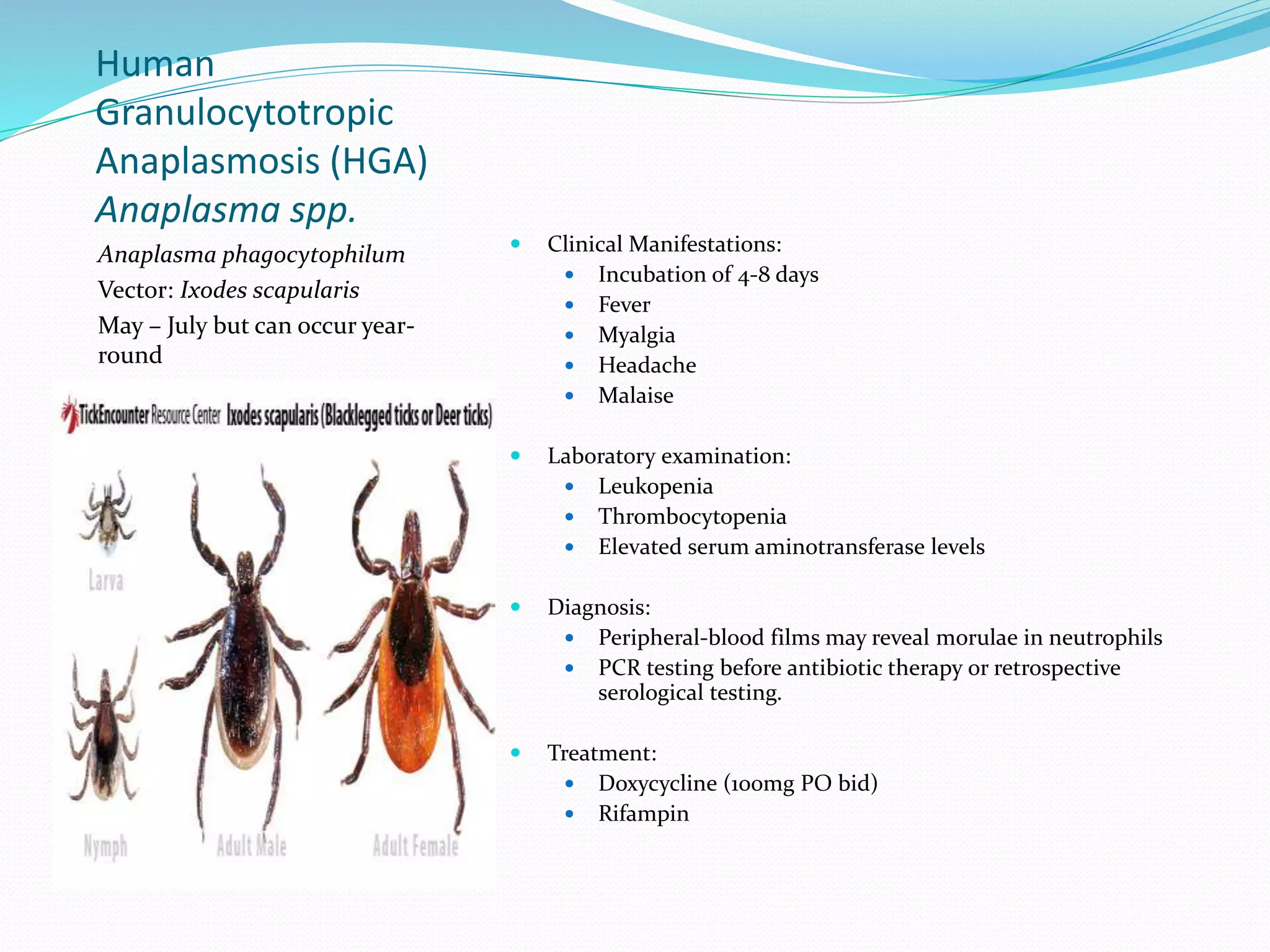
Human
Granulocytotropic
Anaplasmosis (HGA)
Anaplasma spp.
Anaplasma phagocytophilum
Vector: Ixodes scapularis
May – July but can occur year-
round
 Clinical Manifestations:
 Incubation of 4-8 days
 Fever
 Myalgia
 Headache
 Malaise
 Laboratory examination:
 Leukopenia
 Thrombocytopenia
 Elevated serum aminotransferase levels
 Diagnosis:
 Peripheral-blood films may reveal morulae in neutrophils
 PCR testing before antibiotic therapy or retrospective
serological testing.
 Treatment:
 Doxycycline (100mg PO bid)
 Rifampin
Ehrlichiosis
Ehrlichia spp.
(rickettsiale)
A tickborne disease, ehrlichia species are
obligate intracellular microbes that infect
and kill WBCs.
Three species are known to cause disease in
humans:
 ehrlichia ewingii
 ehrlichia chaffeensis
 ehrlichia canis
There are also two non-ehrlichia pathogens
that cause this disease (under the umbrella
term ehrlichiosis):
 anaplasma phagocytophilum
 neorickettsia sennetsu
• Presentation: nonspecific
- headache
- fatigue
- myalgia
- rash, purpura (rare)
- immunosuppression by TNF-alpha
blockade, leading to opportunistic
coinfection (e.g. candida)
Severe cases may feature a toxic
shock-like sydrome and cause death,
but these are almost exclusively in
immunosuppressed patients
developing RDS, hepatitis or
opportunistic coinfection.
• Treatment:
- doxycycline is DOC
- minocycline
- rafampin in patients with
tetracycline allergy
- chloramphenicol has been used,
but susceptible to resistance
Bartonella spp.
Most species of Bartonella have successfully adapted to
surviving in specific domestic or wild animals, creating
a reservoir for human infection.
**B. Bacilliformis and B. quintana are not zoonotic**
Cat-Scratch
Disease(CSD)
Bartonella henselae
Gram-negative bacillus
Contact with apparently healthy
cats, kittens is the primary source
of infection. Patient can scratched,
bitten, or licked.
Approximately 10 cases per 100,000
population.
 Clinical manifestations:
 Localized lesion (papule, vesicle or nodule) at the site of
inoculation
 Painful regional lymphadenopathy 1-3 weeks after cat
contact (Axillary and epitrochlear)
 Low-grade fever
 Malaise
 Anorexia
 Atypical disease involves extranodal manifestations (10-15%)
 Diagnosis:
 Serological testing varies in sensitivity and specificity.
 PCR analysis of lymph node tissue, pus, or the primary
inoculation lesion is highly sensitive and specific
 Culture negative endocarditis: Serologic or PCR testing for
Bartonella in cardiac valve tissue.
 Severe cases may be treated with oral doxycycline or
ciprofloxacin
CSD Continued
 Bacillary angiomatosis: One or more painless skin lesions
that may present as tan, red, or purple in color.
Subcutaneous masses or nodules, ulcurated plaques and
verrucous growths can also accour. Painful osseus lesions
may develop and appear as lytic lesions on radiograph.
 Peliosis: Angioproliferative disorder described as blood-filled
cystic structures that affects primarily the liver.
Pasteurella
multocida
Gram-negative, nonmotile,
penicillin-sensitive coccobacillus
Oxidase +
Catalase +
 Clinical manifestations:
 Rapidly advancing inflammation
 Purulent discharge (few hours after bite)
 Cat bites > dog bites to cause septic arthritis or
osteomyelitis
 Treatment:
 Amoxicillin/clavulanate 250-500mg PO tid
 Ampicillin/sulbactam 1.5-3.0g IV q6h
 Alternative treatment in penicillin-allergic
patients:
 Clindamycin 150-300mg PO qid plus TMP-SMX 1 DS
tablet PO bid or ciprofloxacin 500mg PO bid
Rabies
Rhabdoviridae
Nonsegmented, negative sense,
single-stranded RNA virus
Endemic canine rabies has been
eliminated in the United States but
persists in bats, raccoons, skunks and
foxes.
Incubation period can range from
few days to >1 year but usually 20-90
days
Virus binds to postsynaptic nicotinic
acetylcholine receptors and spreads to
CNS ~250mm/d.
Negri body – eosinophilic
cytoplasmic inclusion composed of
rabies virus proteins and viral RNA
found primarily within Purkinje cells
of the cerebellum and pyramidal
neurons of the hippocampus.
 Clinical manifestations:
 Prodromal symptoms consist of:
 Fever
 Headache
 Malaise
 Nausea
 Vomiting
 Anxiety
 Agitation (2-10 days)
 Paresthesias, pain and pruritus near site of exposure
 Acute neurologic phase:
 Encephalitic (80%): Fever, confusion, hallucinations,
combativeness, seizures. Autonomic dysfunction is
common and presents as hypersalivation, gooseflesh,
cardiac arrhythmia and/or priapism.
 Hydrophobia and aerophobia due to early brainstem
dysfunction
 Paralytic (20%): Unknown muscle weakness
predominates. Begins at bitten extremity and proceeds
to quadriparesis.
 Coma and death: Despite aggressive supportive measures,
recovery is rare. Death occurs usually within 2 weeks.
Rabies
Rabies should be
considered in patients
with acute atypical
encephalitis or acute
flaccid paralysis
(including Guillain-
Barre syndrome
suspected patients)
 Diagnosis: Relatively late (too late) in clinical course.
 Previously unimmunized patient serum neutralizing antibodies
to rabies virus are diagnostic but may not be present until later in
the course of the disease.
 Reverse-transcription PCR can detect fresh virus in saliva, CSF,
skin and brain tissue.
 Direct fluorescent antibody testing is highly sensitive and specific
and can be applied to brain tissue or skin biopsy samples from
the nape of the neck.
 Virus is found in cutaneous nerves at the base of hair follicles
 Prevention:
 Local wound care can greatly reduce risk of rabies infection.
 All previously unvaccinated patients should receive human rabies
immune globulin no later than 7 days!
 Inactivated rabies vaccine should be given as soon as possible.
 Preexposure prophylaxis occasionally given to persons at high
risk.
 Primary vaccine schedule is given on days 0, 7, 21 or 28.
Lyme Disease
Borrelia Burgdorferi
(spirochete)
General characteristics:
Incubation period is 3 to 32 days
Transmission by tick bite, Ixodes,
Ixodidae scapularis
Common in Northeastern United
States
 Stage 1 – early, localized
 Erythema migrans: large, painless, well-demarcated target
shaped lesion
 Commonly seen in thigh, groin or axilla
 Stage 2 – early, disseminated
 Infection may spread through lymphatics and the
bloodstream within days to weeks after the onset of the
“bull’s eye” lesion
 Intermittent flu-like symptoms, headaches, neck stiffness,
fever/chills, fatigue, malaise, musculoskeletal pain
 After several weeks 15% of patients can develop one or
several of the following:
 Meningitis
 Encephalitis
 Cranial neuritis
 Peripheral radiuloneuropathy
 Within weeks to months of onset of symptoms 8% may
present with cardiac manifestations
 Stage 3 – late, persistent
 Arthritis
 Chronic CNS disease
 Acrodermatitis chronica atrophicans
Lyme Disease
Clinical Diagnosis: In early,
localized disease a documented
erythema migrans in a patient with
a history of tick exposure in an
endemic area counters the need for
laboratory confirmation. Treat
empircally.
Serological studies:
ELISA to detect serum IgM and IgG
antibodies during the first month
of illness
Western Blot: confirm positive or
ambiguous results
 Treatment
 Early localized
 Confined to the skin, 14 days of antibiotic therapy is
adequate
 If any indication of spread, extend treatment to 20-30
days.
 Oral Doxycycline (21 days)
 Amoxicillin and cefuroxime are alternative agents
 Erythromycin may be given to pregnant patients with
penicillin allergies
 Treating the complications of facial nerve palsy, arthritis, and
cardiac disease is prolonged therapy (30-60 days). For
meningitis or other CNS complications, treat with IV antibiotics
for 4 weeks.
 Post-Lyme syndrome: For months or years after around 10% of
patients have pain, neurocognitive manifestations, or fatigue
symptoms – indistinguishable from chronic fatigue syndrome
and fibromyalgia.
 Prophylaxis – 200-mg dose of doxycycline, given within 72hr of
the tick bite effectively prevents the disease.
Relapsing Fever
Borrelia recurrentis
(spirochete)
Vector borne disease that can be
spread through human body louse
or ticks.
Louse-borne disease occurs in
epidemics among poor living
conditions, famine and war in
developing countries. Prevalent in
Ethiopia and Sudan.
Poor prognostic signs include
severe jaundice, severe mental
changes, severe bleeding and a
prolonged QT interval on ECG
 Most patients who infected begin to develop symptoms between 5 and 15 days after
the initial bite.
 Sudden fever
 Chills
 Headaches
 Muscle and joint aches
 Nausea
These symptoms continue approximately 2-9 days then disappear.
 Diagnosis:
 May be made on a blood smear as evidence by the presence
of spriochetes. Other spirochete illnesses do not show
spirochetes on blood smear. Once considered the gold standard,
however this method lacks sensitivity and has been replaced by
PCR in many settings if needed.
 Treatment:
 1 – 2 week course of antibiotics
 Most people improve within 24 hours
 Tetracycline-class antibiotics are most effective.
 Jarisch-Herxheimer reaction in over half those treated
 Anxiety
 Diaphoresis
 Fever
 Tachycardia
 Tachypnea
Brucellosis
Brucella spp.
Small, gram negative,
unencapsulated, nonsporulating,
nonmotile rods or coccobacilli that
may persist intracellulary
B. melitensis (sheep, goat, camel)
B. suis (swine)
B. abortus (cattle or buffalo)
B. Canis (dogs)
 Clinical manifestations:
 Incubation period of 1 week to several months
 Undulating fever
 Sweats
 Increasing apathy
 Fatigue
 Lymphadenopathy, hepatosplenomegaly, epididymoorchitis, neurologic involvment,
and focal abscess can also be caused by Brucella infection.
 Diagnosis:
 Cultures using BACTEC system are positive in 7-10 days.
 PCR analysis of blood or tissue is more sensitive, faster, and safer than
culture.
 Treatment:
 Streptomycin 0.75-1 g/d IM for 14-21 days as well as Doxycycline 100mg bid for
6 weeks.
 Substitute Streptomycin for Gentamicin 5-6mg/kg qd
 Rifampin 600-900mg/d plus Doxycycline 100mg bid for 6 weeks in another
alternative method.
 Neurological disease requires 3-6 months of treatment with ceftriaxone.
 Endocarditis requires a four drug regimen for 6 weeks
 Aminoglycoside
 Rifampin
 Tetracycline
 Ceftriaxone or Fluoroquinolone
 Relapse 5-20% depending on antibiotic regimen used. Patients should be monitored
for 2 years.
Campylobacteriosis
Campylobacter jejuni
Motile, curved gram-negative rods
Fecal-oral transmission through
person-person contact or ingestion
of undercooked contaminated
poultry or meat, unpasteurized
milk.
Infected animals such as dogs, cats,
pigs.
 Clinical manifestations:
 Incubation period of 2-4 days
 Fever
 Headache
 Myalgia
 Malaise
 Within the next 12-48hr
 Diarrhea (blood, mucus, leukocytes)
 Cramping
 Abdominal pain
 Most cases are self-limited but may last >1 week
 Diagnosis:
 Culture of stool, blood or other specimens on special media.
 Treatment:
 Fluid and electrolyte replacement
 Antibiotic treatment should be used in patients with high
fever, bloody or severe diarrhea, and persistence for
more than >1 week
 Erythromycin 250mg PO qid 5-7 days
 Azithromycin and fluoroquinolones are alternative
regimens.
 Common complications are Guillain-Barre Syndrome (ascending
paralysis) and reactive arthritis (especially in patients with HLA-
B27 phenotype)
Q Fever
Coxiella burnetii
(G- coccobacillus)
Many animals serve as vectors, but
most human infection results from
occupational exposure (farmers or
abattoir workers with cattle/sheep).
Infections occurs via inhalation.
• Acute Q fever: 3-30 days incubation,
nonspecific symptoms
- fever, chills
- headache
- vomiting and diarrhea
- cough
- pneumonia (visible as multiple opacities on
CXR)
- thrombocytopenia
• Chronic Q fever: almost always causes
endocarditis
- patients are usually immunosuppressed, or
have a pre-existing valvular condition
- vegetations which differ from other bacterial
endocarditis; endothelium-covered valve
nodules
- often coincided with chronic renal failure
 Diagnosis:
- can be made by combination of
patient history (e.g. occupation) and
clinical presentation
- serology looks for antibody
response to c. burnetii, specifically
IFA
- Q fever should be suspected in
all patients with culture-negative
endocarditis
•Treatment:
- doxycycline for acute fever (2 weeks)
- quinolones also effective
- TMP-SMX should be used in pregnant
women until term
- DOC for chronic fever is doxycyline,
used in combination with
hydroxychloroquine for ~18 months
*complications of doxycyline therapy in these patients
can include photosensitivity and renal toxicity
- Patients who cannot recieve this therapy
may be given rifampin plus ciprofloxacin,
which is effective
Chlamydophila
psittaci
Exposure is greatest in poultry
workers and owners of pet birds
(parrots, parakeets)
Present in nasal secretions,
excreta, tissues and feathers of the
infected birds. Aerosol
transmission.
 Clinical manifestations:
 Asymptomatic or mild infections to acute primary atypical
pneumonia to severe chronic pneumonia.
 Incubation period >5-19 days
 Fever
 Chills
 Muscular aches/pains
 Severe headaches
 Hepatomegaly
 Splenomegaly
 GI symptoms
 Cardiac complications
 Endocarditis
 Myocarditis
 Diagnosis:
 Gold standard: MIF test
 Antibody titer >1:16 or a fourfold rise btween paired acute-
and convalescent-phase serum samples
 Treatment:
 Tetracycline 250 mg PO qid for 3 weeks
 Erythromycin 500 mg PO qid (alternative)
Tularemia
Francisella tularensis
(G- bacillus)
Vector-borne illness spread by biting insects
(ticks) or small animals (rabbits, squirrels).
Biting breaks the skin allowing f. tularensis to
enter the body.
Infection can also occur through mucous
membranes, or by inhalantion or ingestion.
There are multiple forms of tularemia, with
different clinical courses.
Ulceroglandular form causes
erythematous nonhealing lesions
with yellow exudate and later
develops a black base
• Common presentation:
- fever, chills
- headache
- myalgia
Oculoglandular form affects ~1%
of patients, who develop purulent
conjunctivits with
lymphadenopathy and extreme
pain.
Pulmonary form is acquired through
inhalation, or by contact with fluid
from another tularemia variety, and
presents with pneumonia-like
symptoms. Chest x-ray shows patchy
lung infiltrates.
• Diagnosis:
- serological testing
*false-negatives seen in up to 30% of patients
- difficult to culture, and may pose a
risk to lab personnel
- PCR used mainly for
ulceroglandular patients
• Treatment:
- DOC is gentamycin, 7-10 day course
- doxycycline may be used, but is only
bacteriostatic and must be used for at least two
weeks
Vector-borne disease affecting
mammals. Mainly carried by
rodents.
Contracted via contact with
infectious material, often
contaminated water.
•Biphasic illness
Leptospiremic phase: first 3-10 days
- fever and headache
- muscle soreness
- hepatospenomegaly
- rash
- jaundice
- lung crackles
- suffusion of the conjunctiva
- inflamed pharynx
Immune phase: next 3-10 days
-Symptoms resolve
•Weil’s Sydrome: severe course of
leptospirosis causing vasculitis → tiad of
hemorrhage, jaundice and acute renal
injury.
Septic shock and multiorgan failure leads
to death in up to half of Weil’s cases.
 Diagnosis:
- leptospires cultured from blood/CSF
during leptospiremic phase, and
from urine during immune phase.
Culture may take weeks-months.
- definitive diagnosis by positive PCR,
or >4-fold rise in antibody titre
 Treatment:
- prompt initiation of doxycycline,
amoxicillin or azithromycin
- doxycycline or azithromycin DOC
where rickettsial diseases co-
endemic
- for severe cases, paraenteral
penicillin, ceftriaxone, cefotaxime or
doxycycline
- due to potentially fatal
complications, severe disease should
be treated empirically before
confirmation of diagnosis
Leprosy
Mycobacterium leprae
(G+ bacillus)
Chronic disease caused by the
obligate intracellular m. leprae,
which only infects humans and
armadillos.
Most infections occur as a result of
contact with infected individuals,
possibly via respiratory droplets .
Insect vectors may also contribute.
Disease of poverty, leprosy is seen
most in developing countries and
rural regions.
• Disease spectrum
The severity of lepromatous disease
varies, but common symptoms are
- skin changes
- increasing bacterial load
- loss of cellular immunity to m. Leprae
Tuberculoid leprosy demonstrates less
severe symptoms:
- macules and plaques, loss of sweat
glands and hair follicles
- hypesthesia
- myopathy
Lepromatous leprosy is more severe and
disfiguring; raised plaques and nodules
and dermal infiltration are seen with
- lionine facies
- dry scaling
- loss of brows and lashes
Bacilli invade the skin, organs and
peripheral nerves, leading to extensive
nerve damage and dysfunction
Blindness, nerve abscesses and
severe neuropathy are later
complications of this form.
 Diagnosis:
- biopsy of skin lesion, or even
normal skin in lepromatous form
- bacilli can be visualised, but are
microscopically indistinguishable
from other mycobacterium
- PCR, serology of little use
 Treatment:
- rifampin is the only effective
bacteriocidal agent
- daspone monotherapy can be
effective, but brings a risk of
anemia
- length of frequency of treatment
depends greatly on the severity of
the disease
Rickettsial Disease
 First 5 days:
 Fever
 Headache
 Myalgias (with or without nausea)
 Vomiting
 Cough
As the course of the disease progresses the clinical symptoms vary from
each subtype (macular, maculopapular, or vesicular rash; eschar;
pneumonitis; meningoencephalitis)
Epidemic Typhus
(Louse Borne)
Rickettsia prowazekii
Eastern flying squirrels and their
lice and fleas maintain the
zoonotic life cycle.
The louse lives in clothing, poor
hygienic conditions, colder
climates, times of war or natural
disaster.
Patient autoinfects themselves
while scratching.
Brill-Zinsser disease is a disease
that reoccurs years after the illness
suggesting that R. prowazekii
remains dormant in the host.
 Clinical manifestations:
 High fevers (abrupt)
 Prostration
 Severe headache
 Cough
 Myalgias
 Photophobia (conjunctival injection and eye pain)
 Rash appears on upper trunk roughly on the 5th day of illness and
spreads to all body surfaces excluding faces, palms and soles.
 Diagnosis
 Often misdiagnosed as “typhoid fever”
 Serology, immunohistochemistry, or detecting the organism
in a louse found on the patient.
 Treatment is a 7 to 15 day course of either
 Doxycycline: 100mg bid
 Chloramphenicol: 500mg qid
 A second treatment is a 3 day course of Azithromycin (500mg qd)
Rickettsia rickettsii
Rocky Mountain
Spotted Fever (RMSF)
•In the United States prevalence is
highest in south-central and
southeastern states. Between May
and September.
•Fulminant RMSF is a rare
presentation most often seen in
black, male patients with G6PD
deficiency.
•Organisms enter the host cells via
the tick bite, multiply in the
vascular endothelium and spread
to different layers of the
vasculature. Damage to the
endothelium results in:
•Increased vascular
permeability
•Activation of complement
•Microhemorrhages
•Microinfarcts
 Clinical manifestations: Onset typically appears 1 week after the
initial tick bite
 Sudden onset of fever
 Chills
 Malaise
 Nausea
 Vomiting
 Myalgias
 Photophobia
 Headache
 4 to 5 days after the onset of fever a papular rash appears. Starts
peripherally (wrists, forearms, palms, ankles, and soles) but
spreads centrally. Eventually becomes maculopapular and lastly
petechial.
 In some cases may lead to interstitial pneumonitis.
 Patients may develop hypovolemia, prerenal azotemia,
hypotension, noncardiogenic pulmonary edema, renal failure,
hepatic injury, and cardiac involvement with dysarrhythmias.
 CNS involvement which can include encephalitis, focal
neurological deficits, or meningoencephalitis.
 In meningoencephalitis CSF findings are notable for
pleocytosis with mononuclear cell or neutrophil
predominance, increased protein levels and normal glucose
levels.
 Laboratory findings may include increased plasma levels of
acute-phase reactants such as C-reactive protein,
hypoalbuminemia, and elevated levels of creatine kinase.
Rickettsia rickettsii
Rocky Mountain
Spotted Fever (RMSF)
Prognosis: Without treatment the
patient usually dies within 8-15
days.
Fulminant RMSF can result in
death within 5 days.
 Diagnosis: primarily clinical but in the first 3 days
recognizing the disease may be difficult since only 3% of
patients present with the classic triad of fever, rash and
known history of tick exposure.
 Immunohistologic examination of a cutaneous biopsy
sample from a rash lesion is only useful during acute illness.
 Serology, indirect immunofluorescence, may be positive 7-10
days after disease onset.
 Laboratory abnormalities may include elevated liver
enzymes and thrombocytopenia
 Treatment:
 Doxycycline (100mg bid PO or IV) DOC
 Chloramphenical
 Given until patient is afebrile and has been improving
for 2-3 days.
Murine typhus
Rickettsia typhi
Reservoir: Rats
Vector: Fleas
Endemic typhus: Lice
Humans become infected by feces
of the flea that is scratched into
the bite.
In the US – southern Texas,
southern California; Globally warm
(coastal) areas.
Risk factors for severe disease
include older age, underlying
disease, and treatment with a
sulfonamide drug.
 Clinical manifestations: Prodromal symptoms 1-3 days before
abrupt onset of chills and fever:
 Headache
 Myalgia
 Arthralgia
 Nausea (vomiting)
 Malaise
 Rash usually appears 4 days after symptoms begin (13%), 2 days
later half of the patients develop maculopapular rash that
involves the trunk more than extremities.
 Pulmonary disease is common.
 Early laboratory abnormalities include anemia and leukopenia
then later leukocytosis.
 Diagnosis: Culture, PCR, serologic studies of acute- and
convalescent-phase sera, or immunohistology, but most patients
are treated empirically.
 Treatment
 Doxycycline: 100mg PO bid for 7-15 days
 Ciprofloxacin if above is contraindicated
Salmonellosis (food poisoning)
Salmonella enteritidis/Salmonella
thyphmurium
Most commonly contracted from a
wide range of improperly prepared
foods, especially poultry, raw eggs.
Also transmitted from animals,
typically pets. Reptiles are prolific
carriers of salmonella.
• Presentation:
- nausea and vomiting
- nonbloody diarrhea
- abdominal cramps
- fever
• Diagnosis:
- primarily made by clinical presentation
- confirmatory diagnosis made via positive
culture of blood or stool. Stool cultures can
remain positive long after illness has resolved.
• Treatment:
- salmonella food poisoning is usually self-
limiting in 6-8 hours
- antibiotics not recommended as they can
cause adverse reactions, and do not improve
the course of the disease
- for severe diarrhea, aggressive rehydration
may be necessary (rare)
- IV fluoroquinolone for HIV patients at risk of
bactermia
Bubonic plague
Yersinia pestis
(G- rod)
Arthropod-borne infection found
primarily in Africa (~80% cases). Fleas
(mostly carried by rats) transfer to
humans and infect.
Y. pestis also has potential for airborne
transmission, via airborne droplets.
This makes it a potential agent of
bioterrorism.
• Presentation:
- Incubation of 2-6 days, followed by sudden
onset
- fever and malaise
- dizziness
- myalgia
- progressive, painful lymphadenitis
(buboes)
Without treatment, patients may progress to
meningitis, secondary pneumonic plague
- nonspecific symptoms (headache,
vomiting)
- productive cough, chest pain, hemoptysis
- penumonitis, >50% mortality rate even
with treatment, and almost 100% without
Septicemic plague is a variation which
occurs primarily in patients >40 years old
- above symptoms
- hemochromatosis
- diabetes
 Diagnosis:
- bubo aspiration, blood or sputum may be used as samples
- visualise bipolar, G- rods of y. pestis with Gram, Wright-Giemsa or Wayson stains
- PCR and/or bacteriophage lysis used as confirmatory tests
- Serological diagnosis can be made in absence of other methods
- Cultures MUST be performed only at specialised labs
 Treatment:
- streptomycin/gentamycin DOC
- Alternatively levoflaxacin, doxycycline or chloramphenicol
- Doxycycline, ciprofloxacin or TMP-SMX used aspostexposure prophylaxis of close-
contacts
 Hospitalised plague patients, especially those with pneumonic variety, must be
treated with extreme caution

Zoonotic and tick-borne diseases

  • 2.
    Human Granulocytotropic Anaplasmosis (HGA) Anaplasma spp. Anaplasmaphagocytophilum Vector: Ixodes scapularis May – July but can occur year- round  Clinical Manifestations:  Incubation of 4-8 days  Fever  Myalgia  Headache  Malaise  Laboratory examination:  Leukopenia  Thrombocytopenia  Elevated serum aminotransferase levels  Diagnosis:  Peripheral-blood films may reveal morulae in neutrophils  PCR testing before antibiotic therapy or retrospective serological testing.  Treatment:  Doxycycline (100mg PO bid)  Rifampin
  • 3.
    Ehrlichiosis Ehrlichia spp. (rickettsiale) A tickbornedisease, ehrlichia species are obligate intracellular microbes that infect and kill WBCs. Three species are known to cause disease in humans:  ehrlichia ewingii  ehrlichia chaffeensis  ehrlichia canis There are also two non-ehrlichia pathogens that cause this disease (under the umbrella term ehrlichiosis):  anaplasma phagocytophilum  neorickettsia sennetsu • Presentation: nonspecific - headache - fatigue - myalgia - rash, purpura (rare) - immunosuppression by TNF-alpha blockade, leading to opportunistic coinfection (e.g. candida) Severe cases may feature a toxic shock-like sydrome and cause death, but these are almost exclusively in immunosuppressed patients developing RDS, hepatitis or opportunistic coinfection. • Treatment: - doxycycline is DOC - minocycline - rafampin in patients with tetracycline allergy - chloramphenicol has been used, but susceptible to resistance
  • 4.
    Bartonella spp. Most speciesof Bartonella have successfully adapted to surviving in specific domestic or wild animals, creating a reservoir for human infection. **B. Bacilliformis and B. quintana are not zoonotic**
  • 5.
    Cat-Scratch Disease(CSD) Bartonella henselae Gram-negative bacillus Contactwith apparently healthy cats, kittens is the primary source of infection. Patient can scratched, bitten, or licked. Approximately 10 cases per 100,000 population.  Clinical manifestations:  Localized lesion (papule, vesicle or nodule) at the site of inoculation  Painful regional lymphadenopathy 1-3 weeks after cat contact (Axillary and epitrochlear)  Low-grade fever  Malaise  Anorexia  Atypical disease involves extranodal manifestations (10-15%)  Diagnosis:  Serological testing varies in sensitivity and specificity.  PCR analysis of lymph node tissue, pus, or the primary inoculation lesion is highly sensitive and specific  Culture negative endocarditis: Serologic or PCR testing for Bartonella in cardiac valve tissue.  Severe cases may be treated with oral doxycycline or ciprofloxacin
  • 6.
    CSD Continued  Bacillaryangiomatosis: One or more painless skin lesions that may present as tan, red, or purple in color. Subcutaneous masses or nodules, ulcurated plaques and verrucous growths can also accour. Painful osseus lesions may develop and appear as lytic lesions on radiograph.  Peliosis: Angioproliferative disorder described as blood-filled cystic structures that affects primarily the liver.
  • 7.
    Pasteurella multocida Gram-negative, nonmotile, penicillin-sensitive coccobacillus Oxidase+ Catalase +  Clinical manifestations:  Rapidly advancing inflammation  Purulent discharge (few hours after bite)  Cat bites > dog bites to cause septic arthritis or osteomyelitis  Treatment:  Amoxicillin/clavulanate 250-500mg PO tid  Ampicillin/sulbactam 1.5-3.0g IV q6h  Alternative treatment in penicillin-allergic patients:  Clindamycin 150-300mg PO qid plus TMP-SMX 1 DS tablet PO bid or ciprofloxacin 500mg PO bid
  • 8.
    Rabies Rhabdoviridae Nonsegmented, negative sense, single-strandedRNA virus Endemic canine rabies has been eliminated in the United States but persists in bats, raccoons, skunks and foxes. Incubation period can range from few days to >1 year but usually 20-90 days Virus binds to postsynaptic nicotinic acetylcholine receptors and spreads to CNS ~250mm/d. Negri body – eosinophilic cytoplasmic inclusion composed of rabies virus proteins and viral RNA found primarily within Purkinje cells of the cerebellum and pyramidal neurons of the hippocampus.  Clinical manifestations:  Prodromal symptoms consist of:  Fever  Headache  Malaise  Nausea  Vomiting  Anxiety  Agitation (2-10 days)  Paresthesias, pain and pruritus near site of exposure  Acute neurologic phase:  Encephalitic (80%): Fever, confusion, hallucinations, combativeness, seizures. Autonomic dysfunction is common and presents as hypersalivation, gooseflesh, cardiac arrhythmia and/or priapism.  Hydrophobia and aerophobia due to early brainstem dysfunction  Paralytic (20%): Unknown muscle weakness predominates. Begins at bitten extremity and proceeds to quadriparesis.  Coma and death: Despite aggressive supportive measures, recovery is rare. Death occurs usually within 2 weeks.
  • 9.
    Rabies Rabies should be consideredin patients with acute atypical encephalitis or acute flaccid paralysis (including Guillain- Barre syndrome suspected patients)  Diagnosis: Relatively late (too late) in clinical course.  Previously unimmunized patient serum neutralizing antibodies to rabies virus are diagnostic but may not be present until later in the course of the disease.  Reverse-transcription PCR can detect fresh virus in saliva, CSF, skin and brain tissue.  Direct fluorescent antibody testing is highly sensitive and specific and can be applied to brain tissue or skin biopsy samples from the nape of the neck.  Virus is found in cutaneous nerves at the base of hair follicles  Prevention:  Local wound care can greatly reduce risk of rabies infection.  All previously unvaccinated patients should receive human rabies immune globulin no later than 7 days!  Inactivated rabies vaccine should be given as soon as possible.  Preexposure prophylaxis occasionally given to persons at high risk.  Primary vaccine schedule is given on days 0, 7, 21 or 28.
  • 10.
    Lyme Disease Borrelia Burgdorferi (spirochete) Generalcharacteristics: Incubation period is 3 to 32 days Transmission by tick bite, Ixodes, Ixodidae scapularis Common in Northeastern United States  Stage 1 – early, localized  Erythema migrans: large, painless, well-demarcated target shaped lesion  Commonly seen in thigh, groin or axilla  Stage 2 – early, disseminated  Infection may spread through lymphatics and the bloodstream within days to weeks after the onset of the “bull’s eye” lesion  Intermittent flu-like symptoms, headaches, neck stiffness, fever/chills, fatigue, malaise, musculoskeletal pain  After several weeks 15% of patients can develop one or several of the following:  Meningitis  Encephalitis  Cranial neuritis  Peripheral radiuloneuropathy  Within weeks to months of onset of symptoms 8% may present with cardiac manifestations  Stage 3 – late, persistent  Arthritis  Chronic CNS disease  Acrodermatitis chronica atrophicans
  • 11.
    Lyme Disease Clinical Diagnosis:In early, localized disease a documented erythema migrans in a patient with a history of tick exposure in an endemic area counters the need for laboratory confirmation. Treat empircally. Serological studies: ELISA to detect serum IgM and IgG antibodies during the first month of illness Western Blot: confirm positive or ambiguous results  Treatment  Early localized  Confined to the skin, 14 days of antibiotic therapy is adequate  If any indication of spread, extend treatment to 20-30 days.  Oral Doxycycline (21 days)  Amoxicillin and cefuroxime are alternative agents  Erythromycin may be given to pregnant patients with penicillin allergies  Treating the complications of facial nerve palsy, arthritis, and cardiac disease is prolonged therapy (30-60 days). For meningitis or other CNS complications, treat with IV antibiotics for 4 weeks.  Post-Lyme syndrome: For months or years after around 10% of patients have pain, neurocognitive manifestations, or fatigue symptoms – indistinguishable from chronic fatigue syndrome and fibromyalgia.  Prophylaxis – 200-mg dose of doxycycline, given within 72hr of the tick bite effectively prevents the disease.
  • 12.
    Relapsing Fever Borrelia recurrentis (spirochete) Vectorborne disease that can be spread through human body louse or ticks. Louse-borne disease occurs in epidemics among poor living conditions, famine and war in developing countries. Prevalent in Ethiopia and Sudan. Poor prognostic signs include severe jaundice, severe mental changes, severe bleeding and a prolonged QT interval on ECG  Most patients who infected begin to develop symptoms between 5 and 15 days after the initial bite.  Sudden fever  Chills  Headaches  Muscle and joint aches  Nausea These symptoms continue approximately 2-9 days then disappear.  Diagnosis:  May be made on a blood smear as evidence by the presence of spriochetes. Other spirochete illnesses do not show spirochetes on blood smear. Once considered the gold standard, however this method lacks sensitivity and has been replaced by PCR in many settings if needed.  Treatment:  1 – 2 week course of antibiotics  Most people improve within 24 hours  Tetracycline-class antibiotics are most effective.  Jarisch-Herxheimer reaction in over half those treated  Anxiety  Diaphoresis  Fever  Tachycardia  Tachypnea
  • 13.
    Brucellosis Brucella spp. Small, gramnegative, unencapsulated, nonsporulating, nonmotile rods or coccobacilli that may persist intracellulary B. melitensis (sheep, goat, camel) B. suis (swine) B. abortus (cattle or buffalo) B. Canis (dogs)  Clinical manifestations:  Incubation period of 1 week to several months  Undulating fever  Sweats  Increasing apathy  Fatigue  Lymphadenopathy, hepatosplenomegaly, epididymoorchitis, neurologic involvment, and focal abscess can also be caused by Brucella infection.  Diagnosis:  Cultures using BACTEC system are positive in 7-10 days.  PCR analysis of blood or tissue is more sensitive, faster, and safer than culture.  Treatment:  Streptomycin 0.75-1 g/d IM for 14-21 days as well as Doxycycline 100mg bid for 6 weeks.  Substitute Streptomycin for Gentamicin 5-6mg/kg qd  Rifampin 600-900mg/d plus Doxycycline 100mg bid for 6 weeks in another alternative method.  Neurological disease requires 3-6 months of treatment with ceftriaxone.  Endocarditis requires a four drug regimen for 6 weeks  Aminoglycoside  Rifampin  Tetracycline  Ceftriaxone or Fluoroquinolone  Relapse 5-20% depending on antibiotic regimen used. Patients should be monitored for 2 years.
  • 14.
    Campylobacteriosis Campylobacter jejuni Motile, curvedgram-negative rods Fecal-oral transmission through person-person contact or ingestion of undercooked contaminated poultry or meat, unpasteurized milk. Infected animals such as dogs, cats, pigs.  Clinical manifestations:  Incubation period of 2-4 days  Fever  Headache  Myalgia  Malaise  Within the next 12-48hr  Diarrhea (blood, mucus, leukocytes)  Cramping  Abdominal pain  Most cases are self-limited but may last >1 week  Diagnosis:  Culture of stool, blood or other specimens on special media.  Treatment:  Fluid and electrolyte replacement  Antibiotic treatment should be used in patients with high fever, bloody or severe diarrhea, and persistence for more than >1 week  Erythromycin 250mg PO qid 5-7 days  Azithromycin and fluoroquinolones are alternative regimens.  Common complications are Guillain-Barre Syndrome (ascending paralysis) and reactive arthritis (especially in patients with HLA- B27 phenotype)
  • 15.
    Q Fever Coxiella burnetii (G-coccobacillus) Many animals serve as vectors, but most human infection results from occupational exposure (farmers or abattoir workers with cattle/sheep). Infections occurs via inhalation. • Acute Q fever: 3-30 days incubation, nonspecific symptoms - fever, chills - headache - vomiting and diarrhea - cough - pneumonia (visible as multiple opacities on CXR) - thrombocytopenia • Chronic Q fever: almost always causes endocarditis - patients are usually immunosuppressed, or have a pre-existing valvular condition - vegetations which differ from other bacterial endocarditis; endothelium-covered valve nodules - often coincided with chronic renal failure
  • 16.
     Diagnosis: - canbe made by combination of patient history (e.g. occupation) and clinical presentation - serology looks for antibody response to c. burnetii, specifically IFA - Q fever should be suspected in all patients with culture-negative endocarditis •Treatment: - doxycycline for acute fever (2 weeks) - quinolones also effective - TMP-SMX should be used in pregnant women until term - DOC for chronic fever is doxycyline, used in combination with hydroxychloroquine for ~18 months *complications of doxycyline therapy in these patients can include photosensitivity and renal toxicity - Patients who cannot recieve this therapy may be given rifampin plus ciprofloxacin, which is effective
  • 17.
    Chlamydophila psittaci Exposure is greatestin poultry workers and owners of pet birds (parrots, parakeets) Present in nasal secretions, excreta, tissues and feathers of the infected birds. Aerosol transmission.  Clinical manifestations:  Asymptomatic or mild infections to acute primary atypical pneumonia to severe chronic pneumonia.  Incubation period >5-19 days  Fever  Chills  Muscular aches/pains  Severe headaches  Hepatomegaly  Splenomegaly  GI symptoms  Cardiac complications  Endocarditis  Myocarditis  Diagnosis:  Gold standard: MIF test  Antibody titer >1:16 or a fourfold rise btween paired acute- and convalescent-phase serum samples  Treatment:  Tetracycline 250 mg PO qid for 3 weeks  Erythromycin 500 mg PO qid (alternative)
  • 18.
    Tularemia Francisella tularensis (G- bacillus) Vector-borneillness spread by biting insects (ticks) or small animals (rabbits, squirrels). Biting breaks the skin allowing f. tularensis to enter the body. Infection can also occur through mucous membranes, or by inhalantion or ingestion. There are multiple forms of tularemia, with different clinical courses. Ulceroglandular form causes erythematous nonhealing lesions with yellow exudate and later develops a black base • Common presentation: - fever, chills - headache - myalgia Oculoglandular form affects ~1% of patients, who develop purulent conjunctivits with lymphadenopathy and extreme pain.
  • 19.
    Pulmonary form isacquired through inhalation, or by contact with fluid from another tularemia variety, and presents with pneumonia-like symptoms. Chest x-ray shows patchy lung infiltrates. • Diagnosis: - serological testing *false-negatives seen in up to 30% of patients - difficult to culture, and may pose a risk to lab personnel - PCR used mainly for ulceroglandular patients • Treatment: - DOC is gentamycin, 7-10 day course - doxycycline may be used, but is only bacteriostatic and must be used for at least two weeks
  • 20.
    Vector-borne disease affecting mammals.Mainly carried by rodents. Contracted via contact with infectious material, often contaminated water. •Biphasic illness Leptospiremic phase: first 3-10 days - fever and headache - muscle soreness - hepatospenomegaly - rash - jaundice - lung crackles - suffusion of the conjunctiva - inflamed pharynx Immune phase: next 3-10 days -Symptoms resolve •Weil’s Sydrome: severe course of leptospirosis causing vasculitis → tiad of hemorrhage, jaundice and acute renal injury. Septic shock and multiorgan failure leads to death in up to half of Weil’s cases.
  • 21.
     Diagnosis: - leptospirescultured from blood/CSF during leptospiremic phase, and from urine during immune phase. Culture may take weeks-months. - definitive diagnosis by positive PCR, or >4-fold rise in antibody titre  Treatment: - prompt initiation of doxycycline, amoxicillin or azithromycin - doxycycline or azithromycin DOC where rickettsial diseases co- endemic - for severe cases, paraenteral penicillin, ceftriaxone, cefotaxime or doxycycline - due to potentially fatal complications, severe disease should be treated empirically before confirmation of diagnosis
  • 22.
    Leprosy Mycobacterium leprae (G+ bacillus) Chronicdisease caused by the obligate intracellular m. leprae, which only infects humans and armadillos. Most infections occur as a result of contact with infected individuals, possibly via respiratory droplets . Insect vectors may also contribute. Disease of poverty, leprosy is seen most in developing countries and rural regions. • Disease spectrum The severity of lepromatous disease varies, but common symptoms are - skin changes - increasing bacterial load - loss of cellular immunity to m. Leprae Tuberculoid leprosy demonstrates less severe symptoms: - macules and plaques, loss of sweat glands and hair follicles - hypesthesia - myopathy Lepromatous leprosy is more severe and disfiguring; raised plaques and nodules and dermal infiltration are seen with - lionine facies - dry scaling - loss of brows and lashes Bacilli invade the skin, organs and peripheral nerves, leading to extensive nerve damage and dysfunction
  • 23.
    Blindness, nerve abscessesand severe neuropathy are later complications of this form.  Diagnosis: - biopsy of skin lesion, or even normal skin in lepromatous form - bacilli can be visualised, but are microscopically indistinguishable from other mycobacterium - PCR, serology of little use  Treatment: - rifampin is the only effective bacteriocidal agent - daspone monotherapy can be effective, but brings a risk of anemia - length of frequency of treatment depends greatly on the severity of the disease
  • 24.
    Rickettsial Disease  First5 days:  Fever  Headache  Myalgias (with or without nausea)  Vomiting  Cough As the course of the disease progresses the clinical symptoms vary from each subtype (macular, maculopapular, or vesicular rash; eschar; pneumonitis; meningoencephalitis)
  • 25.
    Epidemic Typhus (Louse Borne) Rickettsiaprowazekii Eastern flying squirrels and their lice and fleas maintain the zoonotic life cycle. The louse lives in clothing, poor hygienic conditions, colder climates, times of war or natural disaster. Patient autoinfects themselves while scratching. Brill-Zinsser disease is a disease that reoccurs years after the illness suggesting that R. prowazekii remains dormant in the host.  Clinical manifestations:  High fevers (abrupt)  Prostration  Severe headache  Cough  Myalgias  Photophobia (conjunctival injection and eye pain)  Rash appears on upper trunk roughly on the 5th day of illness and spreads to all body surfaces excluding faces, palms and soles.  Diagnosis  Often misdiagnosed as “typhoid fever”  Serology, immunohistochemistry, or detecting the organism in a louse found on the patient.  Treatment is a 7 to 15 day course of either  Doxycycline: 100mg bid  Chloramphenicol: 500mg qid  A second treatment is a 3 day course of Azithromycin (500mg qd)
  • 26.
    Rickettsia rickettsii Rocky Mountain SpottedFever (RMSF) •In the United States prevalence is highest in south-central and southeastern states. Between May and September. •Fulminant RMSF is a rare presentation most often seen in black, male patients with G6PD deficiency. •Organisms enter the host cells via the tick bite, multiply in the vascular endothelium and spread to different layers of the vasculature. Damage to the endothelium results in: •Increased vascular permeability •Activation of complement •Microhemorrhages •Microinfarcts  Clinical manifestations: Onset typically appears 1 week after the initial tick bite  Sudden onset of fever  Chills  Malaise  Nausea  Vomiting  Myalgias  Photophobia  Headache  4 to 5 days after the onset of fever a papular rash appears. Starts peripherally (wrists, forearms, palms, ankles, and soles) but spreads centrally. Eventually becomes maculopapular and lastly petechial.  In some cases may lead to interstitial pneumonitis.  Patients may develop hypovolemia, prerenal azotemia, hypotension, noncardiogenic pulmonary edema, renal failure, hepatic injury, and cardiac involvement with dysarrhythmias.  CNS involvement which can include encephalitis, focal neurological deficits, or meningoencephalitis.  In meningoencephalitis CSF findings are notable for pleocytosis with mononuclear cell or neutrophil predominance, increased protein levels and normal glucose levels.  Laboratory findings may include increased plasma levels of acute-phase reactants such as C-reactive protein, hypoalbuminemia, and elevated levels of creatine kinase.
  • 27.
    Rickettsia rickettsii Rocky Mountain SpottedFever (RMSF) Prognosis: Without treatment the patient usually dies within 8-15 days. Fulminant RMSF can result in death within 5 days.  Diagnosis: primarily clinical but in the first 3 days recognizing the disease may be difficult since only 3% of patients present with the classic triad of fever, rash and known history of tick exposure.  Immunohistologic examination of a cutaneous biopsy sample from a rash lesion is only useful during acute illness.  Serology, indirect immunofluorescence, may be positive 7-10 days after disease onset.  Laboratory abnormalities may include elevated liver enzymes and thrombocytopenia  Treatment:  Doxycycline (100mg bid PO or IV) DOC  Chloramphenical  Given until patient is afebrile and has been improving for 2-3 days.
  • 28.
    Murine typhus Rickettsia typhi Reservoir:Rats Vector: Fleas Endemic typhus: Lice Humans become infected by feces of the flea that is scratched into the bite. In the US – southern Texas, southern California; Globally warm (coastal) areas. Risk factors for severe disease include older age, underlying disease, and treatment with a sulfonamide drug.  Clinical manifestations: Prodromal symptoms 1-3 days before abrupt onset of chills and fever:  Headache  Myalgia  Arthralgia  Nausea (vomiting)  Malaise  Rash usually appears 4 days after symptoms begin (13%), 2 days later half of the patients develop maculopapular rash that involves the trunk more than extremities.  Pulmonary disease is common.  Early laboratory abnormalities include anemia and leukopenia then later leukocytosis.  Diagnosis: Culture, PCR, serologic studies of acute- and convalescent-phase sera, or immunohistology, but most patients are treated empirically.  Treatment  Doxycycline: 100mg PO bid for 7-15 days  Ciprofloxacin if above is contraindicated
  • 29.
    Salmonellosis (food poisoning) Salmonellaenteritidis/Salmonella thyphmurium Most commonly contracted from a wide range of improperly prepared foods, especially poultry, raw eggs. Also transmitted from animals, typically pets. Reptiles are prolific carriers of salmonella. • Presentation: - nausea and vomiting - nonbloody diarrhea - abdominal cramps - fever • Diagnosis: - primarily made by clinical presentation - confirmatory diagnosis made via positive culture of blood or stool. Stool cultures can remain positive long after illness has resolved. • Treatment: - salmonella food poisoning is usually self- limiting in 6-8 hours - antibiotics not recommended as they can cause adverse reactions, and do not improve the course of the disease - for severe diarrhea, aggressive rehydration may be necessary (rare) - IV fluoroquinolone for HIV patients at risk of bactermia
  • 30.
    Bubonic plague Yersinia pestis (G-rod) Arthropod-borne infection found primarily in Africa (~80% cases). Fleas (mostly carried by rats) transfer to humans and infect. Y. pestis also has potential for airborne transmission, via airborne droplets. This makes it a potential agent of bioterrorism. • Presentation: - Incubation of 2-6 days, followed by sudden onset - fever and malaise - dizziness - myalgia - progressive, painful lymphadenitis (buboes) Without treatment, patients may progress to meningitis, secondary pneumonic plague - nonspecific symptoms (headache, vomiting) - productive cough, chest pain, hemoptysis - penumonitis, >50% mortality rate even with treatment, and almost 100% without Septicemic plague is a variation which occurs primarily in patients >40 years old - above symptoms - hemochromatosis - diabetes
  • 31.
     Diagnosis: - buboaspiration, blood or sputum may be used as samples - visualise bipolar, G- rods of y. pestis with Gram, Wright-Giemsa or Wayson stains - PCR and/or bacteriophage lysis used as confirmatory tests - Serological diagnosis can be made in absence of other methods - Cultures MUST be performed only at specialised labs  Treatment: - streptomycin/gentamycin DOC - Alternatively levoflaxacin, doxycycline or chloramphenicol - Doxycycline, ciprofloxacin or TMP-SMX used aspostexposure prophylaxis of close- contacts  Hospitalised plague patients, especially those with pneumonic variety, must be treated with extreme caution