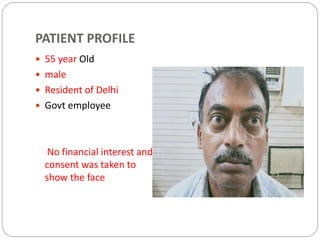
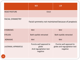
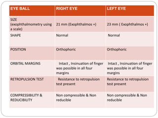
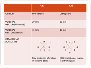
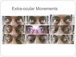
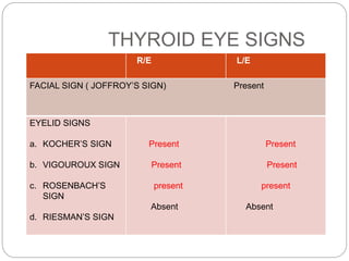
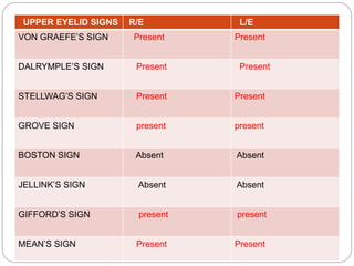
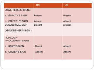
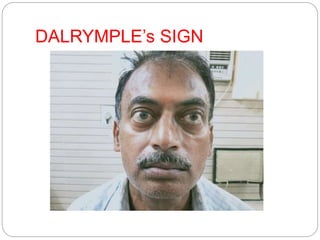
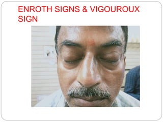
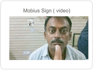
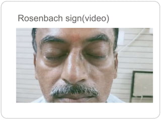
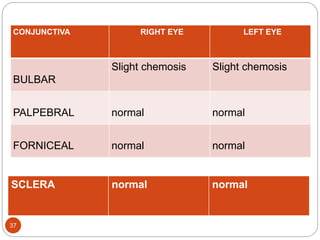
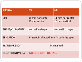
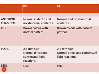
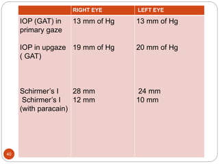
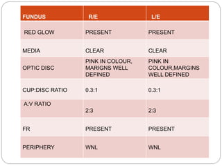
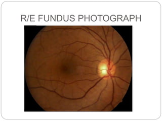
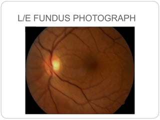
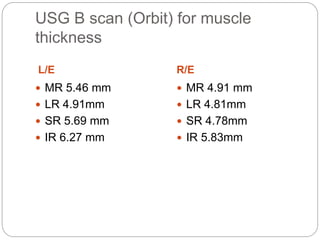
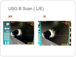
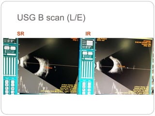
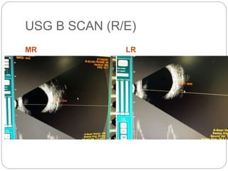
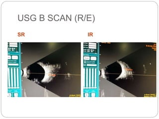
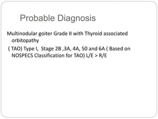
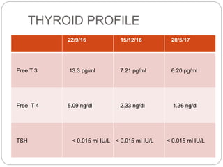
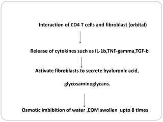
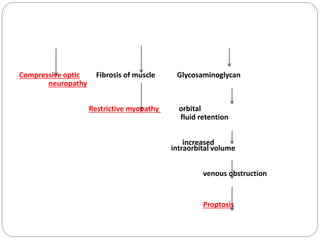
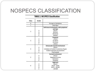
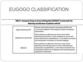
PRESENTER: Dr. Anant Prakash Tripathi
SR: Dr. Ekta
CASE PRESENTATION
PATIENT PROFILE
 55 year Old
 male
 Resident of Delhi
 Govt employee
No financial interest and
consent was taken to
show the face
PRESENTING COMPLAINTS
 Swelling in front of the neck X 3 years
 Bulging of both the eyes (LE > RE) X 2 years
HOPI
Patient was apparently well two years back when he noticed
:
 Swelling in front part of his neck which was :
 Gradual, painless and progressive
 Initially it was size of a pea and has atttained a present size
Patient came to our hospital in july 2019 for the neck
swelling and was diagnosed to have thyroid disease
HOPI continues….
He is on treatment ( oral drugs) for the same for the past one
year
2 years back he noticed :
 Bulging of both of his eyes (LE > RE) which was :
 Insidious in onset
 Gradually progressive
 Associated with heaviness, gritty sensation and watering in
both eyes occasionally
 H/o weight loss, increased sweating present
 Not associated with pain in eyes
 No h/o diplopia
 No h/o increase in proptosis after coughing, sneezing,
crying or in bending forward
 No h/o postural variation
 No h/o any visible pulsations or change in colour of the
overlying skin
 No h/o fever, loss of appetite, arthralgia
 No h/o any other swelling in the body
 No h/o radiation therapy or chemotherapy in the past
 No h/o nasal block, recurrent sinusitis, frequent
respiratory tract infection, epistaxis
 No h/o ocular trauma or surgery
 No h/o headache,, projectile vomiting, seizures
 No h/o cough, haemoptysis, chest pain
PAST HISTORY
 He went to a physician and was prescribed certain
investigations and was diagnosed as a case of toxic
nodular goiter (USG, Tc99 scan, MRI brain n orbit) and
was started on medication.
 No h/o similar episodes in the past
 No h/o any surgery
PAST MEDICAL HISTORY
 k/c/o Hypertension on treatment for 5 years
 No h/o Diabetes mellitus, Tuberculosis or any other
systemic diseases
DRUG HISTORY
 Currently he is taking :
a. NM-20 ( Neo-Mercazole) 20 mg Twice a day
b. Tablet Ciplar 40 mg Thrice a day
FAMILY HISTORY
 No H/o similar ocular complaints in the family
 No H/o thyroid disease in any other family member
PERSONAL HISTORY
 Vegetarian by diet
 Normal sleep and appetite
 Normal bowel and bladder habits
 No H/o allergy/atopy
 Smoker (5-6 cigerettes/day) Stopped 3 months back
GPE
 average built , weight= 65 kgs
 Conscious, well oriented to Time, place, Person
 PR: 102/min, regular, high volume
 BP: 130/80 mmHg in the right arm in sitting position
 Afebrile
 RR: 16/min
 No pallor, icterus, cyanosis or lymphadenopathy ,
pretibial myxedema.
No hand tremors
SYSTEMIC EXAMINATION
 CVS: S1S2 normal, no murmurs
 RS: B/L air entry normal, no adventitious sounds
heard
 P/A : NAD with no organomegaly
 CNS: normal including cranial nerves
EXAMINATION OF NECK
THYROID SWELLING
EXAMINATION OF NECK
 INSPECTION :
 A swelling of approx.4 cm X 3 cm on the anterior aspect
of the neck extending on both the sides from the midline
with well defined borders and nodular surface
 It did’nt move with deglutition or protrusion of the
tongue.
 Skin over the swelling appeared normal
 PALPATION :
 A swelling of 4.5cm X 3 cm was palpated on the anterior
aspect of the neck extending on both the sides from the
midline with well defined borders and nodular surface,
firm in consistency
NECK EXAMINATION continues…..
 It didn’t move with deglutition
 There was no local rise of temperature or tenderness
 No cervical lymphadenopathy
 PERCUSSION :
Percussion over the manubrium gave a resonant note
 AUSCULTATION :
 No bruit heard over the thyroid gland
OPHTHALMIC
EXAMINATION
RE LE
VISUAL ACUITY (DISTANCE)
• UNAIDED
• WITH PH 6/9 p
6/6
6/12 p
6/6
NEAR VISION UNAIDED N 6 N 6
RETINOSCOPY (AT 2/3 rd
METERS WITH 0.8%
TROPICAMIDE AND 5%
PHENYLEPHRINE)
+ 1.0 D
+ 1.0 D
+ 0.50 D
+ 0.50 D
R/E L/E
ACCEPTANCE -0.50 DS 6/6 -1.0 DS 6/6
COLOR VISION Normal Normal
R/E L/E
HEAD POSTURE Erect
FACIAL SYMMETRY
Facial symmetry not maintained because of proptosis
EYEBROWS
EYELIDS
ADNAXAE
Wnl
Both eyelids retracted
Wnl
Wnl
Both eyelids retracted
Wnl
LACRIMAL APPARATUS
Puncta well apposed to
globe
And regurgitation test
negative
Puncta well apposed to
globe and regurgitation test
negative
22
PROPTOSIS WORK UP
EYE BALL RIGHT EYE LEFT EYE
SIZE
(exophthalmometry using
a scale)
21 mm (Exophthalmos +) 23 mm ( Exophthalmos +)
SHAPE Normal Normal
POSITION Orthophoric Orthophoric
ORBITAL MARGINS Intact , Insinuation of finger
was possible in all four
margins
Intact , Insinuation of finger
was possible in all four
margins
RETROPULSION TEST Resistance to retropulsion
test present
Resistance to retropulsion
test present
COMPRESSIBILITY &
REDUCIBILITY
Non compressible & Non
reducible
Non compressible & Non
reducible
R/E L/E
POSITION Orthophoric Orthophoric
PALPEBRAL
APERTURE(horizontal)
29 mm 29 mm
PALPEBRAL
APERTURE(vertical)
15 mm 16 mm
EXTRA-OCULAR
MOVEMENTS -1 0 -1
0 0
-1 0 -1
Mild Limitation of motion
in extreme gazes
-1 0 -1
0 0
-1 -1
0
Mild Limitation of motion
in extreme gazes
Extra-ocular Movements
R/E L/E
EYELID SIGNS
a. KOCHER’S SIGN
b. VIGOUROUX SIGN
c. ROSENBACH’S
SIGN
d. RIESMAN’S SIGN
Present
Present
present
Absent
Present
Present
present
Absent
THYROID EYE SIGNS
FACIAL SIGN ( JOFFROY’S SIGN) Present
UPPER EYELID SIGNS R/E L/E
VON GRAEFE’S SIGN Present Present
DALRYMPLE’S SIGN Present Present
STELLWAG’S SIGN Present Present
GROVE SIGN present present
BOSTON SIGN Absent Absent
JELLINK’S SIGN Absent Absent
GIFFORD’S SIGN present present
MEAN’S SIGN Present Present
R/E L/E
LOWER EYELID SIGNS
a. ENROTH’S SIGN
b. GRIFFITH’S SIGN
Present
Absent
Present
Absent
CONJUCTIVAL SIGN
( GOLDZEIHER’S SIGN )
present present
PUPILLARY
INVOLVEMENT SIGNS
a. KNIES’S SIGN
b. COWEN’S SIGN
Absent
Absent
Absent
Absent
THYROID EYE SIGNS
GOLDZIEGHER’S SIGN
Kocher’s sign
JOEFFROY’s SIGN
DALRYMPLE’s SIGN
ENROTH SIGNS & VIGOUROUX
SIGN
Mobius Sign ( video)
Rosenbach sign(video)
37
CONJUNCTIVA RIGHT EYE LEFT EYE
BULBAR
Slight chemosis Slight chemosis
PALPEBRAL normal normal
FORNICEAL normal normal
SCLERA normal normal
CORNEA R/E L/E
SIZE 11 mm horizontal
10 mm vertical
11 mm horizontal
10 mm vertical
SHAPE/CURVATURE Normal in shape Normal in shape
SENSATION Present in all quadrants in both the eyes
TRANSPARENCY Maintained
BELLS PHENOMENA GOOD IN BOTH THE EYES
R/E L/E
ANTERIOR
CHAMBER
Normal in depth and
no abnormal contents
Normal and no abnormal
contents
IRIS Brown colour with
normal pattern
Brown colour with normal
pattern
PUPIL 2.5 mm size
Normal direct and
consensual light
reactions
2.5 mm size
Normal direct and consensual
light reactions
LENS clear clear
39
40
RIGHT EYE LEFT EYE
IOP (GAT) in
primary gaze
IOP in upgaze
( GAT)
Schirmer’s I
Schirmer’s I
(with paracain)
13 mm of Hg
19 mm of Hg
28 mm
12 mm
13 mm of Hg
20 mm of Hg
24 mm
10 mm
FUNDUS R/E L/E
RED GLOW PRESENT PRESENT
MEDIA CLEAR CLEAR
OPTIC DISC
PINK IN COLOUR,
MARIGNS WELL
DEFINED
PINK IN
COLOUR,MARGINS
WELL DEFINED
CUP:DISC RATIO 0.3:1 0.3:1
A:V RATIO
2:3 2:3
FR PRESENT PRESENT
PERIPHERY WNL WNL
R/E FUNDUS PHOTOGRAPH
L/E FUNDUS PHOTOGRAPH
USG B scan (Orbit) for muscle
thickness
L/E R/E
 MR 5.46 mm
 LR 4.91mm
 SR 5.69 mm
 IR 6.27 mm
 MR 4.91 mm
 LR 4.81mm
 SR 4.78mm
 IR 5.83mm
USG B Scan ( L/E)
MR IR
USG B scan (L/E)
SR IR
USG B SCAN (R/E)
MR LR
USG B SCAN (R/E)
SR IR
Probable Diagnosis
Multinodular goiter Grade II with Thyroid associated
orbitopathy
( TAO) Type I, Stage 2B ,3A, 4A, 50 and 6A ( Based on
NOSPECS Classification for TAO) L/E > R/E
INVESTIGATIONS ( 20/5/17)
Hb-13g/dl
 PCV-48.4%
 RBC- 5.19 million
 MCV/MCH/MCHC- 93.2/29.8/31.9
 TLC- 97
 Blood sugar (Fasting)- 90 mg/dl
 LFT
 Total Bilirubin 0.9 mg /dl
 Direct Bilirubin 0.2 mg /dl
 Indirect Bilirubin 0.9 mg /dl
 SGOT 30 U/L
 SGPT 32 U/L
• KFT
 Urea 36 mg/dl
 Creatinine 0.9 mg/dl
 Urea Acid 3.8 mg/dl
• Urine rountine microscopy- normal
THYROID PROFILE
22/9/16 15/12/16 20/5/17
Free T 3 13.3 pg/ml 7.21 pg/ml 6.20 pg/ml
Free T 4 5.09 ng/dl 2.33 ng/dl 1.36 ng/dl
TSH < 0.015 ml IU/L < 0.015 ml IU/L < 0.015 ml IU/L
 USG Thyroid – Bilateral lobes and isthmus of thyroid gland
showed nodular enlargement left lobe – 18mm, nodule
(8.5x7.5mm) ; right lobe 20mm with increased vascularity.
 Tc 99 scan – suggests of toxic nodular goitre with cold
nodule in left lobe.
 MRI SCAN Brain & ORBIT- Report awaited
DEFINITIVE DIAGNOSIS
Toxic nodular goiter ( MNG) Grade II with Thyroid
associated orbitopathy ( TAO) Type I, stage 2B,3A, 4A,
5 0 and 6 a L/E > R/E
Treatment
 Lubricating e/d CMC 0.5 % both eyes
 Currently he is on anti-thyroid medication
 Reassurance of the patient
 Regular follow up
TAO Pathophysiology
Autoantibody against TSH receptor( TSH-R) in the Thyroid
Cross react with TSH-R in effector cells i.e. fibroblast ,
adipocytes and interstitial tissue between extra ocular
muscles
Inflammatory cellular infiltration ( T lymphocyte, plasma
cells, macrophages and mast cells) in orbital fat,lacrimal
gland and orbital interstitium
Lymphocytes infiltrate in the orbit predominantly of CD4+
and CD8+ T- cells with a few B-cells.
Interaction of CD4 T cells and fibroblast (orbital)
Release of cytokines such as IL-1b,TNF-gamma,TGF-b
Activate fibroblasts to secrete hyaluronic acid,
glycosaminoglycans.
Osmotic imbibition of water ,EOM swollen upto 8 times
Compressive optic Fibrosis of muscle Glycosaminoglycan
neuropathy
Restrictive myopathy orbital
fluid retention
increased
intraorbital volume
venous obstruction
Proptosis
NOSPECS CLASSIFICATION
EUGOGO CLASSIFICATION
CAS (MOURITZ ET AL)
THANK YOU

thyroid eye disease

  • 1.
    PRESENTER: Dr. AnantPrakash Tripathi SR: Dr. Ekta CASE PRESENTATION
  • 2.
    PATIENT PROFILE  55year Old  male  Resident of Delhi  Govt employee No financial interest and consent was taken to show the face
  • 3.
    PRESENTING COMPLAINTS  Swellingin front of the neck X 3 years  Bulging of both the eyes (LE > RE) X 2 years
  • 4.
    HOPI Patient was apparentlywell two years back when he noticed :  Swelling in front part of his neck which was :  Gradual, painless and progressive  Initially it was size of a pea and has atttained a present size Patient came to our hospital in july 2019 for the neck swelling and was diagnosed to have thyroid disease
  • 5.
    HOPI continues…. He ison treatment ( oral drugs) for the same for the past one year 2 years back he noticed :  Bulging of both of his eyes (LE > RE) which was :  Insidious in onset  Gradually progressive  Associated with heaviness, gritty sensation and watering in both eyes occasionally  H/o weight loss, increased sweating present
  • 6.
     Not associatedwith pain in eyes  No h/o diplopia  No h/o increase in proptosis after coughing, sneezing, crying or in bending forward  No h/o postural variation  No h/o any visible pulsations or change in colour of the overlying skin  No h/o fever, loss of appetite, arthralgia  No h/o any other swelling in the body
  • 7.
     No h/oradiation therapy or chemotherapy in the past  No h/o nasal block, recurrent sinusitis, frequent respiratory tract infection, epistaxis  No h/o ocular trauma or surgery  No h/o headache,, projectile vomiting, seizures  No h/o cough, haemoptysis, chest pain
  • 8.
    PAST HISTORY  Hewent to a physician and was prescribed certain investigations and was diagnosed as a case of toxic nodular goiter (USG, Tc99 scan, MRI brain n orbit) and was started on medication.  No h/o similar episodes in the past  No h/o any surgery
  • 9.
    PAST MEDICAL HISTORY k/c/o Hypertension on treatment for 5 years  No h/o Diabetes mellitus, Tuberculosis or any other systemic diseases
  • 10.
    DRUG HISTORY  Currentlyhe is taking : a. NM-20 ( Neo-Mercazole) 20 mg Twice a day b. Tablet Ciplar 40 mg Thrice a day
  • 11.
    FAMILY HISTORY  NoH/o similar ocular complaints in the family  No H/o thyroid disease in any other family member
  • 12.
    PERSONAL HISTORY  Vegetarianby diet  Normal sleep and appetite  Normal bowel and bladder habits  No H/o allergy/atopy  Smoker (5-6 cigerettes/day) Stopped 3 months back
  • 13.
    GPE  average built, weight= 65 kgs  Conscious, well oriented to Time, place, Person  PR: 102/min, regular, high volume  BP: 130/80 mmHg in the right arm in sitting position  Afebrile  RR: 16/min  No pallor, icterus, cyanosis or lymphadenopathy , pretibial myxedema.
  • 14.
  • 15.
    SYSTEMIC EXAMINATION  CVS:S1S2 normal, no murmurs  RS: B/L air entry normal, no adventitious sounds heard  P/A : NAD with no organomegaly  CNS: normal including cranial nerves
  • 16.
  • 17.
    EXAMINATION OF NECK INSPECTION :  A swelling of approx.4 cm X 3 cm on the anterior aspect of the neck extending on both the sides from the midline with well defined borders and nodular surface  It did’nt move with deglutition or protrusion of the tongue.  Skin over the swelling appeared normal  PALPATION :  A swelling of 4.5cm X 3 cm was palpated on the anterior aspect of the neck extending on both the sides from the midline with well defined borders and nodular surface, firm in consistency
  • 18.
    NECK EXAMINATION continues….. It didn’t move with deglutition  There was no local rise of temperature or tenderness  No cervical lymphadenopathy  PERCUSSION : Percussion over the manubrium gave a resonant note  AUSCULTATION :  No bruit heard over the thyroid gland
  • 19.
  • 20.
    RE LE VISUAL ACUITY(DISTANCE) • UNAIDED • WITH PH 6/9 p 6/6 6/12 p 6/6 NEAR VISION UNAIDED N 6 N 6 RETINOSCOPY (AT 2/3 rd METERS WITH 0.8% TROPICAMIDE AND 5% PHENYLEPHRINE) + 1.0 D + 1.0 D + 0.50 D + 0.50 D
  • 21.
    R/E L/E ACCEPTANCE -0.50DS 6/6 -1.0 DS 6/6 COLOR VISION Normal Normal
  • 22.
    R/E L/E HEAD POSTUREErect FACIAL SYMMETRY Facial symmetry not maintained because of proptosis EYEBROWS EYELIDS ADNAXAE Wnl Both eyelids retracted Wnl Wnl Both eyelids retracted Wnl LACRIMAL APPARATUS Puncta well apposed to globe And regurgitation test negative Puncta well apposed to globe and regurgitation test negative 22
  • 23.
  • 24.
    EYE BALL RIGHTEYE LEFT EYE SIZE (exophthalmometry using a scale) 21 mm (Exophthalmos +) 23 mm ( Exophthalmos +) SHAPE Normal Normal POSITION Orthophoric Orthophoric ORBITAL MARGINS Intact , Insinuation of finger was possible in all four margins Intact , Insinuation of finger was possible in all four margins RETROPULSION TEST Resistance to retropulsion test present Resistance to retropulsion test present COMPRESSIBILITY & REDUCIBILITY Non compressible & Non reducible Non compressible & Non reducible
  • 25.
    R/E L/E POSITION OrthophoricOrthophoric PALPEBRAL APERTURE(horizontal) 29 mm 29 mm PALPEBRAL APERTURE(vertical) 15 mm 16 mm EXTRA-OCULAR MOVEMENTS -1 0 -1 0 0 -1 0 -1 Mild Limitation of motion in extreme gazes -1 0 -1 0 0 -1 -1 0 Mild Limitation of motion in extreme gazes
  • 26.
  • 27.
    R/E L/E EYELID SIGNS a.KOCHER’S SIGN b. VIGOUROUX SIGN c. ROSENBACH’S SIGN d. RIESMAN’S SIGN Present Present present Absent Present Present present Absent THYROID EYE SIGNS FACIAL SIGN ( JOFFROY’S SIGN) Present
  • 28.
    UPPER EYELID SIGNSR/E L/E VON GRAEFE’S SIGN Present Present DALRYMPLE’S SIGN Present Present STELLWAG’S SIGN Present Present GROVE SIGN present present BOSTON SIGN Absent Absent JELLINK’S SIGN Absent Absent GIFFORD’S SIGN present present MEAN’S SIGN Present Present
  • 29.
    R/E L/E LOWER EYELIDSIGNS a. ENROTH’S SIGN b. GRIFFITH’S SIGN Present Absent Present Absent CONJUCTIVAL SIGN ( GOLDZEIHER’S SIGN ) present present PUPILLARY INVOLVEMENT SIGNS a. KNIES’S SIGN b. COWEN’S SIGN Absent Absent Absent Absent
  • 30.
  • 31.
  • 32.
  • 33.
  • 34.
    ENROTH SIGNS &VIGOUROUX SIGN
  • 35.
  • 36.
  • 37.
    37 CONJUNCTIVA RIGHT EYELEFT EYE BULBAR Slight chemosis Slight chemosis PALPEBRAL normal normal FORNICEAL normal normal SCLERA normal normal
  • 38.
    CORNEA R/E L/E SIZE11 mm horizontal 10 mm vertical 11 mm horizontal 10 mm vertical SHAPE/CURVATURE Normal in shape Normal in shape SENSATION Present in all quadrants in both the eyes TRANSPARENCY Maintained BELLS PHENOMENA GOOD IN BOTH THE EYES
  • 39.
    R/E L/E ANTERIOR CHAMBER Normal indepth and no abnormal contents Normal and no abnormal contents IRIS Brown colour with normal pattern Brown colour with normal pattern PUPIL 2.5 mm size Normal direct and consensual light reactions 2.5 mm size Normal direct and consensual light reactions LENS clear clear 39
  • 40.
    40 RIGHT EYE LEFTEYE IOP (GAT) in primary gaze IOP in upgaze ( GAT) Schirmer’s I Schirmer’s I (with paracain) 13 mm of Hg 19 mm of Hg 28 mm 12 mm 13 mm of Hg 20 mm of Hg 24 mm 10 mm
  • 41.
    FUNDUS R/E L/E REDGLOW PRESENT PRESENT MEDIA CLEAR CLEAR OPTIC DISC PINK IN COLOUR, MARIGNS WELL DEFINED PINK IN COLOUR,MARGINS WELL DEFINED CUP:DISC RATIO 0.3:1 0.3:1 A:V RATIO 2:3 2:3 FR PRESENT PRESENT PERIPHERY WNL WNL
  • 42.
  • 43.
  • 44.
    USG B scan(Orbit) for muscle thickness L/E R/E  MR 5.46 mm  LR 4.91mm  SR 5.69 mm  IR 6.27 mm  MR 4.91 mm  LR 4.81mm  SR 4.78mm  IR 5.83mm
  • 45.
    USG B Scan( L/E) MR IR
  • 46.
    USG B scan(L/E) SR IR
  • 47.
    USG B SCAN(R/E) MR LR
  • 48.
    USG B SCAN(R/E) SR IR
  • 49.
    Probable Diagnosis Multinodular goiterGrade II with Thyroid associated orbitopathy ( TAO) Type I, Stage 2B ,3A, 4A, 50 and 6A ( Based on NOSPECS Classification for TAO) L/E > R/E
  • 50.
    INVESTIGATIONS ( 20/5/17) Hb-13g/dl PCV-48.4%  RBC- 5.19 million  MCV/MCH/MCHC- 93.2/29.8/31.9  TLC- 97  Blood sugar (Fasting)- 90 mg/dl
  • 51.
     LFT  TotalBilirubin 0.9 mg /dl  Direct Bilirubin 0.2 mg /dl  Indirect Bilirubin 0.9 mg /dl  SGOT 30 U/L  SGPT 32 U/L • KFT  Urea 36 mg/dl  Creatinine 0.9 mg/dl  Urea Acid 3.8 mg/dl • Urine rountine microscopy- normal
  • 52.
    THYROID PROFILE 22/9/16 15/12/1620/5/17 Free T 3 13.3 pg/ml 7.21 pg/ml 6.20 pg/ml Free T 4 5.09 ng/dl 2.33 ng/dl 1.36 ng/dl TSH < 0.015 ml IU/L < 0.015 ml IU/L < 0.015 ml IU/L
  • 53.
     USG Thyroid– Bilateral lobes and isthmus of thyroid gland showed nodular enlargement left lobe – 18mm, nodule (8.5x7.5mm) ; right lobe 20mm with increased vascularity.  Tc 99 scan – suggests of toxic nodular goitre with cold nodule in left lobe.  MRI SCAN Brain & ORBIT- Report awaited
  • 54.
    DEFINITIVE DIAGNOSIS Toxic nodulargoiter ( MNG) Grade II with Thyroid associated orbitopathy ( TAO) Type I, stage 2B,3A, 4A, 5 0 and 6 a L/E > R/E
  • 55.
    Treatment  Lubricating e/dCMC 0.5 % both eyes  Currently he is on anti-thyroid medication  Reassurance of the patient  Regular follow up
  • 56.
    TAO Pathophysiology Autoantibody againstTSH receptor( TSH-R) in the Thyroid Cross react with TSH-R in effector cells i.e. fibroblast , adipocytes and interstitial tissue between extra ocular muscles Inflammatory cellular infiltration ( T lymphocyte, plasma cells, macrophages and mast cells) in orbital fat,lacrimal gland and orbital interstitium Lymphocytes infiltrate in the orbit predominantly of CD4+ and CD8+ T- cells with a few B-cells.
  • 57.
    Interaction of CD4T cells and fibroblast (orbital) Release of cytokines such as IL-1b,TNF-gamma,TGF-b Activate fibroblasts to secrete hyaluronic acid, glycosaminoglycans. Osmotic imbibition of water ,EOM swollen upto 8 times
  • 58.
    Compressive optic Fibrosisof muscle Glycosaminoglycan neuropathy Restrictive myopathy orbital fluid retention increased intraorbital volume venous obstruction Proptosis
  • 59.
  • 60.
  • 61.
  • 62.