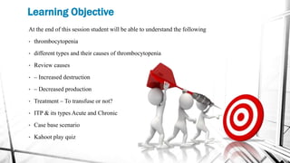
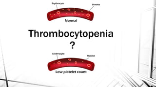
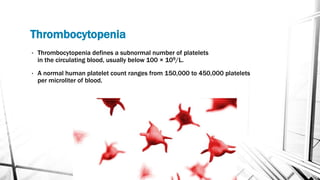
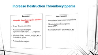
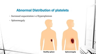
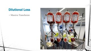
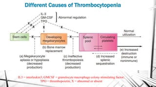
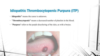
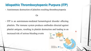
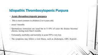
Thrombocytopenia is generally defined as platelet count <150 × 109/L. It can occur due to several reasons, like decreased platelet production (e.g., inherited bone marrow failure syndromes, acquired aplastic anemia, leukemia), ineffective platelet production (myelodysplastic syndrome, megaloblastic anemia), increased destruction (ITP, HLH), increased consumption (DIC, TTP, HUS), sequestration (hypersplenism), or may be due to combination of multiple mechanisms described above.
During evaluating a case of thrombocytopenia, the first step is getting a detailed history and doing a proper clinical examination. Then the next step would be checking the other parameters of complete blood count (CBC), especially hemoglobin (Hb) and the total WBC count, complemented by a peripheral smear (PS) examination, which will clear many doubts and will help us pinpointing our diagnostic approach.
Many a times pseudo-thrombocytopenia is encountered in a PS due to platelet clumping by EDTA and can be rectified by collecting blood samples in a citrate or heparin vials or by doing a direct finger prick smear. Any accompanying cytopenia will expand the differential diagnosis and an isolated thrombocytopenia will further narrow it down. Presence of any additional abnormalities of red cells (megaloblasts) or white cells (presence of hyper-segmented neutrophils, atypical lymphoid/myeloid cells) could be present in megaloblastic anemia/MDS, leukemia respectively, while in the presence of fragmented red cells microangiopathic hemolytic anemia should always be ruled out by doing PT and aPTT (DIC, TTP, HUS). In case of isolated thrombocytopenia, the platelet morphology is also important. In many patients in India, especially in eastern region many people have large platelets with their normal platelet count around 100 × 109/L with normal platelet function (Harris platelet syndrome). However, presence of any abnormal platelet morphology along with a low platelet count may indicate a platelet function disorder (large platelets in Bernard Soulier syndrome/ Glanzmann thrombasthenia or small platelets in Wiskott-Aldrich syndrome), especially if encountered in early part of life during evaluation for bleeding symptoms. In case of isolated thrombocytopenia, presence of additional congenital anomalies may point out towards an inherited marrow failure syndrome, e.g. amegakayocytic thrombocytopenia. Exposure to certain drugs may result in isolated low platelet count, e.g., ceftriaxone, piperacillin, heparin. Presence of toxic changes in neutrophils may indicate sepsis related thrombocytopenia. By excluding all these, immune thrombocytopenia (ITP) to be thought as no specific tests or markers are available for this entity and its diagnosis is largely clinical. A further work up complemented by bone marrow examination and in few cases a platelet function test will definitely help in reaching the final diagnosis.
So, summarizing, in the evaluation of a case of thrombocytopenia, all the
Thrombocytopenia is most frequently encountered Hematological problem in hospitalized patients. The most common causes and differential diagnosis of In-patient and Outpatient presentations of Thrombocytopenia is discussed here. Useful for Internal Medicine Boards . Archer Internal Medicine Board review lectures will be released soon.
This PPT covers pathophysiology of thrombocytopenia which includes causes of thrombocytopenia, symptoms of thrombocytopenia and diagnosis of thrombocytopenia
Simple way to explain primary haemostatic anomalies
Easy to teach
Platelet function as well as disorders of granules and their release reaction. A reader will find a few better resources.
Outline is from introduction to explanation of every single anomaly. Happy reading
Chronic Lypmhocytic leukemia/SLL/B-PLL/T-PLL/ATLL By SOLOMON SUasb by SOLOMON SUASB
the following presenation include
Introduction/Background
• Etiology of CLL
• Symptoms
• Test and Diagnosis
• Staging
• Prognosis
• Treatment
• B Cell diseases BPLL
• T cell diseases
T-PLL
ATLL
LGLL
The white cells granulocytes and their benign disordersSOLOMON SUASB
Dear all this slide will help you to cover the following Contents.
Introduction to leucocytes.
Granulcytes
Granulopoeisis as well as its growth factor
Clinical Application of G-CSF.
Neutrophil and monocyte function
i-e adherence, chemotaxis, phagocytosis, and killing.
Defects in Phagocytic Cell Function.
Benign Disorders of Neutrophils.
Pelger–Huët anomaly
May–Hegglin anomaly
Chédiak–Higashi syndrome
New Drug Discovery and Development .....NEHA GUPTA
The "New Drug Discovery and Development" process involves the identification, design, testing, and manufacturing of novel pharmaceutical compounds with the aim of introducing new and improved treatments for various medical conditions. This comprehensive endeavor encompasses various stages, including target identification, preclinical studies, clinical trials, regulatory approval, and post-market surveillance. It involves multidisciplinary collaboration among scientists, researchers, clinicians, regulatory experts, and pharmaceutical companies to bring innovative therapies to market and address unmet medical needs.
Report Back from SGO 2024: What’s the Latest in Cervical Cancer?bkling
Are you curious about what’s new in cervical cancer research or unsure what the findings mean? Join Dr. Emily Ko, a gynecologic oncologist at Penn Medicine, to learn about the latest updates from the Society of Gynecologic Oncology (SGO) 2024 Annual Meeting on Women’s Cancer. Dr. Ko will discuss what the research presented at the conference means for you and answer your questions about the new developments.
263778731218 Abortion Clinic /Pills In Harare ,sisternakatoto
263778731218 Abortion Clinic /Pills In Harare ,ABORTION WOMEN’S CLINIC +27730423979 IN women clinic we believe that every woman should be able to make choices in her pregnancy. Our job is to provide compassionate care, safety,affordable and confidential services. That’s why we have won the trust from all generations of women all over the world. we use non surgical method(Abortion pills) to terminate…Dr.LISA +27730423979women Clinic is committed to providing the highest quality of obstetrical and gynecological care to women of all ages. Our dedicated staff aim to treat each patient and her health concerns with compassion and respect.Our dedicated group ABORTION WOMEN’S CLINIC +27730423979 IN women clinic we believe that every woman should be able to make choices in her pregnancy. Our job is to provide compassionate care, safety,affordable and confidential services. That’s why we have won the trust from all generations of women all over the world. we use non surgical method(Abortion pills) to terminate…Dr.LISA +27730423979women Clinic is committed to providing the highest quality of obstetrical and gynecological care to women of all ages. Our dedicated staff aim to treat each patient and her health concerns with compassion and respect.Our dedicated group of receptionists, nurses, and physicians have worked together as a teamof receptionists, nurses, and physicians have worked together as a team wwww.lisywomensclinic.co.za/
Ethanol (CH3CH2OH), or beverage alcohol, is a two-carbon alcohol
that is rapidly distributed in the body and brain. Ethanol alters many
neurochemical systems and has rewarding and addictive properties. It
is the oldest recreational drug and likely contributes to more morbidity,
mortality, and public health costs than all illicit drugs combined. The
5th edition of the Diagnostic and Statistical Manual of Mental Disorders
(DSM-5) integrates alcohol abuse and alcohol dependence into a single
disorder called alcohol use disorder (AUD), with mild, moderate,
and severe subclassifications (American Psychiatric Association, 2013).
In the DSM-5, all types of substance abuse and dependence have been
combined into a single substance use disorder (SUD) on a continuum
from mild to severe. A diagnosis of AUD requires that at least two of
the 11 DSM-5 behaviors be present within a 12-month period (mild
AUD: 2–3 criteria; moderate AUD: 4–5 criteria; severe AUD: 6–11 criteria).
The four main behavioral effects of AUD are impaired control over
drinking, negative social consequences, risky use, and altered physiological
effects (tolerance, withdrawal). This chapter presents an overview
of the prevalence and harmful consequences of AUD in the U.S.,
the systemic nature of the disease, neurocircuitry and stages of AUD,
comorbidities, fetal alcohol spectrum disorders, genetic risk factors, and
pharmacotherapies for AUD.
Tom Selleck Health: A Comprehensive Look at the Iconic Actor’s Wellness Journeygreendigital
Tom Selleck, an enduring figure in Hollywood. has captivated audiences for decades with his rugged charm, iconic moustache. and memorable roles in television and film. From his breakout role as Thomas Magnum in Magnum P.I. to his current portrayal of Frank Reagan in Blue Bloods. Selleck's career has spanned over 50 years. But beyond his professional achievements. fans have often been curious about Tom Selleck Health. especially as he has aged in the public eye.
Follow us on: Pinterest
Introduction
Many have been interested in Tom Selleck health. not only because of his enduring presence on screen but also because of the challenges. and lifestyle choices he has faced and made over the years. This article delves into the various aspects of Tom Selleck health. exploring his fitness regimen, diet, mental health. and the challenges he has encountered as he ages. We'll look at how he maintains his well-being. the health issues he has faced, and his approach to ageing .
Early Life and Career
Childhood and Athletic Beginnings
Tom Selleck was born on January 29, 1945, in Detroit, Michigan, and grew up in Sherman Oaks, California. From an early age, he was involved in sports, particularly basketball. which played a significant role in his physical development. His athletic pursuits continued into college. where he attended the University of Southern California (USC) on a basketball scholarship. This early involvement in sports laid a strong foundation for his physical health and disciplined lifestyle.
Transition to Acting
Selleck's transition from an athlete to an actor came with its physical demands. His first significant role in "Magnum P.I." required him to perform various stunts and maintain a fit appearance. This role, which he played from 1980 to 1988. necessitated a rigorous fitness routine to meet the show's demands. setting the stage for his long-term commitment to health and wellness.
Fitness Regimen
Workout Routine
Tom Selleck health and fitness regimen has evolved. adapting to his changing roles and age. During his "Magnum, P.I." days. Selleck's workouts were intense and focused on building and maintaining muscle mass. His routine included weightlifting, cardiovascular exercises. and specific training for the stunts he performed on the show.
Selleck adjusted his fitness routine as he aged to suit his body's needs. Today, his workouts focus on maintaining flexibility, strength, and cardiovascular health. He incorporates low-impact exercises such as swimming, walking, and light weightlifting. This balanced approach helps him stay fit without putting undue strain on his joints and muscles.
Importance of Flexibility and Mobility
In recent years, Selleck has emphasized the importance of flexibility and mobility in his fitness regimen. Understanding the natural decline in muscle mass and joint flexibility with age. he includes stretching and yoga in his routine. These practices help prevent injuries, improve posture, and maintain mobilit
HOT NEW PRODUCT! BIG SALES FAST SHIPPING NOW FROM CHINA!! EU KU DB BK substit...GL Anaacs
Contact us if you are interested:
Email / Skype : kefaya1771@gmail.com
Threema: PXHY5PDH
New BATCH Ku !!! MUCH IN DEMAND FAST SALE EVERY BATCH HAPPY GOOD EFFECT BIG BATCH !
Contact me on Threema or skype to start big business!!
Hot-sale products:
NEW HOT EUTYLONE WHITE CRYSTAL!!
5cl-adba precursor (semi finished )
5cl-adba raw materials
ADBB precursor (semi finished )
ADBB raw materials
APVP powder
5fadb/4f-adb
Jwh018 / Jwh210
Eutylone crystal
Protonitazene (hydrochloride) CAS: 119276-01-6
Flubrotizolam CAS: 57801-95-3
Metonitazene CAS: 14680-51-4
Payment terms: Western Union,MoneyGram,Bitcoin or USDT.
Deliver Time: Usually 7-15days
Shipping method: FedEx, TNT, DHL,UPS etc.Our deliveries are 100% safe, fast, reliable and discreet.
Samples will be sent for your evaluation!If you are interested in, please contact me, let's talk details.
We specializes in exporting high quality Research chemical, medical intermediate, Pharmaceutical chemicals and so on. Products are exported to USA, Canada, France, Korea, Japan,Russia, Southeast Asia and other countries.
Explore natural remedies for syphilis treatment in Singapore. Discover alternative therapies, herbal remedies, and lifestyle changes that may complement conventional treatments. Learn about holistic approaches to managing syphilis symptoms and supporting overall health.
Title: Sense of Smell
Presenter: Dr. Faiza, Assistant Professor of Physiology
Qualifications:
MBBS (Best Graduate, AIMC Lahore)
FCPS Physiology
ICMT, CHPE, DHPE (STMU)
MPH (GC University, Faisalabad)
MBA (Virtual University of Pakistan)
Learning Objectives:
Describe the primary categories of smells and the concept of odor blindness.
Explain the structure and location of the olfactory membrane and mucosa, including the types and roles of cells involved in olfaction.
Describe the pathway and mechanisms of olfactory signal transmission from the olfactory receptors to the brain.
Illustrate the biochemical cascade triggered by odorant binding to olfactory receptors, including the role of G-proteins and second messengers in generating an action potential.
Identify different types of olfactory disorders such as anosmia, hyposmia, hyperosmia, and dysosmia, including their potential causes.
Key Topics:
Olfactory Genes:
3% of the human genome accounts for olfactory genes.
400 genes for odorant receptors.
Olfactory Membrane:
Located in the superior part of the nasal cavity.
Medially: Folds downward along the superior septum.
Laterally: Folds over the superior turbinate and upper surface of the middle turbinate.
Total surface area: 5-10 square centimeters.
Olfactory Mucosa:
Olfactory Cells: Bipolar nerve cells derived from the CNS (100 million), with 4-25 olfactory cilia per cell.
Sustentacular Cells: Produce mucus and maintain ionic and molecular environment.
Basal Cells: Replace worn-out olfactory cells with an average lifespan of 1-2 months.
Bowman’s Gland: Secretes mucus.
Stimulation of Olfactory Cells:
Odorant dissolves in mucus and attaches to receptors on olfactory cilia.
Involves a cascade effect through G-proteins and second messengers, leading to depolarization and action potential generation in the olfactory nerve.
Quality of a Good Odorant:
Small (3-20 Carbon atoms), volatile, water-soluble, and lipid-soluble.
Facilitated by odorant-binding proteins in mucus.
Membrane Potential and Action Potential:
Resting membrane potential: -55mV.
Action potential frequency in the olfactory nerve increases with odorant strength.
Adaptation Towards the Sense of Smell:
Rapid adaptation within the first second, with further slow adaptation.
Psychological adaptation greater than receptor adaptation, involving feedback inhibition from the central nervous system.
Primary Sensations of Smell:
Camphoraceous, Musky, Floral, Pepperminty, Ethereal, Pungent, Putrid.
Odor Detection Threshold:
Examples: Hydrogen sulfide (0.0005 ppm), Methyl-mercaptan (0.002 ppm).
Some toxic substances are odorless at lethal concentrations.
Characteristics of Smell:
Odor blindness for single substances due to lack of appropriate receptor protein.
Behavioral and emotional influences of smell.
Transmission of Olfactory Signals:
From olfactory cells to glomeruli in the olfactory bulb, involving lateral inhibition.
Primitive, less old, and new olfactory systems with different path
New Directions in Targeted Therapeutic Approaches for Older Adults With Mantl...i3 Health
i3 Health is pleased to make the speaker slides from this activity available for use as a non-accredited self-study or teaching resource.
This slide deck presented by Dr. Kami Maddocks, Professor-Clinical in the Division of Hematology and
Associate Division Director for Ambulatory Operations
The Ohio State University Comprehensive Cancer Center, will provide insight into new directions in targeted therapeutic approaches for older adults with mantle cell lymphoma.
STATEMENT OF NEED
Mantle cell lymphoma (MCL) is a rare, aggressive B-cell non-Hodgkin lymphoma (NHL) accounting for 5% to 7% of all lymphomas. Its prognosis ranges from indolent disease that does not require treatment for years to very aggressive disease, which is associated with poor survival (Silkenstedt et al, 2021). Typically, MCL is diagnosed at advanced stage and in older patients who cannot tolerate intensive therapy (NCCN, 2022). Although recent advances have slightly increased remission rates, recurrence and relapse remain very common, leading to a median overall survival between 3 and 6 years (LLS, 2021). Though there are several effective options, progress is still needed towards establishing an accepted frontline approach for MCL (Castellino et al, 2022). Treatment selection and management of MCL are complicated by the heterogeneity of prognosis, advanced age and comorbidities of patients, and lack of an established standard approach for treatment, making it vital that clinicians be familiar with the latest research and advances in this area. In this activity chaired by Michael Wang, MD, Professor in the Department of Lymphoma & Myeloma at MD Anderson Cancer Center, expert faculty will discuss prognostic factors informing treatment, the promising results of recent trials in new therapeutic approaches, and the implications of treatment resistance in therapeutic selection for MCL.
Target Audience
Hematology/oncology fellows, attending faculty, and other health care professionals involved in the treatment of patients with mantle cell lymphoma (MCL).
Learning Objectives
1.) Identify clinical and biological prognostic factors that can guide treatment decision making for older adults with MCL
2.) Evaluate emerging data on targeted therapeutic approaches for treatment-naive and relapsed/refractory MCL and their applicability to older adults
3.) Assess mechanisms of resistance to targeted therapies for MCL and their implications for treatment selection
Knee anatomy and clinical tests 2024.pdfvimalpl1234
This includes all relevant anatomy and clinical tests compiled from standard textbooks, Campbell,netter etc..It is comprehensive and best suited for orthopaedicians and orthopaedic residents.
Pulmonary Thromboembolism - etilogy, types, medical- Surgical and nursing man...VarunMahajani
Disruption of blood supply to lung alveoli due to blockage of one or more pulmonary blood vessels is called as Pulmonary thromboembolism. In this presentation we will discuss its causes, types and its management in depth.
TEST BANK for Operations Management, 14th Edition by William J. Stevenson, Ve...kevinkariuki227
TEST BANK for Operations Management, 14th Edition by William J. Stevenson, Verified Chapters 1 - 19, Complete Newest Version.pdf
2. Learning Objective
At the end of this session student will be able to understand the following
• thrombocytopenia
• different types and their causes of thrombocytopenia
• Review causes
• – Increased destruction
• – Decreased production
• Treatment – To transfuse or not?
• ITP & its types Acute and Chronic
• Case base scenario
• Kahoot play quiz
4. Thrombocytopenia
• Thrombocytopenia defines a subnormal number of platelets
in the circulating blood, usually below 100 × 109/L.
• A normal human platelet count ranges from 150,000 to 450,000 platelets
per microliter of blood.
5. Sign and Symptoms
The Symptoms of thrombocytopenia is not oftenly seen until the Platelets
level fall down below 50 x109/Ltr but upon falling down from the said level
clinical declaration occur.
A level less than 30 x109/Ltr can raise to possible symptoms like
Petechiae, menorrhagia, or spontaneous bruising.
A level less than 10 x109/Ltr can raise to the high-grade bleeding.
• Spontaneous Skin Purpura.
• Mucosal Hemorrhage.
• Prolonged Bleeding After Trauma.
12. Different Causes of Thrombocytopenia
IL3 = interleukin3; GMCSF = granulocyte macrophage colony stimulating factor;
TPO = thrombopoietin; X = abnormal or absent
13. • We will only Discuss the Following
• Thrombocytopenia Due to Decrease Production of Thrombocytes i-e
1. Bone marrow Failure/suppression.
• Increased destruction of platelets i-e
1. Disseminated intravascular coagulation.
2. Increased splenic pooling
3. Massive transfusion syndrome
4. ITP
5. Drug Induced Thrombocytopenia.
6. Post-Transfusion Purpura.
14. Thrombocytopenia Due to Failure Production of
Platelets
• Most common cause of thrombocytopenia and occur due to Selective Bone
Marrow Suppression for producing Megakaryocytes Fragments.
• Bone marrow Failure can be occurred due to the Following Reasons.
• Cytotoxic Drugs & Radiotherapy (Direct effect the Bone marrow)
• Aplastic Anemias
• Leukemia
• Myelofibrosis
• Marrow Infiltration (e.g. Carcinoma, Lymphoma, Gaucher’s Disease)
• Megaloblastic Anemias
• HIV Infection (Direct effect the Megakaryocyte)
15. Thrombocytopenia Due to Failure Production of
Platelets CONT…….
• Bone Marrow Depression is rarely Congenital
• Occur by Mutation in c‐MPL thrombopoietin
receptor or of the RBM8A gene (RNA Binding
Motif Protein 8A) is a Protein Coding gene..
• The c-MPL gene provides instructions for making
the thrombopoietin receptor protein, which
promotes the growth and division (proliferation)
of cells.
16. Diagnosis of BM suppressed Thrombocytopenia
Diagnosis of these causes of thrombocytopenia is made from the
• Clinical history,
• Peripheral blood count,
• The blood film
• Bone marrow examination.
17. Thrombocytopenia Due to Increased
destruction/Usage of platelets
1. Disseminated intravascular coagulation:
• Disseminated intravascular coagulation (DIC) involves abnormal, excessive
generation of thrombin and fibrin in the circulating blood.
• During the process, increased platelet aggregation and coagulation factor
consumption occur.
18. 2. Increased spleenic pooling:
• Normally, the spleen stores one-third of the platelets that are produced by the
bone marrow and the remaining two-thirds of the platelets produced by the bone
marrow are in circulation.
• No platelets are stored in the bone marrow.
• If a condition causes the spleen to enlarge (splenomegaly), the spleen will
function abnormally, sequestering up to 90% of the total platelet mass in the
spleen.
• The result is a decrease in circulating platelets (thrombocytopenia).
• Conditions Include hepatic cirrhosis, Gaucher's disease(read comment), some
leukemias (like hairy cell leukemia).
Spleenic pooling Thrombocytopenia
19.
20. 3. Massive Transfusion Syndrome:
• Platelets are unstable in blood Stored at Room Temperature i-e 24°C
• Platelets count rapidly fall stored for more than 24 hrs.
• Patient Transfused with Massive amount of stored blood such as 10 units in
24 hrs period will show abnormal clotting and thrombocytopenia.
• Patient with such condition can be Treated with Packed Platelets bag and
Fresh Frozen Plasma.(FFP)
Dilutional Thrombocytopenia
21. 4. Drug Induced Thrombocytopenia.
• Certain drugs can cause thrombocytopenia via immunological Response.
• Quinine (including that in tonic water), quinidine and heparin are
particularly common causes.
• Drug‐dependent antibodies against platelets may be demonstrated in the sera
of some patients.
• antibody–drug–protein complex is deposited on the platelet surface.
• If complement is attached and the sequence goes to completion, the platelet
may be lysed directly.
• Treatment: Can be Treated by Stop the administered Drug which is
suspected but in case of severe bleeding a patient must be given a platelet
concentration
Thrombocytopenia Due to Increased
destruction/Usage of platelets
23. 6. Post-Transfusion Purpura.
• Rare but potentially lethal complication
• 7–10 days after transfusion of a platelet‐containing product, usually red cells.
• Caused by Allo-antibody in the recipient (resulting from previous transfusion or
pregnancy)
• which is usually directed against a platelet‐specific antigen HPA‐Ia (PIAI).
• Both the transfused and recipient platelets are destroyed by the immune complexes.
• Treatment It is usually self ‐ limiting but immunoglobulin or plasma exchange may be
needed
Thrombocytopenia Due to Increased
destruction/Usage of platelets
27. • Idiopathic" means the cause is unknown.
• "Thrombocytopenia" means a decreased number of platelets in the blood.
• "Purpura" refers to the purple discoloring of the skin, as with a bruise.
Idiopathic Thrombocytopenic Purpura (ITP)
28. • Autoimmune destruction of platelets resulting thrombocytopenia
Or
• ITP is an autoimmune-mediated hematological disorder affecting
platelets. The immune system produces antibodies directed against
platelet antigens, resulting in platelet destruction and leading to an
increased risk of serious bleeding events
Idiopathic Thrombocytopenic Purpura (ITP)
29.
30. Idiopathic Thrombocytopenic Purpura
• Acute thrombocytopenic purpura
• This is most common in children (2 to 6 years old).
• onset< 6months
• Spontaneous remissions are usual but in 5–10% of cases the disease becomes
chronic, lasting more than 6 months.
• Fortunately, morbidity and mortality in acute ITP is very low.
• The symptoms may follow a viral illness, such as chickenpox, EBV, H.pylori .
31. Acute ITP Diagnosis
• The diagnosis is one of exclusion. If the platelet count is over
30 x109/Ltr no treatment is necessary unless the bleeding is severe.
• Indeed many doctors do not treat even with platelet counts
< 10 x109/Ltr if there is no hemorrhage.
• Treatment is with steroids because they are immunosuppressant and/or intravenous
immunoglobulin(they inhibit macrophages activity), especially if there is significant
bleeding i-e Life threatening condition then go for platelet transfusion.
32. Idiopathic Thrombocytopenic Purpura
• Chronic thrombocytopenic purpura —
• Most common Disorder.
• The highest incidence has been considered to be in women aged 15–50 years.
• Chronic ITP implies disease that has been present for 12 months or more from
diagnosis
• The onset of the disorder can happen at any age, and the symptoms can last a
minimum of six months to several years.
33. Chronic ITP CONT.
• Usually idiopathic but may have seen associated with
• System Lupus Erythematosus (SLE)
• Human Immunodeficiency Virus (HIV) Infection,
• Viral Hepatitis,
• Chronic Lymphocytic Leukemia (CLL),
• Hodgkin Lymphoma Or Autoimmune Hemolytic Anemia.
Idiopathic Thrombocytopenic Purpura
34. Chronic ITP Pathogenesis
• Platelet auto antibodies, usually IgG, result in the premature removal of platelets from the
circulation by macrophages of the reticuloendothelial system, especially the spleen.
• In many cases, the antibody is directed against the glycoprotein (GP) IIb/IIIa or Ib
complex.
• The normal lifespan of a platelet is 10 days but in ITP this is reduced to a few hours.
• Total megakaryocyte mass and platelet turnover are increased in parallel to approximately
five times normal.
35. Platelets coated by antibodies are phagocytosed
by macrophages.
• Fc receptors of the splenic macrophages (M)
recognize antibody-sensitized platelets and
eliminate them from the circulation.
Pathogenesis
36. Clinical Feature for Chronic ITP
• Petechial hemorrhage,
• Easy Bruising
• In woman cause menorrhagia
• Mucosal bleeding (e.g. epistaxis or gum bleeding)
occurs in severe cases but intracranial hemorrhage is
rare.
• The main risk is of cerebral hemorrhage.
37. Characteristic Acute ITP Chronic ITP
Age at onset 2- 6 yr 20- 50 yr
Sex predilection None Female over male, 3: 1
Platelet count < 20,000/mcL 30,000- 80,000/mcL
Duration 2-6 weeks Months to years
Spontaneous remission 90% of patients Uncommon
Seasonal pattern
Higher incidence in winter and
spring
None
Prognosis Self limited Relapse
Antibody Auto-antibody against GpIIb/IIIa or Ib Auto-antibody against GpIIb/IIIa or Ib
38. Diagnosis of ITP
• The blood film shows reduced numbers of platelets, those present often being large.
• The bone marrow shows normal or increased numbers of megakaryocytes
• Sensitive tests are able to demonstrate specific anti‐glycoprotein GPIIb/IIIa or GPIb
antibodies on the platelet surface or in the serum in most patients.
• Platelet‐associated IgG assays are less specific. These tests are not usually used in
clinical practice.
40. Chronic ITP Treatment
In general aim for treatment is to maintain the Platelet Count.
1. Corticosteroids 80%of patients remit on high‐ dose corticosteroid therapy. Prednisolone 1
mg/kg/day is the usual initial therapy in adults and the dosage is gradually reduced after 10–14
days. In poor responders the dosage is reduced more slowly but alternative immunosuppression or
splenectomy is considered.
2. High‐dose intravenous immunoglobulin therapy is able to produce a rapid rise in
platelet count in the majority of patients. A regimen of 400 mg/kg/day for 5 days or 1 g/kg/day for
2 days is used. The mechanism of action may be blockage of Fc receptors on macrophages or
modification of auto-antibody production.
3. Monoclonal antibody Rituximab (anti‐CD20) produces responses in approximately 50%,
which are often durable, and it is now usually tried before splenectomy.
4. Platelet transfusions Platelet concentrates are beneficial in patients with acute life‐threatening
bleeding, but their benefit will only last for a few hours.
41. References
• Post graduate haematology by Victor Hoffbrand 7th edition
• WinTrobe's Clinical Hematology 13th Edition
• William McKenzie Hematology.
• https://www.youtube.com/watch?v=iXsoxzx-uMw @medicosis_perfectionalis.
43. Case Based Scenario 1
A 32 years female presented to emergency of a hospital with
random bruises and bleeding from gums on brushing teeth since one
week. Physical examination was normal. Hb 10.5 , MCV 70, TLC 7
and PLT 10 x 10˄9/Ltr. LFT were normal.
1. What are the likely peripheral blood film findings in this
case. What is the probable diagnosis?
ITP
44. A 32 weeks pregnant female presented with bruises and
ecchymosis spots on arms and trunk. Her peripheral blood
examination revealed TLC 8000, Hb 10.9 and PLT 7x10˄9/Ltr.
Also there were large megakaryocyte in bone marrow
examination.
Most probable Diagnosis????
Chronic ITP
Case Based Scenario 2
SLE is an autoimmune disease in which the immune system attacks its own tissues,
Interleukin 3 (IL-3) is a species-specific pleiotropic cytokine that promotes the survival and proliferation of pluripotent hematopoietic stem cells
Disseminated intravascular coagulation (DIC) involves abnormal, excessive generation of thrombin and fibrin in the circulating blood. During the process, increased platelet aggregation and coagulation factor consumption occur. DIC that evolves slowly (over weeks or months) causes primarily venous thrombotic and embolic manifestations; DIC that evolves rapidly (over hours or days) causes primarily bleeding. Severe, rapidly evolving DIC is diagnosed by demonstrating thrombocytopenia, an elevated partial thromboplastin time and prothrombin time, increased levels of plasma D-dimers (or serum fibrin degradation products), and a decreasing plasma fibrinogen level. Treatment includes correction of the cause and replacement of platelets, coagulation factors (in fresh frozen plasma), and fibrinogen (in cryoprecipitate) to control severe bleeding. Heparin is used as therapy (or prophylaxis) in patients with slowly evolving DIC who have (or are at risk of) venous thromboembolism.
Gaucher (go-SHAY) disease is the result of a buildup of certain fatty substances in certain organs, particularly your spleen and liver. This causes these organs to enlarge and can affect their function.
AlloAntibody : an antibody formed in response to pregnancy, transfusion, or transplantation targeted against a blood group antigen that is not present on the person's red blood cells.
SLE is an autoimmune disease in which the immune system attacks its own tissues, causing widespread inflammation and tissue damage in the affected organs.
Petechial Hemorrhage
A petechial hemorrhage is a tiny pinpoint red mark that is an important sign of asphyxia caused by some external means of obstructing the airways.
A bruise, also known as a contusion, is a type of hematoma of tissue, the most common cause being capillaries damaged by trauma, causing localized bleeding that extravasates into the surrounding interstitial tissues.
Menorrhagia is the medical term for menstrual periods with abnormally heavy or prolonged bleeding.