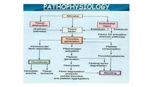
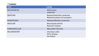
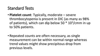
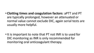
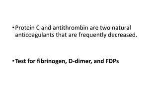
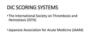
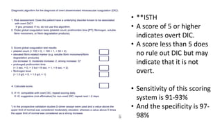
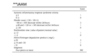
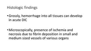
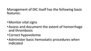
DISSEMINATED
INTRAVASCULAR
COAGULOPATHY
Dr. ABIMBOLA B. O.
OUTLINE
• INTRODUCTION
• TYPES
• PATHOPHYSIOLOGY
• AETIOLOGY
• SIGNS AND SYMPTOMS
• DIFFERENTIAL DIAGNOSES
• DIAGNOSIS
• TREATMENT
• REFERRENCES
INTRODUCTION
•Disseminated Intravascular coagulopathy (DIC) is
an acquired clinicopathologic syndrome
characterized by widespread intravascular fibrin
formation in response to excessive blood
protease activity that overcomes the natural
anticoagulant mechanisms.
•Derangement of the fibrinolytic system further
contributes to intravascular clot formation, but
in some cases accelerated fibrinolysis may cause
severe bleeding. Hence, a patient with DIC can
present with a simultaneously occurring
thrombotic and bleeding problem
•DIC is estimated to be present in 1% of
hospitalized patients.
TYPES
•ACUTE DIC
•CHRONIC DIC
•ACUTE DIC develops when sudden exposure of blood
to procoagulants (e.g. Tissue factor, Tissue
thromboplastin) generates intravascular coagulation.
•Compensatory hemostatic mechanisms are
overwhelmed quickly resulting in a consumptive
coagulopathy leading to hemorrhage.
•Abnormalities of blood coagulation parameters are
readily identified and organ failure frequently results.
•Whereas, CHRONIC DIC reflects a compensated state
when blood is continuously or intermittently
exposed to small amounts of TF.
•Compensatory mechanisms in the liver and bone
marrow are not overwhelmed, and there may be
little obvious clinical or laboratory indication.
•It is more frequently observed in patients with solid
tumors and those with large aortic aneurysms.
PATHOPHYSIOLOGY
•Four simultaneously occurring mechanisms are
known to occur, to include:
•Increased Thrombin generation. Mediated
predominantly by tissue factor/factor VIIa
pathway.
• Impaired function of physiological anticoagulant
pathway.
a) Reduction of antithrombin levels, the result of a
combination of increased consumption, enzyme
degradation, impaired liver synthesis and vascular
leakage.
b) Depression of protein C system, the result of a
combination of increased consumption, impaired liver
synthesis, vascular leakage and down leakage of
thrombodulin.
c) Insufficient Tissue Factor Pathway Inhibitor (TFPI)
•Impaired fibrinolysis. Mediated by release of
plasminogen activators from endothelial cells
immediately followed by an increase in the plasma
levels of plasminogen inhibitor type 1 (PAI-1)
•Activation of inflammatory response. Mediated
by activated coagulation proteins and by
depression of the protein C system
AETIOLOGY
Occurs generally via one of the following pathways:
• A systemic inflammatory response leading to
activation of the cytokine network and subsequent
activation of coagulation (sepsis, major trauma).
• Release or exposure of pro coagulant material in the
bloodstream (cancer, obstetric cases)
TYPE CAUSE
INFECTIOUS Bacterial
Viral
Fungal
Parasitic
MALIGNANCY Hematologic
Metastatic
OBSTETRIC Amniotic fluid embolism
Abruptio placentae
PreEclampsia/Eclampsia/HELLP syndrome
Septic abortion/Retained stillbirth
TRAUMA Burns
RTA
Snake envenomation
TRANSFUSION Hemolytic reactions
OTHER Prosthetic devices
Shunts
Ventricular assist devices
**ACUTE
**CHRONIC
TYPE CAUSE
MALIGNANCIES Solid tumors
Leukemias
OBSTETRIC Retained dead fetus syndrome
Retained products of conception
HEMATOLOGIC Myeloproliferative syndromes
VASCULAR Rheumatoid arthritis
Reynaud's disease
CARDIOVASCULAR Myocardial infarction
INFLAMMATORY Ulcerative colitis
Chron disease
Sarcoidosis
SIGNS AND SYMPTOMS
•Symptoms are often those of the underlying
condition.
•In addition there is typically a history of blood
loss through bleeding in areas such as the
gingivae and GI system.
• Acutely presenting DIC often manifests as petechiae
and ecchymosis along with blood loss from IV Lines
and Catheters.
FEATURES AFFECTED PATIENTS
Bleeding 64% (bleeding from at least 3 unrelated sites is particularly
suggestive of DIC)
Renal dysfunction 25%
Hepatic dysfunction 19%
Respiratory dysfunction 16%
Shock 14%
CNS dysfunction 2%
•Physical findings in acute DIC are usually those of
underlying condition, however there could also be
petechiae on the soft palate and extremities
(thrombocytopenia). and ecchymosis at
venipuncture or traumatized sites.
•In chronic DIC, primary manifestation is
thrombosis, and the signs of venous
thromboembolism may be present.
DIFFERENTIAL DIAGNOSES
•Dysfibrinogenemia
•Hemolytic-Uremic syndrome
•Heparin-Induced Thrombocytopenia
•Thrombotic Thrombocytopenic Purpura
DIAGNOSIS
• Could be difficult especially in cases of chronic DIC
where clinical and laboratory abnormalities may be
subtle.
• No single routinely available test is sufficiently specific
or sensitive.
• Diagnosis is made by combining clinical impression
with any laboratory abnormalities noted.
Standard Tests
• Platelet count: Typically, moderate – severe
thrombocytopenia is present in DIC (as many as 98%
of patients), which can dip below 50 * 103/cmm in up
to 50% patients.
• Repeated counts are often necessary, as single
measurement can be within normal range whereas
trend values might show precipitous drop from
previous levels.
•Clotting times and coagulation factors: aPTT and PT
are typically prolonged, however an attenuated or
normal value cannot exclude DIC, again serial tests are
usually more helpful.
•It is important to note that PT not INR is to used for
DIC monitoring as INR is only recommended for
monitoring oral anticoagulant therapy.
•Protein C and antithrombin are two natural
anticoagulants that are frequently decreased.
•Test for fibrinogen, D-dimer, and FDPs
DIC SCORING SYSTEMS
•The International Society on Thrombosis and
Hemostasis (ISTH)
•Japanese Association for Acute Medicine (JAAM)
• **ISTH
• A score of 5 or higher
indicates overt DIC.
• A score less than 5 does
no rule out DIC but may
indicate that it is not
overt.
• Sensitivity of this scoring
system is 91-93%
• And the specificity is 97-
98%
**JAAM
Histologic findings
•Grossly, hemorrhage into all tissues can develop
in acute DIC
•Microscopically, presence of ischemia and
necrosis due to fibrin deposition in small and
medium sized vessels of various organs
TREATMENT AND MANAGEMENT
•Treatment should primarily focus on addressing
the underlying disorder.
Management of DIC itself has the following basic
features:
•Monitor vital signs
•Assess and document the extent of hemorrhage
and thrombosis
•Correct hypovolemia
•Administer basic hemostatic procedures when
indicated
•Platelet and coagulation factor replacement
should not be instituted on the basis of
laboratory results alone; such therapy is
indicated only in patients with active bleeding,
those requiring an invasive procedure or
otherwise at risk for bleeding complications.
Platelets
•Platelets transfusion may be considered in
patients with DIC and severe thrombocytopenia,
with active bleeding or the risk of bleeding.
•Most clinicians provide platelet replacement in
non bleeding patients if count drops below
20*103/cmm (1 or 2 U/kg/day)
Coagulation factors
• Specific deficiencies in coagulation factors e.g.
fibrinogen can be corrected by administration of
cryoprecipitate or purified fibrinogen concentrate
with FFP (the suggested starting dose is 15mg/kg).
• In case of a relative Vit. K deficiency in the face of
consumption, administration of Vitamin K may be
required.
Anticoagulation
• Effectiveness of heparin therapy will be limited
without concomitant replacement of antithrombin.
• However use of heparin in chronic DIC where there is
preponderance of coagulation without consumptive
coagulopathy is well established.
• (a dose of 4-5U/kg constant infusion is safe)
Tissue factor pathway inhibitor
•TFPI mechanism of coagulation has received
attention as potential therapy in sepsis
associated DIC.
Long-Term Monitoring
• May include anti-platelet agents for those with low-
grade DIC, antibiotics appropriate with the primary
diagnosis or both.
• Chronic DIC in patients with cancer can be managed
with subcutaneous heparin or LMWH.
• Refer to a hematologist on outpatient basis after
initial assessment and stabilization.
CONCLUSION
•DIC alongside other coagulopathies and even
infectious diseases like VHFs could be life
threatening, therefore a sound clinical acumen
and an in-depth theoretical knowledge might be
required to differentiate one from the other and
take measures early enough to reduce mortality.
REFERENCES
• Costello RA, Nehring SM. Disseminated Intravascular Coagulation
(DIC). 2018 Jan
• Papageorgiou C, Jourdi G, Adjambari E, Walborn A, Patel P, Fareed J,
et al. Disseminated Intravascular Coagulation: An Update on
Pathogenesis, Diagnosis and Therapeutic Strategies. Clin Appl Thromb
Hemost. 2018 Oct 8. 1076029618806424.

Disseminated Intravascular Coagulopathy.pdf

  • 1.
  • 2.
    OUTLINE • INTRODUCTION • TYPES •PATHOPHYSIOLOGY • AETIOLOGY • SIGNS AND SYMPTOMS • DIFFERENTIAL DIAGNOSES • DIAGNOSIS • TREATMENT • REFERRENCES
  • 3.
    INTRODUCTION •Disseminated Intravascular coagulopathy(DIC) is an acquired clinicopathologic syndrome characterized by widespread intravascular fibrin formation in response to excessive blood protease activity that overcomes the natural anticoagulant mechanisms.
  • 4.
    •Derangement of thefibrinolytic system further contributes to intravascular clot formation, but in some cases accelerated fibrinolysis may cause severe bleeding. Hence, a patient with DIC can present with a simultaneously occurring thrombotic and bleeding problem •DIC is estimated to be present in 1% of hospitalized patients.
  • 5.
  • 6.
    •ACUTE DIC developswhen sudden exposure of blood to procoagulants (e.g. Tissue factor, Tissue thromboplastin) generates intravascular coagulation. •Compensatory hemostatic mechanisms are overwhelmed quickly resulting in a consumptive coagulopathy leading to hemorrhage. •Abnormalities of blood coagulation parameters are readily identified and organ failure frequently results.
  • 7.
    •Whereas, CHRONIC DICreflects a compensated state when blood is continuously or intermittently exposed to small amounts of TF. •Compensatory mechanisms in the liver and bone marrow are not overwhelmed, and there may be little obvious clinical or laboratory indication. •It is more frequently observed in patients with solid tumors and those with large aortic aneurysms.
  • 8.
    PATHOPHYSIOLOGY •Four simultaneously occurringmechanisms are known to occur, to include: •Increased Thrombin generation. Mediated predominantly by tissue factor/factor VIIa pathway.
  • 9.
    • Impaired functionof physiological anticoagulant pathway. a) Reduction of antithrombin levels, the result of a combination of increased consumption, enzyme degradation, impaired liver synthesis and vascular leakage. b) Depression of protein C system, the result of a combination of increased consumption, impaired liver synthesis, vascular leakage and down leakage of thrombodulin.
  • 10.
    c) Insufficient TissueFactor Pathway Inhibitor (TFPI) •Impaired fibrinolysis. Mediated by release of plasminogen activators from endothelial cells immediately followed by an increase in the plasma levels of plasminogen inhibitor type 1 (PAI-1)
  • 11.
    •Activation of inflammatoryresponse. Mediated by activated coagulation proteins and by depression of the protein C system
  • 13.
    AETIOLOGY Occurs generally viaone of the following pathways: • A systemic inflammatory response leading to activation of the cytokine network and subsequent activation of coagulation (sepsis, major trauma). • Release or exposure of pro coagulant material in the bloodstream (cancer, obstetric cases)
  • 14.
    TYPE CAUSE INFECTIOUS Bacterial Viral Fungal Parasitic MALIGNANCYHematologic Metastatic OBSTETRIC Amniotic fluid embolism Abruptio placentae PreEclampsia/Eclampsia/HELLP syndrome Septic abortion/Retained stillbirth TRAUMA Burns RTA Snake envenomation TRANSFUSION Hemolytic reactions OTHER Prosthetic devices Shunts Ventricular assist devices **ACUTE
  • 15.
    **CHRONIC TYPE CAUSE MALIGNANCIES Solidtumors Leukemias OBSTETRIC Retained dead fetus syndrome Retained products of conception HEMATOLOGIC Myeloproliferative syndromes VASCULAR Rheumatoid arthritis Reynaud's disease CARDIOVASCULAR Myocardial infarction INFLAMMATORY Ulcerative colitis Chron disease Sarcoidosis
  • 16.
    SIGNS AND SYMPTOMS •Symptomsare often those of the underlying condition. •In addition there is typically a history of blood loss through bleeding in areas such as the gingivae and GI system.
  • 17.
    • Acutely presentingDIC often manifests as petechiae and ecchymosis along with blood loss from IV Lines and Catheters. FEATURES AFFECTED PATIENTS Bleeding 64% (bleeding from at least 3 unrelated sites is particularly suggestive of DIC) Renal dysfunction 25% Hepatic dysfunction 19% Respiratory dysfunction 16% Shock 14% CNS dysfunction 2%
  • 18.
    •Physical findings inacute DIC are usually those of underlying condition, however there could also be petechiae on the soft palate and extremities (thrombocytopenia). and ecchymosis at venipuncture or traumatized sites. •In chronic DIC, primary manifestation is thrombosis, and the signs of venous thromboembolism may be present.
  • 19.
  • 20.
    DIAGNOSIS • Could bedifficult especially in cases of chronic DIC where clinical and laboratory abnormalities may be subtle. • No single routinely available test is sufficiently specific or sensitive. • Diagnosis is made by combining clinical impression with any laboratory abnormalities noted.
  • 21.
    Standard Tests • Plateletcount: Typically, moderate – severe thrombocytopenia is present in DIC (as many as 98% of patients), which can dip below 50 * 103/cmm in up to 50% patients. • Repeated counts are often necessary, as single measurement can be within normal range whereas trend values might show precipitous drop from previous levels.
  • 22.
    •Clotting times andcoagulation factors: aPTT and PT are typically prolonged, however an attenuated or normal value cannot exclude DIC, again serial tests are usually more helpful. •It is important to note that PT not INR is to used for DIC monitoring as INR is only recommended for monitoring oral anticoagulant therapy.
  • 23.
    •Protein C andantithrombin are two natural anticoagulants that are frequently decreased. •Test for fibrinogen, D-dimer, and FDPs
  • 24.
    DIC SCORING SYSTEMS •TheInternational Society on Thrombosis and Hemostasis (ISTH) •Japanese Association for Acute Medicine (JAAM)
  • 25.
    • **ISTH • Ascore of 5 or higher indicates overt DIC. • A score less than 5 does no rule out DIC but may indicate that it is not overt. • Sensitivity of this scoring system is 91-93% • And the specificity is 97- 98%
  • 26.
  • 27.
    Histologic findings •Grossly, hemorrhageinto all tissues can develop in acute DIC •Microscopically, presence of ischemia and necrosis due to fibrin deposition in small and medium sized vessels of various organs
  • 28.
    TREATMENT AND MANAGEMENT •Treatmentshould primarily focus on addressing the underlying disorder.
  • 29.
    Management of DICitself has the following basic features: •Monitor vital signs •Assess and document the extent of hemorrhage and thrombosis •Correct hypovolemia •Administer basic hemostatic procedures when indicated
  • 30.
    •Platelet and coagulationfactor replacement should not be instituted on the basis of laboratory results alone; such therapy is indicated only in patients with active bleeding, those requiring an invasive procedure or otherwise at risk for bleeding complications.
  • 31.
    Platelets •Platelets transfusion maybe considered in patients with DIC and severe thrombocytopenia, with active bleeding or the risk of bleeding. •Most clinicians provide platelet replacement in non bleeding patients if count drops below 20*103/cmm (1 or 2 U/kg/day)
  • 32.
    Coagulation factors • Specificdeficiencies in coagulation factors e.g. fibrinogen can be corrected by administration of cryoprecipitate or purified fibrinogen concentrate with FFP (the suggested starting dose is 15mg/kg). • In case of a relative Vit. K deficiency in the face of consumption, administration of Vitamin K may be required.
  • 33.
    Anticoagulation • Effectiveness ofheparin therapy will be limited without concomitant replacement of antithrombin. • However use of heparin in chronic DIC where there is preponderance of coagulation without consumptive coagulopathy is well established. • (a dose of 4-5U/kg constant infusion is safe)
  • 34.
    Tissue factor pathwayinhibitor •TFPI mechanism of coagulation has received attention as potential therapy in sepsis associated DIC.
  • 35.
    Long-Term Monitoring • Mayinclude anti-platelet agents for those with low- grade DIC, antibiotics appropriate with the primary diagnosis or both. • Chronic DIC in patients with cancer can be managed with subcutaneous heparin or LMWH. • Refer to a hematologist on outpatient basis after initial assessment and stabilization.
  • 36.
    CONCLUSION •DIC alongside othercoagulopathies and even infectious diseases like VHFs could be life threatening, therefore a sound clinical acumen and an in-depth theoretical knowledge might be required to differentiate one from the other and take measures early enough to reduce mortality.
  • 37.
    REFERENCES • Costello RA,Nehring SM. Disseminated Intravascular Coagulation (DIC). 2018 Jan • Papageorgiou C, Jourdi G, Adjambari E, Walborn A, Patel P, Fareed J, et al. Disseminated Intravascular Coagulation: An Update on Pathogenesis, Diagnosis and Therapeutic Strategies. Clin Appl Thromb Hemost. 2018 Oct 8. 1076029618806424.