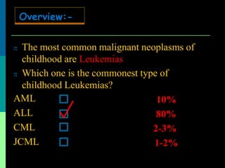
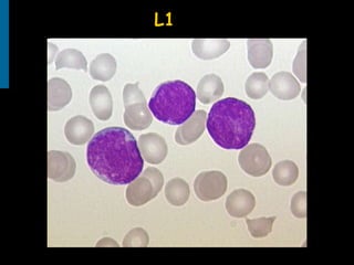
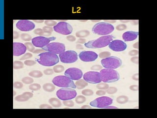
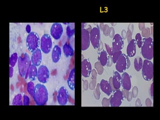
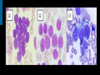
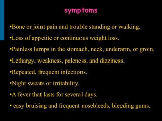
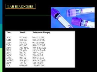
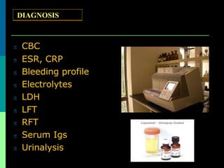
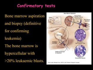
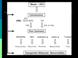
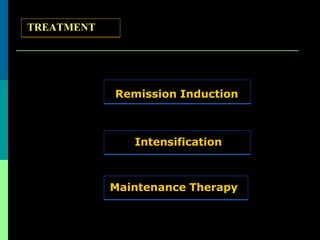
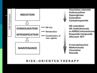
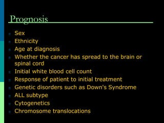
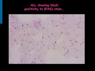
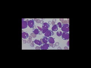
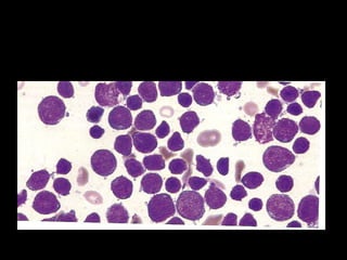
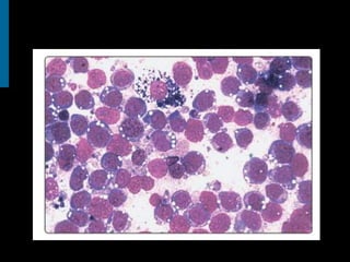
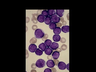
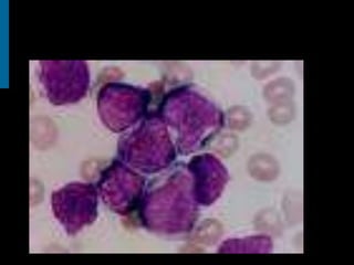
Acute lymphoblastic Leukemias(ALL)
By the end of this lecture everybody should be
aware about:
Objectives
WHAT IS ALL??
TREATMENT
CLASSIFICATION
EITIOLOGY
DIAGNOSIS
CLINICAL
PRESENTATION
 It is the malignant tumor of haematopoietic
precursor cells of lymphoid lineage.
Definition:-
 The most common malignant neoplasms of
childhood ..
Overview:-
 The most common malignant neoplasms of
childhood are Leukemias.
 Which one is the commonest type of
childhood Leukemias?
AML
ALL
CML
JCML
Overview:-
 The most common malignant neoplasms of
childhood are Leukemias
 Which one is the commonest type of
childhood Leukemias?
AML
ALL
CML
JCML
…………….%
…………….%
.……………%
…………….%
Overview:-
 The most common malignant neoplasms of
childhood are Leukemias
 Which one is the commonest type of
childhood Leukemias?
AML
ALL
CML
JCML
…………….%
80%
.……………%
…………….%
Overview:-
 The most common malignant neoplasms of
childhood are Leukemias
 Which one is the commonest type of
childhood Leukemias?
AML
ALL
CML
JCML
10%
80%
2-3%
1-2%
Overview:-
Epidemiology
•Approximately 1 in 50,000 (ages 0 to 19 years).
•It has a peak incident rate of 3–7 years old, decreasing in
incidence with increasing age.
•common in males than females.
• There is an increased incidence in people with Down
Syndrome, Fanconi anemia, Ataxia telangiectasia,…etc
Etiology
•ALL is associated with exposure to radiation and
chemicals as found by studies of survivors of
atom bomb exposure in Hiroshima and Nagasaki.
• Exposure to benzene and other chemicals .
• Secondary leukemia can develop in individuals
treated for other cancers with radiation and
chemotherapy.
Pathophysiology
•The malignant lymphoid precursor cells (ie, lymphoblasts) are
arrested in an early stage of development.
•This arrest is caused by an abnormal expression of genes, often as
a result of chromosomal translocations. The lymphoblasts replace
the normal marrow elements, resulting in a marked decrease in the
production of normal blood cells.
•Consequently, anemia, thrombocytopenia, and neutropenia occur
to varying degrees. The lymphoblasts also proliferate in organs
other than the marrow, particularly the liver, spleen, and lymph
nodes.
Classification
FAB
WHO
Classification
• French-American-British Classification
(based on morphology and cytochemistry)
• World Health Organization Classification
·( (based on molecular, immunophenotypic, imorphologic, and clinical
features)
FAB -classification
L1: show uniform, small blast cells with
scanty cytoplasm.
L2: larger blast cells with more prominent nucleoli
and cytoplasm and with more heterogeneity.
L3: blasts are large with prominent nucleoli,
strongly basophilic cytoplasm and cytoplasmic
vacuoles.
L1
L2
L3
World Health Organization
•The recent WHO instead of morphology use of the immunophenotypic
classification mentioned below:
•1. Acute lymphoblastic leukemia/lymphoma. (L1/L2)
• i. Precursor B acute lymphoblastic leukemia/lymphoma.
•ii. Precursor T acute lymphoblastic leukemia/lymphoma
•2. Burkitt's leukemia/lymphoma. (L3)
•3. Biphenotypic acute leukemia.
CLINICAL
PRESENTATION
CLINICAL
PRESENTATION
 Common presentations of allacute
Leukemias?
1) Fever
2) Fatigue
3) Bleeding
CLINICAL
PRESENTATION
•Bone marrow failure Anaemia (pallor, lethargy and dyspnoea);
neutropenia (fever, malaise, features of mouth ulcer, respiratory,
perianal or other infectionsand thrombombocytopenia
•(spontaneous bruises, purpura, bleeding gums and menorrhagia;
•Organ infiltration Tender bones, lymphadenopathy, moderate
splenomegaly, hepatomegaly and meningeal syndrome (headache,
nausea and vomiting, blurring of vision and diplopia)..
•Less common manifestations include testicular swelling.
symptoms
•Bone or joint pain and trouble standing or walking.
•Loss of appetite or continuous weight loss.
•Painless lumps in the stomach, neck, underarm, or groin.
•Lethargy, weakness, paleness, and dizziness.
•Repeated, frequent infections.
•Night sweats or irritability.
•A fever that lasts for several days.
• easy bruising and frequent nosebleeds, bleeding gums.
LAB DIAGNOSIS
 CBC
 ESR, CRP
 Bleeding profile
 Electrolytes
 LDH
 LFT
 RFT
 Serum Igs
 Urinalysis
DIAGNOSIS
DIAGNOSIS
• Normochromic, Normocytic anaemia with
thrombocytopenia in most cases.
•The TWBCs count may be decreased, normal or
increased to 200 x 109/L or more.
•Blood film examination typically shows a variable
numbers of blast cells(>20% ).
Confirmatory tests
Bone marrow aspiration
and biopsy (definitive
for confirming
leukemia)
The bone marrow is
hypercellular with
>20% leukaemic blasts.
•Cytochemical stains(SBB, MPO, PAS,…etc).
•Immunohistochemistry.
•Flow cytometry.
•Cytogenetic analysis.
•Polymerase chain reaction.
•Gene expression profiling.
Differential Diagnosis
 Aplastic anemia
 Idiopathic thrombocytopenic purpura
 Recent viral infection
 Infectious mononucleosis
 Pertussis or parapertussis
 Small round cell tumors involving the bone
marrow, including neuroblastoma,
rhabdomyosarcoma, and retinoblastoma
 Juvenile rheumatoid arthritis, rheumatic
fever, other collagen diseases, or
osteomyelitis
 How much is the curerate?????????
TREATMENT
 Around85%
TREATMENT
TREATMENT
Remission Induction
Intensification
Maintenance Therapy
Prognosis
 Sex
 Ethnicity
 Age at diagnosis
 Whether the cancer has spread to the brain or
spinal cord
 Initial white blood cell count
 Response of patient to initial treatment
 Genetic disorders such as Down's Syndrome
 ALL subtype
 Cytogenetics
 Chromosome translocations
ALL showing block
positivity to (PAS) stain..
Thank
you

ALL

  • 1.
  • 2.
    By the endof this lecture everybody should be aware about: Objectives WHAT IS ALL?? TREATMENT CLASSIFICATION EITIOLOGY DIAGNOSIS CLINICAL PRESENTATION
  • 3.
     It isthe malignant tumor of haematopoietic precursor cells of lymphoid lineage. Definition:-
  • 4.
     The mostcommon malignant neoplasms of childhood .. Overview:-
  • 5.
     The mostcommon malignant neoplasms of childhood are Leukemias.  Which one is the commonest type of childhood Leukemias? AML ALL CML JCML Overview:-
  • 6.
     The mostcommon malignant neoplasms of childhood are Leukemias  Which one is the commonest type of childhood Leukemias? AML ALL CML JCML …………….% …………….% .……………% …………….% Overview:-
  • 7.
     The mostcommon malignant neoplasms of childhood are Leukemias  Which one is the commonest type of childhood Leukemias? AML ALL CML JCML …………….% 80% .……………% …………….% Overview:-
  • 8.
     The mostcommon malignant neoplasms of childhood are Leukemias  Which one is the commonest type of childhood Leukemias? AML ALL CML JCML 10% 80% 2-3% 1-2% Overview:-
  • 9.
    Epidemiology •Approximately 1 in50,000 (ages 0 to 19 years). •It has a peak incident rate of 3–7 years old, decreasing in incidence with increasing age. •common in males than females. • There is an increased incidence in people with Down Syndrome, Fanconi anemia, Ataxia telangiectasia,…etc
  • 11.
    Etiology •ALL is associatedwith exposure to radiation and chemicals as found by studies of survivors of atom bomb exposure in Hiroshima and Nagasaki. • Exposure to benzene and other chemicals . • Secondary leukemia can develop in individuals treated for other cancers with radiation and chemotherapy.
  • 12.
    Pathophysiology •The malignant lymphoidprecursor cells (ie, lymphoblasts) are arrested in an early stage of development. •This arrest is caused by an abnormal expression of genes, often as a result of chromosomal translocations. The lymphoblasts replace the normal marrow elements, resulting in a marked decrease in the production of normal blood cells. •Consequently, anemia, thrombocytopenia, and neutropenia occur to varying degrees. The lymphoblasts also proliferate in organs other than the marrow, particularly the liver, spleen, and lymph nodes.
  • 13.
  • 14.
    Classification • French-American-British Classification (basedon morphology and cytochemistry) • World Health Organization Classification ·( (based on molecular, immunophenotypic, imorphologic, and clinical features)
  • 15.
    FAB -classification L1: showuniform, small blast cells with scanty cytoplasm. L2: larger blast cells with more prominent nucleoli and cytoplasm and with more heterogeneity. L3: blasts are large with prominent nucleoli, strongly basophilic cytoplasm and cytoplasmic vacuoles.
  • 16.
  • 17.
  • 18.
  • 20.
    World Health Organization •Therecent WHO instead of morphology use of the immunophenotypic classification mentioned below: •1. Acute lymphoblastic leukemia/lymphoma. (L1/L2) • i. Precursor B acute lymphoblastic leukemia/lymphoma. •ii. Precursor T acute lymphoblastic leukemia/lymphoma •2. Burkitt's leukemia/lymphoma. (L3) •3. Biphenotypic acute leukemia.
  • 22.
  • 23.
    CLINICAL PRESENTATION  Common presentationsof allacute Leukemias? 1) Fever 2) Fatigue 3) Bleeding
  • 24.
    CLINICAL PRESENTATION •Bone marrow failureAnaemia (pallor, lethargy and dyspnoea); neutropenia (fever, malaise, features of mouth ulcer, respiratory, perianal or other infectionsand thrombombocytopenia •(spontaneous bruises, purpura, bleeding gums and menorrhagia; •Organ infiltration Tender bones, lymphadenopathy, moderate splenomegaly, hepatomegaly and meningeal syndrome (headache, nausea and vomiting, blurring of vision and diplopia).. •Less common manifestations include testicular swelling.
  • 25.
    symptoms •Bone or jointpain and trouble standing or walking. •Loss of appetite or continuous weight loss. •Painless lumps in the stomach, neck, underarm, or groin. •Lethargy, weakness, paleness, and dizziness. •Repeated, frequent infections. •Night sweats or irritability. •A fever that lasts for several days. • easy bruising and frequent nosebleeds, bleeding gums.
  • 26.
  • 27.
     CBC  ESR,CRP  Bleeding profile  Electrolytes  LDH  LFT  RFT  Serum Igs  Urinalysis DIAGNOSIS
  • 28.
    DIAGNOSIS • Normochromic, Normocyticanaemia with thrombocytopenia in most cases. •The TWBCs count may be decreased, normal or increased to 200 x 109/L or more. •Blood film examination typically shows a variable numbers of blast cells(>20% ).
  • 29.
    Confirmatory tests Bone marrowaspiration and biopsy (definitive for confirming leukemia) The bone marrow is hypercellular with >20% leukaemic blasts.
  • 30.
    •Cytochemical stains(SBB, MPO,PAS,…etc). •Immunohistochemistry. •Flow cytometry. •Cytogenetic analysis. •Polymerase chain reaction. •Gene expression profiling.
  • 31.
    Differential Diagnosis  Aplasticanemia  Idiopathic thrombocytopenic purpura  Recent viral infection  Infectious mononucleosis  Pertussis or parapertussis  Small round cell tumors involving the bone marrow, including neuroblastoma, rhabdomyosarcoma, and retinoblastoma  Juvenile rheumatoid arthritis, rheumatic fever, other collagen diseases, or osteomyelitis
  • 35.
     How muchis the curerate????????? TREATMENT
  • 36.
  • 37.
  • 39.
    Prognosis  Sex  Ethnicity Age at diagnosis  Whether the cancer has spread to the brain or spinal cord  Initial white blood cell count  Response of patient to initial treatment  Genetic disorders such as Down's Syndrome  ALL subtype  Cytogenetics  Chromosome translocations
  • 43.
  • 51.