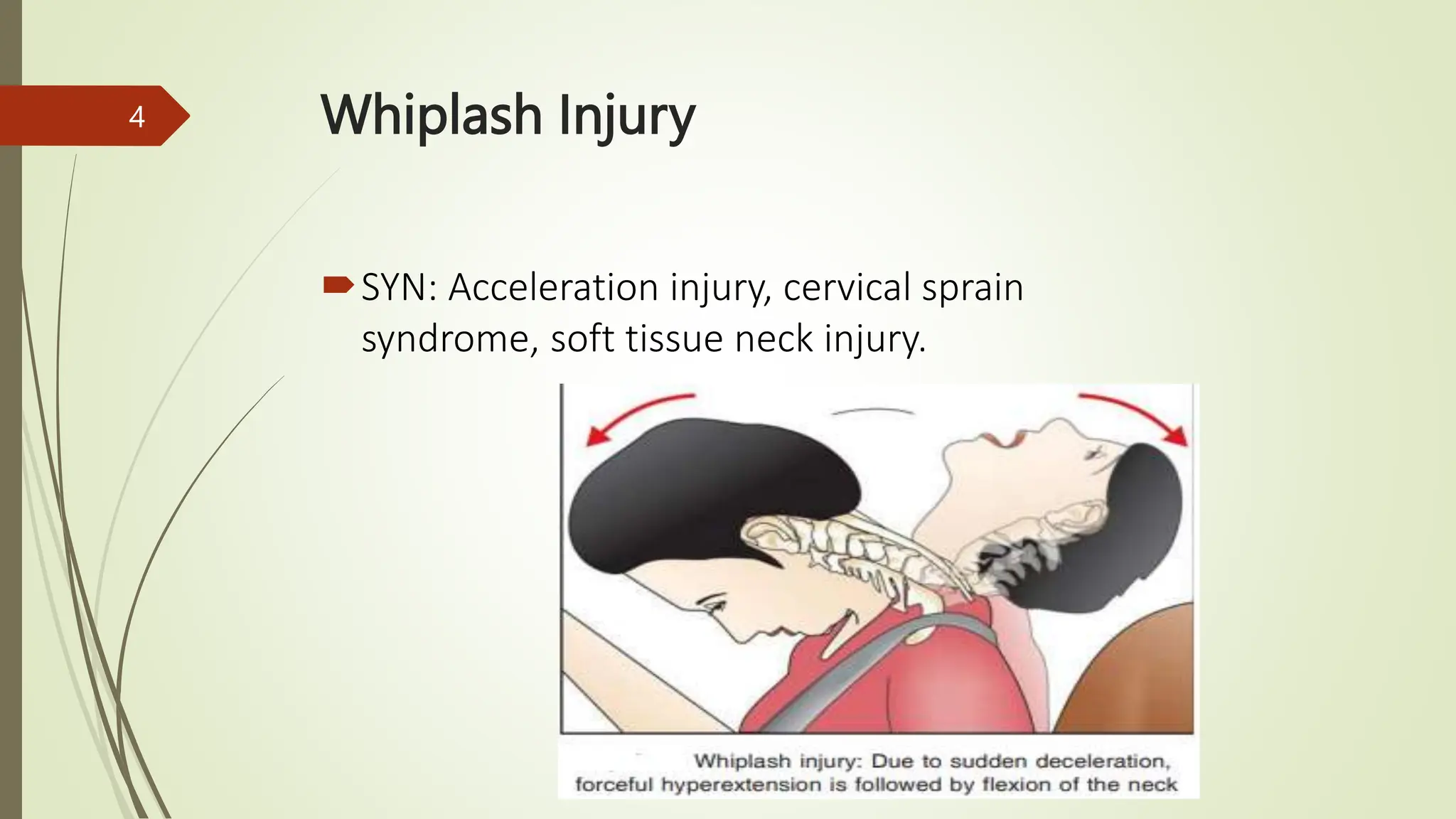
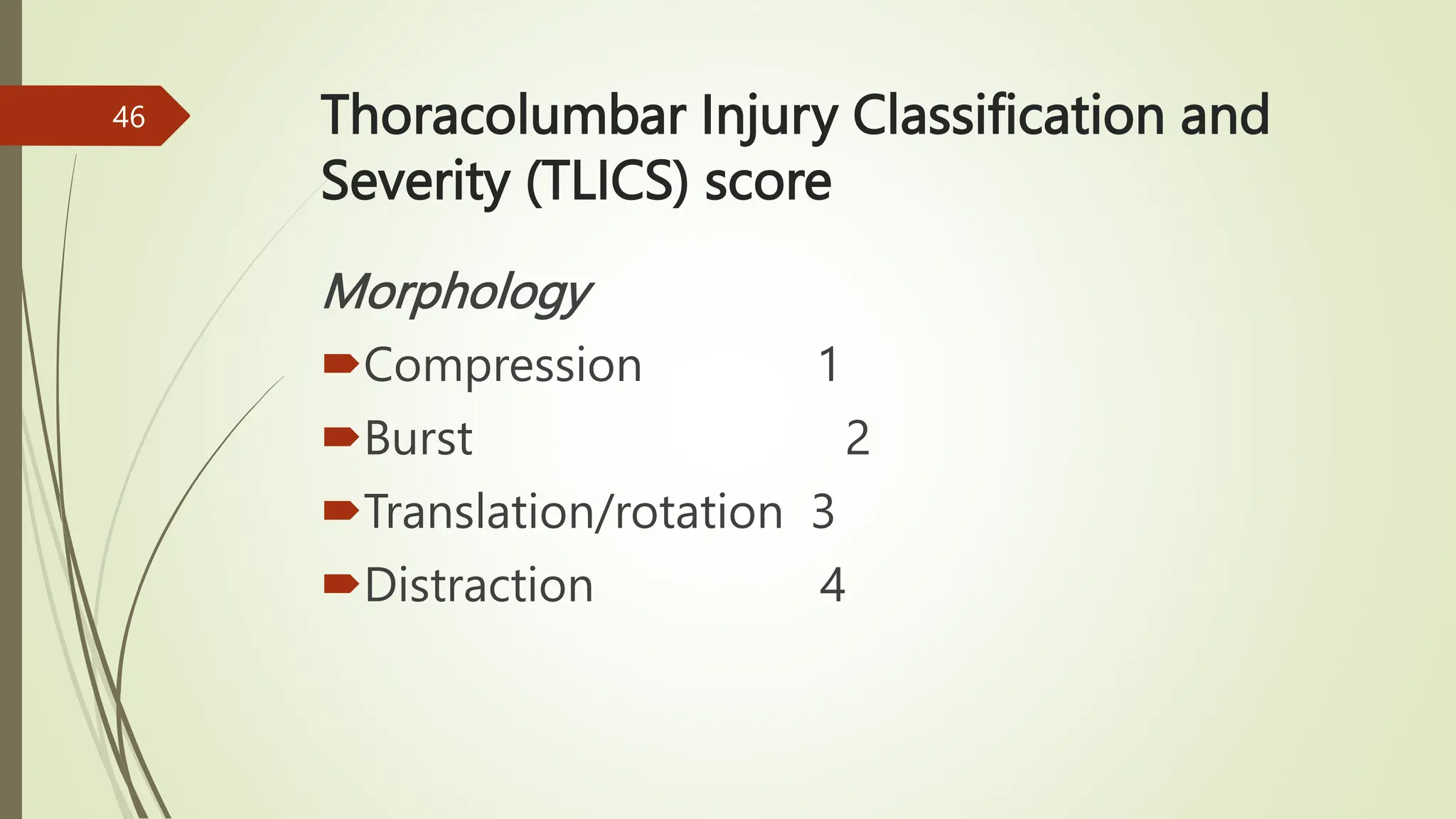
Kenya Medical training college
Faculty of Clinical Sciences
Department Of Orthopaedics and Trauma Medicine
Year Two Semester Two
Traumatology II
By
Gideon Sifirino
Class: Diploma September 2021
1
Learning outcomes
By the end of this lesson the
learner should be able to;
Discuss the anatomy of the thoracolumbar
Discuss common injuries of thoracolumbar
Discuss management thoracolumbar i njuries
2
3
Whiplash Injury
SYN: Acceleration injury, cervical sprain
syndrome, soft tissue neck injury.
4
Definition
It is an unconventional and inconsequential
ligamentous injury of the cervical spine
allegedly due to an extension injury following
a rear-end collision in an RTA.
5
Incidence
 It is seen in about 25 percent
of rear-end collision of RTAs.
Seventy percent of those affected
are women.
It is common in the 3rd or 4th
decades
6
Clinical Features
Upper neck pain that becomes worse
With movement.
Occipital headache.
Neck stiffness.
Rarely vertigo, auditory or visual disturbances,
etc.
7
Cont'd
Hoarseness of voice, (involvement of recurrent
laryngeal nerve).
Difficulty in swallowing, (stretching and contusion of
esophagus).
8
Cont'd
Pain in the back and the shoulder, (radiating pain).
Pain and paresthesia (due to involvement of nerves).
9
Cont'd
 Decreased range of neck movements
Neck muscle spasm
10
Cont'd
Symptoms may develop as early as
within 2 hours to as late
as 8 days.
11
Investigations
X-rays are usually normal.
MRI helps to make a diagnosis
12
Treatment
 Drugs: NSAIDs, muscle relaxants.
Collars: These are recommended for the
first three days.
Short arc active movements are slowly
begun.
Active ROM exercises are slowly commenced.
13
Cont'd
After the pain subsides, isometric strengthening
exercises are slowly commenced.
Other modalities take ultrasound, traction,
manipulation, massage, etc. also helps.
14
Clay Shoveller’s Fracture
An avulsion of the tip of
the C7 spinous process may occur
due a sudden pull of the
trapezius muscle.
15
Cont'd
This historically happened when shovelling heavy
unrelenting clay.
The fracture is painful but stable
and can be treated conservatively.
16
Fractures Of The Thoracic And Lumbar
(Thoracolumbar) Spine
Young patients; high-energy trauma
Elderly; bony insufficiency
17
Risk Factors
Rheumatoid arthritis
Osteoporosis
History of long-term steroid
 Skeletal metastases
18
Vulnerability
The thoracolumbar transitional zone is particularly
vulnerable, with 40–60% of all spinal fractures
involving;
T12
L1
L2
19
Classification
A diagnostic algorithm for this classification is most
easily approached in reverse order
by exclusion of the most severe injuries.
20
C-type: displacement or dislocation injuries
Rare
severe spinal disruption with dissociation between
cranial and caudal spinal segments
Cord injury is common
21
22
B-type: tension band injuries
Anterior tension band failure in extension
B3 Hyperextension
23
Cont'd
Posterior tension band failure in flexion
B2 Osseoligamentous disruption
24
Cont'd
B1 Osseous disruption (Chance fractures)
These are characterized by failure of
the posterior bony elements (spinous process, lamina)
in tension.
25
26
A-type: compression injuries
The anterior structures fail under compression
27
Cont'd
A4 Burst fractures
The entire vertebral body fails under compression.
28
29
Cont'd
Radiological Features
Loss of vertebral height
Loss of cortical integrity of the posterior
vertebral body on the lateral radiograph
Widened pedicles on the AP view.
30
31
Cont'd
On CT scan, retropulsion of fragments
into the vertebral canal.
This may result in spinal cord injury
32
33
Cont'd
A3 Incomplete burst fractures
Involve the posterior bony wall but
only one endplate.
34
35
Cont'd
A2 Pincer fractures
Both endplates fail but both the
posterior and anterior walls of the
vertebral body.
36
37
Cont'd
A1 Simple wedge compression fractures
Most commonly encountered spinal fractures.
The posterior elements are intact but the anterior
vertebral body fails in flexion
and compression.
Spinal cord injury is uncommon.
38
39
Cont'd
A0 Minor fractures (non-structural)
Minor and not associated with instability (e.g.
transverse process).
40
41
Note that these fractures may be
markers of other injuries, such as
unstable pelvic fractures or renal injuries.
42
AO neurological score
 N0: neurologically intact
N1: transient neurological deficit, which is
no longer present
N2: radicular symptoms
N3: incomplete spinal cord injury or any
degree of cauda equina injury
43
Cont'd
N4: complete spinal cord injury
NX: neurological status unknown due to
sedation or head injury.
44
Assessing stability
Morphology
Neurological status
Integrity of posterior elements
45
Thoracolumbar Injury Classification and
Severity (TLICS) score
Morphology
Compression 1
Burst 2
Translation/rotation 3
Distraction 4
46
Cont'd
Neurological status
Intact 0
Nerve root 2
Complete cord injury 2
Incomplete cord injury 3
Cauda equina injury 3
47
Cont'd
Integrity of posterior elements
Intact 0
Indeterminate 2
Injured 3
48
Radiological features
AP and lateral radiographs
CT
MRI
49
Cont'd
AP view; coronal deformity or widening of the
pedicles
Middle column disruption
50
Cont'd
The lateral view ;loss of normal
Vertebral shape or loss of sagittal
alignment (such as a kyphotic deformity).
51
Cont'd
CT: This is indicated where a neurological
deficit clinically or inadequate plain films. Axial
imaging provides a particularly good
indication of whether there is retropulsion of
bone into the canal.
52
Cont'd
MRI: spinal cord and soft tissues
for signs of injury.
Posterior ligament complex injury.
53
Orthopaedic management
Non-operative
Stable fractures
For those patients admitted to the ward,
the aim is to provide analgesia and
bed rest initially, then progressive mobilization
once trunk control returns.
54
Cont'd
Patients are said to have trunk
control once they can comfortably roll
themselves around the bed, are able
to tense their abdominal musculature and feel
as if they can sit up.
55
Cont'd
Bracing may help early mobilization, provide
pain relief and prevention of fractures from
deteriorating.
56
Operative
Posterior surgical decompression and
instrumented spinal fusion
57
Cont'd
Decompression with laminectomy alone
should be avoided, as it will
further destabilize the spine by compromising
the posterior supporting structures.
58
Cont'd
Posterior fusion alone (no decompression)
may be undertaken where there are no signs
of neurological compromise.
59
Cont'd
Anterior decompression and stabilization may
also be considered and has the potential
advantage of providing direct visualization of
fracture fragments and therefore better canal
clearance and better anterior column support.
60
61
Questions???
62
Assignment
 To be Provided…
63
References
 Ebnezer, J, (2012): Textbook of Orthopedics Fifth Edition:,
Jaypee Brothers Medical Publishers (P) Ltd.
 Blom, A.W, Warwick, D and Whitehouse, M. R.: Apley and
Solomon’s System of Orthopaedics and Trauma Tenth Edition:
CRC Press Taylor & Francis Group.
64
The end
Thank you
65

Trauma - Thoracic and chest injuries.pptx

Editor's Notes

  • #24 In these injuries, the anterior tension band fails under tension.
  • #25 These injuries are important, as they are often unstable, and the extent of the injury may not be apparent initially on plain radiographs.
  • #44 Neurological injury is recorded in addition to the morphological classification above