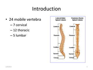
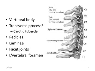
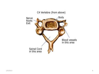
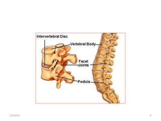
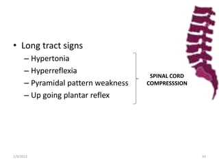
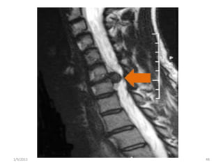
DEGENERATIVE SPINAL
DISORDERS
BY DR RUHAMA YOSEPH
SURGICAL RESIDENT
OUTLINE
• Introduction & Anatomy
• Cervical degenerative disc diseases
– Cervical spondylosis
– Cervical disc disorder
• Lumbar degenerative disc diseases
– Lumbar disc herniation
– Spinal stenosis
– Spondylolisthesis
1/9/2013 2
Introduction
• 24 mobile vertebra
– 7 cervical
– 12 thoracic
– 5 lumbar
1/9/2013 3
• Anatomy
– Function of inter-vertebral disc is to permit stable
motion of the spine
– Nucleus Pulposus
Central part of the disc
– Annulus Fibrosus
Multi-laminated ligament encompassing periphery
of the disc space
1/9/2013 4
1/9/2013 5
Nucleus
Pulposus
Annulus
Fibrosus
• Vertebral body
• Transverse process*
– Carotid tubercle
• Pedicles
• Laminae
• Facet joints
• I/vertebral foramen
1/9/2013 6
• Anterior longitudinal ligament
• Posterior longitudinal ligament
• Ligamentum nuchae
• I/spinous & supraspinous
• Ligamentum flavum
1/9/2013 7
1/9/2013 8
1/9/2013 9
• Definitions
– Radiculopathy
Dysfunction of nerve root with manifestation of;
pain, dermatomal sensory loss, muscle weakness,
hypoactive DTR along its distribution
– Degeneration
Desiccation, fusion, narrowing of disc space,
fissuring, bulging of annulus
1/9/2013 10
• Definitions…
– Disc Degeneration
Clinical syndromes associated with degeneration
inside and outside the I/vertebral disc
– Herniation
Localized displacement of disc material (<50% of
the disc circumference)
1/9/2013 11
Cervical Degenerative Disc Diseases
1/9/2013 12
• Includes:
– Cervical spondylosis
– Cervical disc herniation
1/9/2013 13
• At rest and in a neutral position the cervical
spine has gentle lordotic configuration
• The balance point between flexion and
extension extends from the odontoid process
to the cervico-thoracic junction passing
through the centre of C2’s vertebral body
1/9/2013 14
• Cervical facet joints allow :
– Flexion/ extension movement (subaxial)
– Lateral bending
– Rotational movement
• 50% of rotational movement is allowed at C1-C2
junction upon the odontoid process
1/9/2013 15
• Stability of cervical spine motion segments
depends on:
– Ligamenteous structures
• Facet joint capsule, longitudinal ligaments and annulus
fibrosus
– Paraspinal musculature
1/9/2013 16
Cervical Spondylosis
• Degeneration of cervical mobile segments is
an inevitable consequence of aging
• It is a degenerative arthritic condition
affecting the cervical spine, I/vertebral discs
and facet joints
• Radiological suggestive finding found in 75%
of those above 50, who may not have
symptoms
1/9/2013 17
• Causes of degeneration are:
– Repetitive movement
– Stress and strain of osseoligamenteous structures
– Genetic and developmental factors
• Subaxial cervical spine is most often and
severely affected
– Because widest degree of range of motion and
maximal stress is bore by those levels (C5-C6, C6-
C7, C4-C5)
1/9/2013 18
• Pathogenesis:
– Disc changes are the initial event followed by
degeneration of facet, ligaments, reactive bony
changes,…
– In normal disc weight bearing concentrates at the
center, whereas in degeneration it becomes more
diffuse
• Stress on underlying structures of adjacent vertebra
1/9/2013 19
• Pathogenesis…
– In normal condition movement is pivoted across
stationary focal instantaneous axis , whereas in
degeneration it is across a wide range of axis
which is also variable
• Laxity of longitudinal ligaments and annulus
• Accumulation oseophytes and formation of bone spurs
– These affect the facet articulations located
posteriorly
1/9/2013 20
Disc changes
•Volume
•Biomechanical characteristics
Laxity of ligaments and
altered motion
dynamics
Stress on facet joints
Synovial tissue and
bony reactive changes
Osteophyte accumulation
& Traction spurs
1/9/2013 21
• Pathogenesis…
– OPLL (Ossification of Posterior Ligament)
• Preferential accumulation of calcium in posterior
ligament
• Common in Asian population
– DISH ( Diffuse Idiopathic Skeletal Hypertrophy)
• Exuberant accumulation of anterior osteophytes and
calcification along vertebral bodies
• African-American males
1/9/2013 22
1/9/2013 23
OPLL
1/9/2013 24
DISH
• Clinical picture:
1. Decreased motion of neck
• Due to the degenerative structural changes
2. Pain
• Most common manifestation
3. Neurologic deficit
1/9/2013 25
• Pain can be axial or appendicular, continuous
or activity oriented…
• Appendicular pain
– Pain radiating to distal part of arm is strongly
suggestive of encroachment onto nerve root
1/9/2013 26
• Axial pain
– Can be referred or from inflammation and muscle
fatigue (from posterior rami)
– Usually posterior neck area sometimes extending
cephald or caudad
1/9/2013 27
• Neurological manifestation
– Radiculopathy (nerve root compression)
– Myelopathy (spinal cord compression)
– Sensory changes
1/9/2013 28
• Cervical cord compression:
– Static: result in narrowing of spinal canal by
accumulation of osteophytes and disk herniation
• Signs and symptoms of myelopathy manifest at AP
diameter <12 mm
– Dynamic: micro or overt instability of motion
segments as the ligamenteous structures
function become impaired
1/9/2013 29
• DDX:
– Peripheral nerve entrapment syndrome
• Carpal tunnel syndrome
• Ulnar neuropathy
– Neural degenerative disorders
• Multiple Sclerosis
• Amyotrophic Lateral Sclerosis
• GBS, Transverse Myelitis
– Thoracic outlet compression
– Peripheral neuropathy…
1/9/2013 30
• Imaging:
– Plain neck x-ray
– Spinal canal study
– MRI
– CT myelography
– Nerve conduction studies
– Diskography
Presence of degenerative change doesn’t always correlate
with presence of symptoms
1/9/2013 31
• Neck x-ray:
– Helps to see alignment and stability of the spine
– Bone changes such as osteophyte accumulation
and sclerosis of endplates and encroachment to
foramina can be seen
• Spinal canal study:
– By MRI, myelography, post-myelogram CT
1/9/2013 32
1/9/2013 33
• MRI: *
Shows spinal cord in relation to surrounding
osseoligamenteous structures
• CT-myelogram:
To see extent of nerve root or spinal cord compression
• Myelogram:
AP x-ray on flexion/extension to identify foci of
compression
1/9/2013 34
1/9/2013 35
ANTERIOR
OSTEOPHYTE
1/9/2013 36
FLEXION
Cervical Disc Prolapse
• Usually involve C6-C7 level, less commonly
C5-C6
• Degeneration starts due to repetitive force
applied at these points which are fulcrum for
the mobility of head and spine
1/9/2013 37
• Causes:
– Degeneration process
– Repetitive excessive stress acting as an
exacerbating factor
– Minor traumas on top of these can result in acute
disc prolapse
1/9/2013 38
• Direction of herniation is usually
posterolateral
• Nerve roots compressed as they pass through
inter-vertebral foramen
• Same level nerve roots affected (unlike
lumbar)
– C5-C6: =>C6 nerve root affected
– C6-C7: =>C7 affected
– C7-T1: =>C8 affected
1/9/2013 39
• Clinical picture:
– Neck, arm pain
– Neurologic manifestation of nerve root
compression
– Brachial Neuralgia
– Pain follows sclerotomes and is widespread
– Sensory dermatomal disturbance includes
numbness, tingling
1/9/2013 40
• Cl/P…
– Location of sensory disturbance more useful than
pain distribution
• Thumb & index => C6
• Middle finger & index => C7
• Little & ring finger => C8
– Muscle weakness
• Of elbow extension => C7
• Of shoulder abduction elevation of arm =>C5
• Mild elbow flexion => C6
1/9/2013 41
• Restricted cervical spine movement
– Head rigidly held to one side, moderately flexed and
tilted to the painful side
• In long standing prolapse there will be wasting of
muscles
– In C7 root compression wasting of Triceps M.
• Diminished DTR
• Biceps => C5
• Brachioradialis => C6
• Triceps =>C7
1/9/2013 42
• Long tract signs
– Hypertonia
– Hyperreflexia
– Pyramidal pattern weakness
– Up going plantar reflex
SPINAL CORD
COMPRESSSION
1/9/2013 43
Clinical localization
C6/C7 prolapsed intervertebral disc
(C7 nerve root)
Weakness of elbow extension
Absent triceps jerk
Numbness or tingling in the middle or index
finger
C5/6 prolapsed intervertebral disc
(C6 nerve root)
Depressed supinator reflex
Numbness or tingling in the thumb or index
finger
Occasionally mild weakness of elbow flexion
C7/T1 prolapsed intervertebral disc
(C8 nerve root)
Weakness may involve long flexor muscles,
triceps, finger extensors and intrinsic muscles
Diminished sensation in ring and little finger
and on the medial border of the hand and
forearm
Triceps jerk may be depressed
1/9/2013 44
• DDX:
– Cervical spondylosis
– Spinal tumor (meningioma, neurofibroma)- root
compression effect
– Thoracic outlet syndrome
– Pancoast’s tumor infiltrating roots of brachial
plexus
– Peripheral nerve entrapment
1/9/2013 45
• Investigation:
– MRI
– CT
– Myelography with water based media
1/9/2013 46
1/9/2013 47
1/9/2013 48
1/9/2013 49
1/9/2013 50
1/9/2013 51
Management
For majority pain relief and conservative
treatment suffices
– Bed rest
– Cervical collar
– Analgesics- NSAIDS
– Muscle relaxant
1/9/2013 52
Indications for further investigation &
surgery:
1. Pain
• Severe, continuous arm pain for >10 days
• Chronic/ relapsing pain
2. Significant weakness of U/L unresponsive to
conservative treatment
3. Evidence of central disc prolapse and cord
compression
1/9/2013 53
• Operative management:
1. Cervical Foraminotomy
2. Anterior cervical Discetomy
1/9/2013 54
• Cervical Foraminotomy
– To gain access, bone fenestrated posteriorly
 Bone from lateral margin of foramina and articular
facet removed
 Nerve root identified in foramen
 Neural canal enlarged
 Herniated disc excised while retracting nerve root
Advantage: Nerve directly decompressed
Disadvantage: recurrence
1/9/2013 55
• Anterior cervical Discetomy
– Cervical disc removed together with prolapse
– Fusion: usually with bone graft supplemented by
a plate bridging disc spaces
– For central disc prolapse this is a mandatory
procedure
– Disadvantage: fusion results in additional stress
on adjacent cervical levels predisposing them for
degeneration
1/9/2013 56
• Post-op care:
– Mobilization encouraged
– Soft cervical collar in the 1st wk after
foraminotomy
– Firm collar for 4-6 wks after anterior discetomy
1/9/2013 57
LUMBAR DEGENERATIVE DISC
DISEASE
1/9/2013 58
Lumbar Degenerative Disc Disease
• Three types:
1. Lumbar Spinal Stenosis
2. Lumbar Spondylolisthesis
3. Lumbar Disc Herniation
1/9/2013 59
• Epidemiology:
– Life time prevalence of LDDD is 60-90%
– Peak incidence at 40 years age
– 90% get better in 1 month time
– Radiologic evidences can be seen as early as 2nd
decade of life
– Only 1% present with nerve root symptoms, and
1-3% with lumbar disc herniation
1/9/2013 60
• Risk factors of LDDD’s:
– Smoking
• Nicotine’s atherosclerotic and vasoconstrctive effects
• Industrial vibrations
1/9/2013 61
Lumbar Disc Herniation
• Kirkaldy-Willis has described 3 anatomic
phases
– Dysfunctional phase
– Unstable phase
– Stabilization phase, osteophyte formation
1/9/2013 62
1/9/2013 63
• Posterolateral
herniation
• 48% at L5-S1 joint
• 45% at L4-L5 joint
1/9/2013 64
• Pain:
– Low back, later becomes radicular
– Symmetrical
– Radiating pain, starts at buttock, radiates to
posterior part of thigh and leg
– In severe L5-S1 disc herniation radiates to foot and
toes
1/9/2013 65
• Pain…
– Relieved by bending forward
– Patient avoids excessive movement but changes
position frequently
– ‘cough effect’
1/9/2013 66
• Bladder symptoms:
– Voiding dysfunction in1 -18%
– Difficulty of voiding, straining, acute urinary
retention
– Later irritative symptoms
1/9/2013 67
• Weakness:
– L4-5: dorsiflexion weakness
– L5-S1: plantarflexion weakness
– Usualy the nerve root that leaves at next disc level
is compressed
• Lateral compression: same level weakness
• Medial compression: nerve roots that leave farther
down will be affected
1/9/2013 68
• Postural deformity
– A reflex mechanism evident with scoliosis and
diminished lumbar lordosis
– Protective muscle spasm that puts to a position
with less pressure on nerve roots
1/9/2013 69
• Straight leg raising test (Lasegues sign)
– Tightens sciatic nerve roots in the spinal canal and
moves it downward
– Severe pain in the presence of protruded disc*
At Supine position, raise the afflicted limb by the
ankle
Pain elicited, parasthesia, hip extension
– Worse with L5-S1
1/9/2013 70
• Crossed Straight leg raising test (Fajersztaj
sign)
At Supine position, raise the normal limb by the
ankle
Pain elicited, parasthesia, hip extension
– Contra lateral leg pain felt
1/9/2013 71
• Cram test
At Supine position, raise the afflicted limb with
knee slightly flexed, then extend knee
Pain elicited, parasthesia, hip extension
• Femoral stretch test
– Patient placed prone and knee maximally
dorsiflexed
1/9/2013 72
• Nerve root irritation
Manifested by:
1. Loss of muscle power
2. Tendon reflexes
3. Sensory change
1/9/2013 73
1. Loss of muscle power (in 28%)
L4-L5:
– In majority loss of dorsiflexion of great toe
– In severe cases loss of dorsiflexion of foot and
also eversion
– Test: standing on the heel of diseased leg only
(with forefoot lifted)
L5-S1: plantar flexion weakness
– Test: walking with toe only
1/9/2013 74
2. Tendon reflexes (in 51%):
If ankle reflex is
– Perfectly normal => protrusion at L4-L5
– Completely absent => protrusion at L5-S1
– Diminished => uncertain
1/9/2013 75
3. Sensory changes (45%)
– Gives hypalgia or analgesia
– Lacks objectivity
L4-L5: anterolateral leg and dorsum of foot
L5-S1: posterolateral leg and outer aspect of
foot
1/9/2013 76
• L3-L4 disc herniation:
– Pain radiating to anterolateral aspect of thigh
– Less prominent finding on straight leg raising test
– Local tenderness
– Diminished knee reflex
– Quadriceps muscle weakness
– Sensory loss rarely
1/9/2013 77
• L1-L2 disc herniation
– characterized by pain radiating to inguinal and
subinguinal regions
1/9/2013 78
• Diagnosis:
– X-ray, CT, Myelogram, MRI
1/9/2013 79
• DDx:
– Tumors of lumbar spine and extra-dural space
– Carcinomatosis of sciatic plexus
– Intra-pelvic tumors
– Spondylolisthesis
– TB
1/9/2013 80
• Conservative Treatment:
– Bed rest for 2-4 days
– Activity modification
– Low stress exercise in the 1st month
– Analgesics; acetaminophen, NSAIDS
• Opoids for sever pain for maximum 2-3 wks
– Muscle relaxant for 2-3 wks
– Spinal manipulation therapy
– Epidural injections
1/9/2013 81
• Operative Treatment:
1. Failure of conservative management after 6 wks
2. Emergency indications
1. Cauda Equina Syndrome
2. Acute development or progression of motor weakness
3. Intolerable pain despite adequate narcotic medication
4. Frequent attacks when conservative treatment not
tolerated
1/9/2013 82
• Operative Treatment…
– Weaknesses include drop foot, paraplegia in
massive prolapse; the sooner the management
the more satisfactory result
1/9/2013 83
• Operative Treatment…
– CES (Cauda Equina Syndrome)
• Sphincter disturbance
• ‘saddle anesthesia’
• Motor weakness- significant
• LBP or sciatica
• Absence of Archilles tendon reflex bilaterally
=> Massive herniated lumbar disc
1/9/2013 84
• Operative Treatment…
– Discography
• For pre-operative planning
• Provocative testing of concordant pain
• Reserved for equivocal MRI
1/9/2013 85
• Operative treatment…
– After localization by imaging
– Removal of extruded disc with curettage of disc
space
– Only one disc operated at a time
1/9/2013 86
• Surgical options:
1. Trans-canal approach
Open lumbar laminectomy and discectomy
2. Intradiscal procedure
Relies on decreased pressure inside the disc space to
decompress herniated part
E.g Chemonucleolysis, thermal Rx, laser, endoscopic
discectomy
Controversial
1/9/2013 87
• Surgical options…
– Open lumbar laminectomy and discectomy done
routinely
1/9/2013 88
• Surgical options…
– Fusion: indicated for
disabling LBP with one or 2 level degeneration
Lumbar spine deformity and instability
• Posterior Fusion: bilateral laminectomy, aggressive
discectomy & placement of bone graft
• Anterior Fusion - not an option for males
1/9/2013 89
Anterior Lumbar Inter-body Fusion, L5-S1
1/9/2013 90
• Surgical options…
– Lumbar laminectomy
• Complications:
– Infection
– Increased motor deficit
– Unintended durotomy=> CSF fistula & pseudomeningeocele
– Recurrence
– Others; nerve injury, injury of anterior structures, hematoma
1/9/2013 91
• Post –operative care
– The routines
– Stool softeners
– Anti-biotics
– Acetaminophen
– Follow-up with neurology examination
1/9/2013 92
Spinal Stenosis
• Is narrowing of spinal canal
• Pressure on spinal nerves leads
to inflammation & symptomatic
manifestation
• Common above 50 years age
1/9/2013 93
• Etiology:
– Primary stenosis (Congenital):
• Spinal Dysraphism
• Osteopetrosis
– Secondary Stenosis (Acquired):
• Degenerative
• Iatrogenic
• Systemic: neoplasia, TB
• Trauma
1/9/2013 94
• In degenerative spinal stenosis bone and joint
changes occur:
 Disc herniation
 Articular facet hypertrophy
 Osteophyte formation
 Instability
1/9/2013 95
• Slipping of one vertebra onto another may
also occur
• Results in spinal canal
narrowing and
compression of nerves
1/9/2013 96
• Symptoms:
– Pain
– Numbness
– Weakness
– Neurologic claudication
• Pain radiating proximal to distal alleviated
by bending forward
1/9/2013 97
• Diagnosis:
– X-ray 1st line, CT, Myelogram, MRI*
1/9/2013 98
• Treatment:
– Epidural steroid injection
• 60-80% report pain relief
• In 25% long term relief
– Surgical
• Laminectomy for uncomplicated central stenosis
• Fusion when instability and deformity present
1/9/2013 99
Spondylolisthesis
• Is slipping of one vertebra over lower one
• Causes are:
– Congenital
– Degenerative
– spondylolysis
• Occurs in >50yrs
• Commonly female
• L4-L5
1/9/2013 100
• Meyerding (1932) grading:
• Grade 1…….25%
• Grade 2…….25-50%
• Grade 3…….50-75%
• Grade 4…….75-100%
• Spondyloptosis…..100%
1/9/2013 101
• Clinical picture
– Typical LBP
– Pain from mechanical strian or nerve
impingement
– Can also result in CES
• Diagnosis:
– X-ray, CT, Myelogram, MRI
1/9/2013 102
• Treatment:
– Conservative
Rigid Back brace for 2-3 mo if pars fracture
present
Epidural steroid injection
Physiotherapy
1/9/2013 103
• Surgery:
– When the slip is severe and not relieved
conservatively
• Laminectomy
• Posterior Fusion with or without instrumentation
• Posterior inter-body fusion
1/9/2013 104
• Post-opertively
– Rigid brace worn for 4 months
– Rehabilitation physical exercise starts afterwards
1/9/2013 105
THANKYOU!
1/9/2013 106
REFERENCES
• Mullan,Sean. ESSENTIALS OF NEUROSURGERY.
1961.
• Rothman and Simeone. THE SPINE.1982
• Greenberg. HANDBOOK OF
NEUROSURGERY,6th Ed.2006.
1/9/2013 107

Degenerative spinal disorders

  • 1.
    DEGENERATIVE SPINAL DISORDERS BY DRRUHAMA YOSEPH SURGICAL RESIDENT
  • 2.
    OUTLINE • Introduction &Anatomy • Cervical degenerative disc diseases – Cervical spondylosis – Cervical disc disorder • Lumbar degenerative disc diseases – Lumbar disc herniation – Spinal stenosis – Spondylolisthesis 1/9/2013 2
  • 3.
    Introduction • 24 mobilevertebra – 7 cervical – 12 thoracic – 5 lumbar 1/9/2013 3
  • 4.
    • Anatomy – Functionof inter-vertebral disc is to permit stable motion of the spine – Nucleus Pulposus Central part of the disc – Annulus Fibrosus Multi-laminated ligament encompassing periphery of the disc space 1/9/2013 4
  • 5.
  • 6.
    • Vertebral body •Transverse process* – Carotid tubercle • Pedicles • Laminae • Facet joints • I/vertebral foramen 1/9/2013 6
  • 7.
    • Anterior longitudinalligament • Posterior longitudinal ligament • Ligamentum nuchae • I/spinous & supraspinous • Ligamentum flavum 1/9/2013 7
  • 8.
  • 9.
  • 10.
    • Definitions – Radiculopathy Dysfunctionof nerve root with manifestation of; pain, dermatomal sensory loss, muscle weakness, hypoactive DTR along its distribution – Degeneration Desiccation, fusion, narrowing of disc space, fissuring, bulging of annulus 1/9/2013 10
  • 11.
    • Definitions… – DiscDegeneration Clinical syndromes associated with degeneration inside and outside the I/vertebral disc – Herniation Localized displacement of disc material (<50% of the disc circumference) 1/9/2013 11
  • 12.
    Cervical Degenerative DiscDiseases 1/9/2013 12
  • 13.
    • Includes: – Cervicalspondylosis – Cervical disc herniation 1/9/2013 13
  • 14.
    • At restand in a neutral position the cervical spine has gentle lordotic configuration • The balance point between flexion and extension extends from the odontoid process to the cervico-thoracic junction passing through the centre of C2’s vertebral body 1/9/2013 14
  • 15.
    • Cervical facetjoints allow : – Flexion/ extension movement (subaxial) – Lateral bending – Rotational movement • 50% of rotational movement is allowed at C1-C2 junction upon the odontoid process 1/9/2013 15
  • 16.
    • Stability ofcervical spine motion segments depends on: – Ligamenteous structures • Facet joint capsule, longitudinal ligaments and annulus fibrosus – Paraspinal musculature 1/9/2013 16
  • 17.
    Cervical Spondylosis • Degenerationof cervical mobile segments is an inevitable consequence of aging • It is a degenerative arthritic condition affecting the cervical spine, I/vertebral discs and facet joints • Radiological suggestive finding found in 75% of those above 50, who may not have symptoms 1/9/2013 17
  • 18.
    • Causes ofdegeneration are: – Repetitive movement – Stress and strain of osseoligamenteous structures – Genetic and developmental factors • Subaxial cervical spine is most often and severely affected – Because widest degree of range of motion and maximal stress is bore by those levels (C5-C6, C6- C7, C4-C5) 1/9/2013 18
  • 19.
    • Pathogenesis: – Discchanges are the initial event followed by degeneration of facet, ligaments, reactive bony changes,… – In normal disc weight bearing concentrates at the center, whereas in degeneration it becomes more diffuse • Stress on underlying structures of adjacent vertebra 1/9/2013 19
  • 20.
    • Pathogenesis… – Innormal condition movement is pivoted across stationary focal instantaneous axis , whereas in degeneration it is across a wide range of axis which is also variable • Laxity of longitudinal ligaments and annulus • Accumulation oseophytes and formation of bone spurs – These affect the facet articulations located posteriorly 1/9/2013 20
  • 21.
    Disc changes •Volume •Biomechanical characteristics Laxityof ligaments and altered motion dynamics Stress on facet joints Synovial tissue and bony reactive changes Osteophyte accumulation & Traction spurs 1/9/2013 21
  • 22.
    • Pathogenesis… – OPLL(Ossification of Posterior Ligament) • Preferential accumulation of calcium in posterior ligament • Common in Asian population – DISH ( Diffuse Idiopathic Skeletal Hypertrophy) • Exuberant accumulation of anterior osteophytes and calcification along vertebral bodies • African-American males 1/9/2013 22
  • 23.
  • 24.
  • 25.
    • Clinical picture: 1.Decreased motion of neck • Due to the degenerative structural changes 2. Pain • Most common manifestation 3. Neurologic deficit 1/9/2013 25
  • 26.
    • Pain canbe axial or appendicular, continuous or activity oriented… • Appendicular pain – Pain radiating to distal part of arm is strongly suggestive of encroachment onto nerve root 1/9/2013 26
  • 27.
    • Axial pain –Can be referred or from inflammation and muscle fatigue (from posterior rami) – Usually posterior neck area sometimes extending cephald or caudad 1/9/2013 27
  • 28.
    • Neurological manifestation –Radiculopathy (nerve root compression) – Myelopathy (spinal cord compression) – Sensory changes 1/9/2013 28
  • 29.
    • Cervical cordcompression: – Static: result in narrowing of spinal canal by accumulation of osteophytes and disk herniation • Signs and symptoms of myelopathy manifest at AP diameter <12 mm – Dynamic: micro or overt instability of motion segments as the ligamenteous structures function become impaired 1/9/2013 29
  • 30.
    • DDX: – Peripheralnerve entrapment syndrome • Carpal tunnel syndrome • Ulnar neuropathy – Neural degenerative disorders • Multiple Sclerosis • Amyotrophic Lateral Sclerosis • GBS, Transverse Myelitis – Thoracic outlet compression – Peripheral neuropathy… 1/9/2013 30
  • 31.
    • Imaging: – Plainneck x-ray – Spinal canal study – MRI – CT myelography – Nerve conduction studies – Diskography Presence of degenerative change doesn’t always correlate with presence of symptoms 1/9/2013 31
  • 32.
    • Neck x-ray: –Helps to see alignment and stability of the spine – Bone changes such as osteophyte accumulation and sclerosis of endplates and encroachment to foramina can be seen • Spinal canal study: – By MRI, myelography, post-myelogram CT 1/9/2013 32
  • 33.
  • 34.
    • MRI: * Showsspinal cord in relation to surrounding osseoligamenteous structures • CT-myelogram: To see extent of nerve root or spinal cord compression • Myelogram: AP x-ray on flexion/extension to identify foci of compression 1/9/2013 34
  • 35.
  • 36.
  • 37.
    Cervical Disc Prolapse •Usually involve C6-C7 level, less commonly C5-C6 • Degeneration starts due to repetitive force applied at these points which are fulcrum for the mobility of head and spine 1/9/2013 37
  • 38.
    • Causes: – Degenerationprocess – Repetitive excessive stress acting as an exacerbating factor – Minor traumas on top of these can result in acute disc prolapse 1/9/2013 38
  • 39.
    • Direction ofherniation is usually posterolateral • Nerve roots compressed as they pass through inter-vertebral foramen • Same level nerve roots affected (unlike lumbar) – C5-C6: =>C6 nerve root affected – C6-C7: =>C7 affected – C7-T1: =>C8 affected 1/9/2013 39
  • 40.
    • Clinical picture: –Neck, arm pain – Neurologic manifestation of nerve root compression – Brachial Neuralgia – Pain follows sclerotomes and is widespread – Sensory dermatomal disturbance includes numbness, tingling 1/9/2013 40
  • 41.
    • Cl/P… – Locationof sensory disturbance more useful than pain distribution • Thumb & index => C6 • Middle finger & index => C7 • Little & ring finger => C8 – Muscle weakness • Of elbow extension => C7 • Of shoulder abduction elevation of arm =>C5 • Mild elbow flexion => C6 1/9/2013 41
  • 42.
    • Restricted cervicalspine movement – Head rigidly held to one side, moderately flexed and tilted to the painful side • In long standing prolapse there will be wasting of muscles – In C7 root compression wasting of Triceps M. • Diminished DTR • Biceps => C5 • Brachioradialis => C6 • Triceps =>C7 1/9/2013 42
  • 43.
    • Long tractsigns – Hypertonia – Hyperreflexia – Pyramidal pattern weakness – Up going plantar reflex SPINAL CORD COMPRESSSION 1/9/2013 43
  • 44.
    Clinical localization C6/C7 prolapsedintervertebral disc (C7 nerve root) Weakness of elbow extension Absent triceps jerk Numbness or tingling in the middle or index finger C5/6 prolapsed intervertebral disc (C6 nerve root) Depressed supinator reflex Numbness or tingling in the thumb or index finger Occasionally mild weakness of elbow flexion C7/T1 prolapsed intervertebral disc (C8 nerve root) Weakness may involve long flexor muscles, triceps, finger extensors and intrinsic muscles Diminished sensation in ring and little finger and on the medial border of the hand and forearm Triceps jerk may be depressed 1/9/2013 44
  • 45.
    • DDX: – Cervicalspondylosis – Spinal tumor (meningioma, neurofibroma)- root compression effect – Thoracic outlet syndrome – Pancoast’s tumor infiltrating roots of brachial plexus – Peripheral nerve entrapment 1/9/2013 45
  • 46.
    • Investigation: – MRI –CT – Myelography with water based media 1/9/2013 46
  • 47.
  • 48.
  • 49.
  • 50.
  • 51.
  • 52.
    Management For majority painrelief and conservative treatment suffices – Bed rest – Cervical collar – Analgesics- NSAIDS – Muscle relaxant 1/9/2013 52
  • 53.
    Indications for furtherinvestigation & surgery: 1. Pain • Severe, continuous arm pain for >10 days • Chronic/ relapsing pain 2. Significant weakness of U/L unresponsive to conservative treatment 3. Evidence of central disc prolapse and cord compression 1/9/2013 53
  • 54.
    • Operative management: 1.Cervical Foraminotomy 2. Anterior cervical Discetomy 1/9/2013 54
  • 55.
    • Cervical Foraminotomy –To gain access, bone fenestrated posteriorly  Bone from lateral margin of foramina and articular facet removed  Nerve root identified in foramen  Neural canal enlarged  Herniated disc excised while retracting nerve root Advantage: Nerve directly decompressed Disadvantage: recurrence 1/9/2013 55
  • 56.
    • Anterior cervicalDiscetomy – Cervical disc removed together with prolapse – Fusion: usually with bone graft supplemented by a plate bridging disc spaces – For central disc prolapse this is a mandatory procedure – Disadvantage: fusion results in additional stress on adjacent cervical levels predisposing them for degeneration 1/9/2013 56
  • 57.
    • Post-op care: –Mobilization encouraged – Soft cervical collar in the 1st wk after foraminotomy – Firm collar for 4-6 wks after anterior discetomy 1/9/2013 57
  • 58.
  • 59.
    Lumbar Degenerative DiscDisease • Three types: 1. Lumbar Spinal Stenosis 2. Lumbar Spondylolisthesis 3. Lumbar Disc Herniation 1/9/2013 59
  • 60.
    • Epidemiology: – Lifetime prevalence of LDDD is 60-90% – Peak incidence at 40 years age – 90% get better in 1 month time – Radiologic evidences can be seen as early as 2nd decade of life – Only 1% present with nerve root symptoms, and 1-3% with lumbar disc herniation 1/9/2013 60
  • 61.
    • Risk factorsof LDDD’s: – Smoking • Nicotine’s atherosclerotic and vasoconstrctive effects • Industrial vibrations 1/9/2013 61
  • 62.
    Lumbar Disc Herniation •Kirkaldy-Willis has described 3 anatomic phases – Dysfunctional phase – Unstable phase – Stabilization phase, osteophyte formation 1/9/2013 62
  • 63.
  • 64.
    • Posterolateral herniation • 48%at L5-S1 joint • 45% at L4-L5 joint 1/9/2013 64
  • 65.
    • Pain: – Lowback, later becomes radicular – Symmetrical – Radiating pain, starts at buttock, radiates to posterior part of thigh and leg – In severe L5-S1 disc herniation radiates to foot and toes 1/9/2013 65
  • 66.
    • Pain… – Relievedby bending forward – Patient avoids excessive movement but changes position frequently – ‘cough effect’ 1/9/2013 66
  • 67.
    • Bladder symptoms: –Voiding dysfunction in1 -18% – Difficulty of voiding, straining, acute urinary retention – Later irritative symptoms 1/9/2013 67
  • 68.
    • Weakness: – L4-5:dorsiflexion weakness – L5-S1: plantarflexion weakness – Usualy the nerve root that leaves at next disc level is compressed • Lateral compression: same level weakness • Medial compression: nerve roots that leave farther down will be affected 1/9/2013 68
  • 69.
    • Postural deformity –A reflex mechanism evident with scoliosis and diminished lumbar lordosis – Protective muscle spasm that puts to a position with less pressure on nerve roots 1/9/2013 69
  • 70.
    • Straight legraising test (Lasegues sign) – Tightens sciatic nerve roots in the spinal canal and moves it downward – Severe pain in the presence of protruded disc* At Supine position, raise the afflicted limb by the ankle Pain elicited, parasthesia, hip extension – Worse with L5-S1 1/9/2013 70
  • 71.
    • Crossed Straightleg raising test (Fajersztaj sign) At Supine position, raise the normal limb by the ankle Pain elicited, parasthesia, hip extension – Contra lateral leg pain felt 1/9/2013 71
  • 72.
    • Cram test AtSupine position, raise the afflicted limb with knee slightly flexed, then extend knee Pain elicited, parasthesia, hip extension • Femoral stretch test – Patient placed prone and knee maximally dorsiflexed 1/9/2013 72
  • 73.
    • Nerve rootirritation Manifested by: 1. Loss of muscle power 2. Tendon reflexes 3. Sensory change 1/9/2013 73
  • 74.
    1. Loss ofmuscle power (in 28%) L4-L5: – In majority loss of dorsiflexion of great toe – In severe cases loss of dorsiflexion of foot and also eversion – Test: standing on the heel of diseased leg only (with forefoot lifted) L5-S1: plantar flexion weakness – Test: walking with toe only 1/9/2013 74
  • 75.
    2. Tendon reflexes(in 51%): If ankle reflex is – Perfectly normal => protrusion at L4-L5 – Completely absent => protrusion at L5-S1 – Diminished => uncertain 1/9/2013 75
  • 76.
    3. Sensory changes(45%) – Gives hypalgia or analgesia – Lacks objectivity L4-L5: anterolateral leg and dorsum of foot L5-S1: posterolateral leg and outer aspect of foot 1/9/2013 76
  • 77.
    • L3-L4 discherniation: – Pain radiating to anterolateral aspect of thigh – Less prominent finding on straight leg raising test – Local tenderness – Diminished knee reflex – Quadriceps muscle weakness – Sensory loss rarely 1/9/2013 77
  • 78.
    • L1-L2 discherniation – characterized by pain radiating to inguinal and subinguinal regions 1/9/2013 78
  • 79.
    • Diagnosis: – X-ray,CT, Myelogram, MRI 1/9/2013 79
  • 80.
    • DDx: – Tumorsof lumbar spine and extra-dural space – Carcinomatosis of sciatic plexus – Intra-pelvic tumors – Spondylolisthesis – TB 1/9/2013 80
  • 81.
    • Conservative Treatment: –Bed rest for 2-4 days – Activity modification – Low stress exercise in the 1st month – Analgesics; acetaminophen, NSAIDS • Opoids for sever pain for maximum 2-3 wks – Muscle relaxant for 2-3 wks – Spinal manipulation therapy – Epidural injections 1/9/2013 81
  • 82.
    • Operative Treatment: 1.Failure of conservative management after 6 wks 2. Emergency indications 1. Cauda Equina Syndrome 2. Acute development or progression of motor weakness 3. Intolerable pain despite adequate narcotic medication 4. Frequent attacks when conservative treatment not tolerated 1/9/2013 82
  • 83.
    • Operative Treatment… –Weaknesses include drop foot, paraplegia in massive prolapse; the sooner the management the more satisfactory result 1/9/2013 83
  • 84.
    • Operative Treatment… –CES (Cauda Equina Syndrome) • Sphincter disturbance • ‘saddle anesthesia’ • Motor weakness- significant • LBP or sciatica • Absence of Archilles tendon reflex bilaterally => Massive herniated lumbar disc 1/9/2013 84
  • 85.
    • Operative Treatment… –Discography • For pre-operative planning • Provocative testing of concordant pain • Reserved for equivocal MRI 1/9/2013 85
  • 86.
    • Operative treatment… –After localization by imaging – Removal of extruded disc with curettage of disc space – Only one disc operated at a time 1/9/2013 86
  • 87.
    • Surgical options: 1.Trans-canal approach Open lumbar laminectomy and discectomy 2. Intradiscal procedure Relies on decreased pressure inside the disc space to decompress herniated part E.g Chemonucleolysis, thermal Rx, laser, endoscopic discectomy Controversial 1/9/2013 87
  • 88.
    • Surgical options… –Open lumbar laminectomy and discectomy done routinely 1/9/2013 88
  • 89.
    • Surgical options… –Fusion: indicated for disabling LBP with one or 2 level degeneration Lumbar spine deformity and instability • Posterior Fusion: bilateral laminectomy, aggressive discectomy & placement of bone graft • Anterior Fusion - not an option for males 1/9/2013 89
  • 90.
    Anterior Lumbar Inter-bodyFusion, L5-S1 1/9/2013 90
  • 91.
    • Surgical options… –Lumbar laminectomy • Complications: – Infection – Increased motor deficit – Unintended durotomy=> CSF fistula & pseudomeningeocele – Recurrence – Others; nerve injury, injury of anterior structures, hematoma 1/9/2013 91
  • 92.
    • Post –operativecare – The routines – Stool softeners – Anti-biotics – Acetaminophen – Follow-up with neurology examination 1/9/2013 92
  • 93.
    Spinal Stenosis • Isnarrowing of spinal canal • Pressure on spinal nerves leads to inflammation & symptomatic manifestation • Common above 50 years age 1/9/2013 93
  • 94.
    • Etiology: – Primarystenosis (Congenital): • Spinal Dysraphism • Osteopetrosis – Secondary Stenosis (Acquired): • Degenerative • Iatrogenic • Systemic: neoplasia, TB • Trauma 1/9/2013 94
  • 95.
    • In degenerativespinal stenosis bone and joint changes occur:  Disc herniation  Articular facet hypertrophy  Osteophyte formation  Instability 1/9/2013 95
  • 96.
    • Slipping ofone vertebra onto another may also occur • Results in spinal canal narrowing and compression of nerves 1/9/2013 96
  • 97.
    • Symptoms: – Pain –Numbness – Weakness – Neurologic claudication • Pain radiating proximal to distal alleviated by bending forward 1/9/2013 97
  • 98.
    • Diagnosis: – X-ray1st line, CT, Myelogram, MRI* 1/9/2013 98
  • 99.
    • Treatment: – Epiduralsteroid injection • 60-80% report pain relief • In 25% long term relief – Surgical • Laminectomy for uncomplicated central stenosis • Fusion when instability and deformity present 1/9/2013 99
  • 100.
    Spondylolisthesis • Is slippingof one vertebra over lower one • Causes are: – Congenital – Degenerative – spondylolysis • Occurs in >50yrs • Commonly female • L4-L5 1/9/2013 100
  • 101.
    • Meyerding (1932)grading: • Grade 1…….25% • Grade 2…….25-50% • Grade 3…….50-75% • Grade 4…….75-100% • Spondyloptosis…..100% 1/9/2013 101
  • 102.
    • Clinical picture –Typical LBP – Pain from mechanical strian or nerve impingement – Can also result in CES • Diagnosis: – X-ray, CT, Myelogram, MRI 1/9/2013 102
  • 103.
    • Treatment: – Conservative RigidBack brace for 2-3 mo if pars fracture present Epidural steroid injection Physiotherapy 1/9/2013 103
  • 104.
    • Surgery: – Whenthe slip is severe and not relieved conservatively • Laminectomy • Posterior Fusion with or without instrumentation • Posterior inter-body fusion 1/9/2013 104
  • 105.
    • Post-opertively – Rigidbrace worn for 4 months – Rehabilitation physical exercise starts afterwards 1/9/2013 105
  • 106.
  • 107.
    REFERENCES • Mullan,Sean. ESSENTIALSOF NEUROSURGERY. 1961. • Rothman and Simeone. THE SPINE.1982 • Greenberg. HANDBOOK OF NEUROSURGERY,6th Ed.2006. 1/9/2013 107

Editor's Notes

  • #10 Pars, more rigid arrangment
  • #15 Neutral position- minimal msc contraction to maintain head upright
  • #20 Disc changes- loss of water content, change in rleationship of glucose, aminoglycans
  • #27 Referred from osseoligamenteous degeneration Posterior ramus commonly innervates vertebral B, annulus, facet joint, ligament structures Continous isunaltered by position and caused by inflammation; activity pain is position related and from mechanical cause
  • #28 Referred from osseoligamenteous degeneration Posterior ramus commonly innervates vertebral B, annulus, facet joint, ligament structures Continous isunaltered by position and caused by inflammation; activity pain is position related and from mechanical cause
  • #29 Compression can be at lateral aspect of canl, I/vertebral foramen, disc space
  • #30 Compression can be at lateral aspect of canl, I/vertebral foramen, disc space
  • #31 CTS- exacerbated at night. Ulnar-pain, numbness of medial aspect of forearm and hand,hand intrinsic weakness, similar with C8 radiculopathy but the later prefers proximal arm involvement and also can be confirmed by provocative test. MS- assymetrical, patchy, with multiple foci, multiple flare-ups. Can be confirmed with LP and MRI ( to R/O spinal disorders) ALS- motor neuron disease of mised upper and lower MNL. Sensory phenomenon is absent. Muscle wasting with faciculation, also bulbar M involvement
  • #32 Helps to localize compression sites and identify other pain generating changes. N conduction study for radiculopathy Diskgraphy- to elicit presence of pain
  • #35 Disk, facet jts, ligamnets
  • #41 Pain originates from cervical region and radiates to peri-scapular area, shoulder, arm Sclerotomes:- segmental distribution to muscle and bone
  • #42 C8=> weakness of long flexors , triceps, finger extensors, intrinsic muscles
  • #43 Tilting helps to put the nerve roots in a relaxed position. The flexion helps to decrease tension by separatinf the i/vertebral spaces posteriorly
  • #46 Eg carpal tunnel syndrome, tardy ulnar nerve palsy
  • #47 CT- Iodine based contraast
  • #48 Cervical axial T1-weighted image (arrow shows disc prolapse
  • #49 Sagittal MRI showing disc prolapse compressing the spinal theca and distorting the cervical cord.
  • #50 Sagittal MRI showing disc prolapse compressing the spinal theca and distorting the cervical cord.
  • #51 Myelogram- posterolateral
  • #52 CT myelogram image- posterolateral compression
  • #53 * Manipulation of neck is hazardous
  • #54 * Manipulation of neck is hazardous
  • #55 * Manipulation of neck is hazardous
  • #58 *
  • #61 Doesn’t corelate with LBP. Annual incidence 5%
  • #63 1-radial teaars, 2- annular tear, 3-resorption
  • #71 *differentiates sciatica from hip pathology
  • #72 *differentiates sciatica from hip pathology
  • #73 *differentiates sciatica from hip pathology
  • #75 L4-5: Wasting of belly/ or decreased tone of peroneus quadratus muscle elicited by dorsifelsion of toes. Test 12x
  • #77 Sensory- depends on level of prick, patient and examiner factors
  • #78 Sensory if present to anterolateral aspect of thigh
  • #82 Epidural injections- corticosteroid, no advantage beyond 6wks
  • #85 Saddle A- anus, lower genitals, perinuem, buttock, supr postr thigh
  • #88 Saddle A- anus, lower genitals, perinuem, buttock, supr postr thigh
  • #90 Saddle A- anus, lower genitals, perinuem, buttock, supr postr thigh
  • #93 Saddle A- anus, lower genitals, perinuem, buttock, supr postr thigh
  • #101 Female because of estrogen L5-S1 in < 20yrs