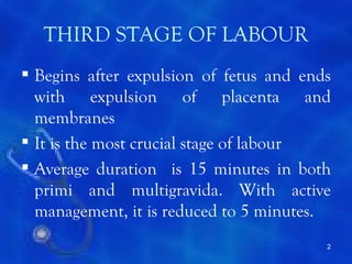
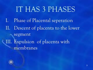
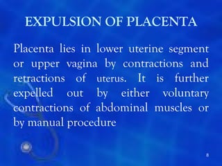
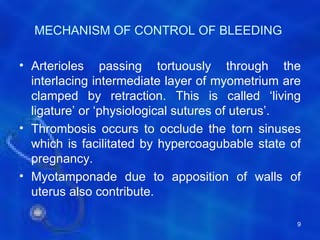
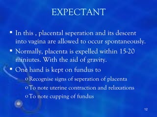
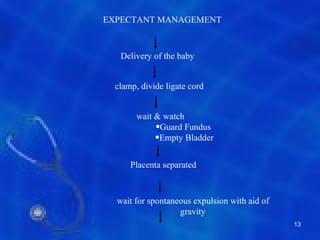
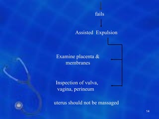
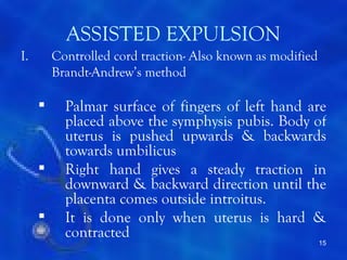
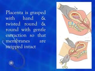
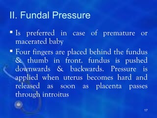
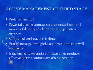
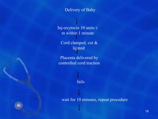
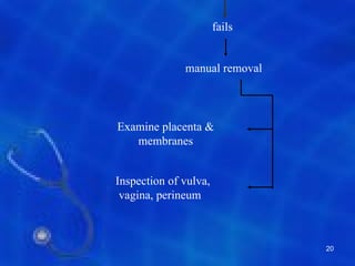
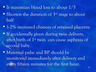
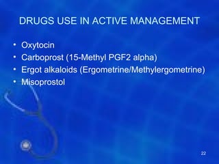
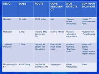
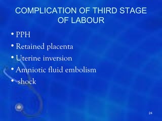
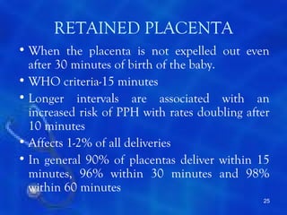
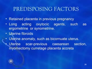
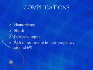
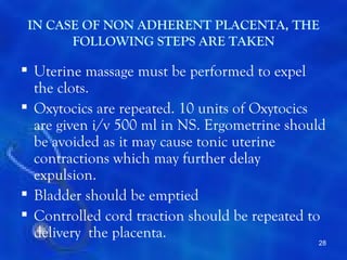
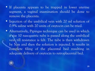
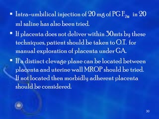
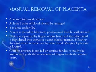
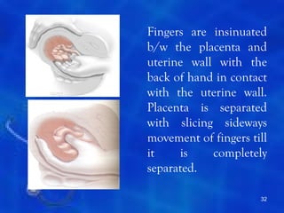
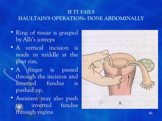
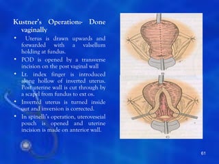
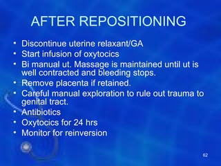
This document provides information on the management of the third stage of labour and complications. It discusses the three phases of the third stage, signs of placental separation, and mechanisms of controlling bleeding. It describes expectant and active management approaches. For retained placenta, steps include uterine massage, oxytocics, and controlled cord traction. Manual removal under anesthesia may be needed. Complications include postpartum hemorrhage, retained placenta, uterine inversion, and shock. Risk factors, diagnosis, and conservative management are outlined for morbidly adherent placenta such as placenta accreta.
The placenta is said to be retained when it is not expelled from the uterus even 30 minutes after the delivery of the baby
Manual placenta removal is a procedure to remove a retained placenta from the uterus after childbirth.
Mannual removal of placenta is done under GA.
Patient placed in lithotomy position
Bladder is catheterized
The second stage of labor begins when the cervix is completely dilated (open), and ends with the birth of your baby. Contractions push the baby down the birth canal, and you may feel intense pressure, similar to an urge to have a bowel movement. Your health care provider may ask you to push with each contraction.
INTRODUCTION
DEFINITION
TYPES
CAUSES
MANAGEMENT-Management of 3rd stage bleeding
Actual management
MANAGEMENT OF 3RD STAGE BLEEDING
Steps of management
1. Placental site bleeding-
To palpate the fundus and massage the uterus to make it hard. The massage is to be done by placing four fingers behind the uterus and thumb in front.
To start crystalloid solution (NS or RL) with oxytocin (1L with 20 units) at 60 drops per minute and to arrange for blood transfusion if necessary.
Oxytocin 10 unit IM or methergine 0.2 mg is given intravenously.
To catheterize the bladder.
To give antibiotics (Ampicillin 2gm and Metronidazole 500mg IV)
2. Management of traumatic bleed
The uterovaginal canal is to be explored under general anesthesia after the placenta is expelled and haemostatic sutures are placed on the offending sites.
STEPS OF MANUAL REMOVAL OF PLACENTA
The patient is placed in lithotomy position. With all aseptic measures, the bladder is catheterized.
One hand is introduced into the uterus in cone shaped manner following the cord. While introducing the hand, the labia are separated by the fingers at the other hand.
Counter pressure on the uterine fundus is applied by the hand placed over the abdomens. The abdominal hand should steady the fundus and guide the movement of the fingers inside the uterine cavity till the placenta is completely separated.
Labour induction
Induction of labour
Guidelines on induction of labour
Guidelines on labour induction
induction of labour is not risk free
prostaglandins for induction of labour
Bishop score
Cervical ripening techniques
mechanical and pharmacological induction of labour
Post dates induction
options for cervical ripening
oral vs. vaginal misoprostol
advantages diadvantages and techniques for induction of labour
gynecology & obstetrics
Labour induction methods
review of guidelines for labour induction
When fetal head is delivered, but shoulders are stuck and cannot be delivered it is known as shoulder dystocia.
The anterior shoulder becomes trapped behind on the symphysis pubis, whilst the posterior shoulder may be in the hollow of the sacrum or high above the sacral promontory.
Retained placenta can be defined as lack of placental expulsion within 30 minutes of delivery of an infant. it is more common in preterm. Retained Placenta can lead to massive PPH and increase maternal morbidity and mortality.
complcations of third stage of labour, includes PPH, Inversion of uterus, retained placenta, placenta accreta, increta, percreta, amniotic fluid embolism
Series of events that takes place in the genital organ in an effort to expel the viable products of conception out of the womb through the vagina into the outer world is called labour.
there are four stages of labour.
about the process of third stage of labor and management of post Partum Hemorrhage ,which is one of the major causes of blood loss in a pregnant women that needs active management.
THIRD STAGE OF LABOUR AND ITS MANAGEMENT.pdfDolisha Warbi
definition, duration, events, (placenta separation, descend of placenta, expulsion of placenta , the Schultz mechanisms, Mathew Duncan mechanisms, signs of separation, expectant management, active management, complexion , examination of placenta and its membrane, complication.
Pathophysiology of Normal Labor:
A series of events that take place in female genital organs to expel the product of conception that are fetus, placenta, membranes) out of womb through the vagina into the outer world. We further describe pathogenesis and features of different stages of labor
Ozempic: Preoperative Management of Patients on GLP-1 Receptor Agonists Saeid Safari
Preoperative Management of Patients on GLP-1 Receptor Agonists like Ozempic and Semiglutide
ASA GUIDELINE
NYSORA Guideline
2 Case Reports of Gastric Ultrasound
TEST BANK for Operations Management, 14th Edition by William J. Stevenson, Ve...kevinkariuki227
TEST BANK for Operations Management, 14th Edition by William J. Stevenson, Verified Chapters 1 - 19, Complete Newest Version.pdf
Anti ulcer drugs and their Advance pharmacology ||
Anti-ulcer drugs are medications used to prevent and treat ulcers in the stomach and upper part of the small intestine (duodenal ulcers). These ulcers are often caused by an imbalance between stomach acid and the mucosal lining, which protects the stomach lining.
||Scope: Overview of various classes of anti-ulcer drugs, their mechanisms of action, indications, side effects, and clinical considerations.
Lung Cancer: Artificial Intelligence, Synergetics, Complex System Analysis, S...Oleg Kshivets
RESULTS: Overall life span (LS) was 2252.1±1742.5 days and cumulative 5-year survival (5YS) reached 73.2%, 10 years – 64.8%, 20 years – 42.5%. 513 LCP lived more than 5 years (LS=3124.6±1525.6 days), 148 LCP – more than 10 years (LS=5054.4±1504.1 days).199 LCP died because of LC (LS=562.7±374.5 days). 5YS of LCP after bi/lobectomies was significantly superior in comparison with LCP after pneumonectomies (78.1% vs.63.7%, P=0.00001 by log-rank test). AT significantly improved 5YS (66.3% vs. 34.8%) (P=0.00000 by log-rank test) only for LCP with N1-2. Cox modeling displayed that 5YS of LCP significantly depended on: phase transition (PT) early-invasive LC in terms of synergetics, PT N0—N12, cell ratio factors (ratio between cancer cells- CC and blood cells subpopulations), G1-3, histology, glucose, AT, blood cell circuit, prothrombin index, heparin tolerance, recalcification time (P=0.000-0.038). Neural networks, genetic algorithm selection and bootstrap simulation revealed relationships between 5YS and PT early-invasive LC (rank=1), PT N0—N12 (rank=2), thrombocytes/CC (3), erythrocytes/CC (4), eosinophils/CC (5), healthy cells/CC (6), lymphocytes/CC (7), segmented neutrophils/CC (8), stick neutrophils/CC (9), monocytes/CC (10); leucocytes/CC (11). Correct prediction of 5YS was 100% by neural networks computing (area under ROC curve=1.0; error=0.0).
CONCLUSIONS: 5YS of LCP after radical procedures significantly depended on: 1) PT early-invasive cancer; 2) PT N0--N12; 3) cell ratio factors; 4) blood cell circuit; 5) biochemical factors; 6) hemostasis system; 7) AT; 8) LC characteristics; 9) LC cell dynamics; 10) surgery type: lobectomy/pneumonectomy; 11) anthropometric data. Optimal diagnosis and treatment strategies for LC are: 1) screening and early detection of LC; 2) availability of experienced thoracic surgeons because of complexity of radical procedures; 3) aggressive en block surgery and adequate lymph node dissection for completeness; 4) precise prediction; 5) adjuvant chemoimmunoradiotherapy for LCP with unfavorable prognosis.
Ethanol (CH3CH2OH), or beverage alcohol, is a two-carbon alcohol
that is rapidly distributed in the body and brain. Ethanol alters many
neurochemical systems and has rewarding and addictive properties. It
is the oldest recreational drug and likely contributes to more morbidity,
mortality, and public health costs than all illicit drugs combined. The
5th edition of the Diagnostic and Statistical Manual of Mental Disorders
(DSM-5) integrates alcohol abuse and alcohol dependence into a single
disorder called alcohol use disorder (AUD), with mild, moderate,
and severe subclassifications (American Psychiatric Association, 2013).
In the DSM-5, all types of substance abuse and dependence have been
combined into a single substance use disorder (SUD) on a continuum
from mild to severe. A diagnosis of AUD requires that at least two of
the 11 DSM-5 behaviors be present within a 12-month period (mild
AUD: 2–3 criteria; moderate AUD: 4–5 criteria; severe AUD: 6–11 criteria).
The four main behavioral effects of AUD are impaired control over
drinking, negative social consequences, risky use, and altered physiological
effects (tolerance, withdrawal). This chapter presents an overview
of the prevalence and harmful consequences of AUD in the U.S.,
the systemic nature of the disease, neurocircuitry and stages of AUD,
comorbidities, fetal alcohol spectrum disorders, genetic risk factors, and
pharmacotherapies for AUD.
- Video recording of this lecture in English language: https://youtu.be/lK81BzxMqdo
- Video recording of this lecture in Arabic language: https://youtu.be/Ve4P0COk9OI
- Link to download the book free: https://nephrotube.blogspot.com/p/nephrotube-nephrology-books.html
- Link to NephroTube website: www.NephroTube.com
- Link to NephroTube social media accounts: https://nephrotube.blogspot.com/p/join-nephrotube-on-social-media.html
Title: Sense of Smell
Presenter: Dr. Faiza, Assistant Professor of Physiology
Qualifications:
MBBS (Best Graduate, AIMC Lahore)
FCPS Physiology
ICMT, CHPE, DHPE (STMU)
MPH (GC University, Faisalabad)
MBA (Virtual University of Pakistan)
Learning Objectives:
Describe the primary categories of smells and the concept of odor blindness.
Explain the structure and location of the olfactory membrane and mucosa, including the types and roles of cells involved in olfaction.
Describe the pathway and mechanisms of olfactory signal transmission from the olfactory receptors to the brain.
Illustrate the biochemical cascade triggered by odorant binding to olfactory receptors, including the role of G-proteins and second messengers in generating an action potential.
Identify different types of olfactory disorders such as anosmia, hyposmia, hyperosmia, and dysosmia, including their potential causes.
Key Topics:
Olfactory Genes:
3% of the human genome accounts for olfactory genes.
400 genes for odorant receptors.
Olfactory Membrane:
Located in the superior part of the nasal cavity.
Medially: Folds downward along the superior septum.
Laterally: Folds over the superior turbinate and upper surface of the middle turbinate.
Total surface area: 5-10 square centimeters.
Olfactory Mucosa:
Olfactory Cells: Bipolar nerve cells derived from the CNS (100 million), with 4-25 olfactory cilia per cell.
Sustentacular Cells: Produce mucus and maintain ionic and molecular environment.
Basal Cells: Replace worn-out olfactory cells with an average lifespan of 1-2 months.
Bowman’s Gland: Secretes mucus.
Stimulation of Olfactory Cells:
Odorant dissolves in mucus and attaches to receptors on olfactory cilia.
Involves a cascade effect through G-proteins and second messengers, leading to depolarization and action potential generation in the olfactory nerve.
Quality of a Good Odorant:
Small (3-20 Carbon atoms), volatile, water-soluble, and lipid-soluble.
Facilitated by odorant-binding proteins in mucus.
Membrane Potential and Action Potential:
Resting membrane potential: -55mV.
Action potential frequency in the olfactory nerve increases with odorant strength.
Adaptation Towards the Sense of Smell:
Rapid adaptation within the first second, with further slow adaptation.
Psychological adaptation greater than receptor adaptation, involving feedback inhibition from the central nervous system.
Primary Sensations of Smell:
Camphoraceous, Musky, Floral, Pepperminty, Ethereal, Pungent, Putrid.
Odor Detection Threshold:
Examples: Hydrogen sulfide (0.0005 ppm), Methyl-mercaptan (0.002 ppm).
Some toxic substances are odorless at lethal concentrations.
Characteristics of Smell:
Odor blindness for single substances due to lack of appropriate receptor protein.
Behavioral and emotional influences of smell.
Transmission of Olfactory Signals:
From olfactory cells to glomeruli in the olfactory bulb, involving lateral inhibition.
Primitive, less old, and new olfactory systems with different path
For Better Surat #ℂall #Girl Service ❤85270-49040❤ Surat #ℂall #Girls
Third stage labour complication
1. MANAGEMENT OF THIRD STAGE OF
LABOUR & COMPLICATIONS
DR RAJEEV SOOD
ASTT. PROF OBG
IGMC SHIMLA
1
2. THIRD STAGE OF LABOUR
Begins after expulsion of fetus and ends
with expulsion of placenta and
membranes
It is the most crucial stage of labour
Average duration is 15 minutes in both
primi and multigravida. With active
management, it is reduced to 5 minutes.
2
3. IT HAS 3 PHASES
I. Phase of Placental seperation
II. Descent of placenta to the lower
segment
III. Expulsion of placenta with
membranes
3
4. PHASE OF PLACENTAL SEPERATION :-
For some time after delivery of the foetus, patient
experiences no pain. Intermittent discomfort coinciding
with uterine contractions occurs. After the birth of baby
uterus measures 20 cm vertically and 10cm antero
posteriorialy, discoid in shape .
Surface area of placental site is reduced due to retraction.
Placenta is inelastic cannot contract simultaneously,
hence buckling occurs.
Plane of seperation is through the deep spongy layer of
decudia basalis .
4
5. THERE ARE TWO WAYS OF seperation
1. Central (SCHULTZE)
:-detachment starts from
centre, uterine sinuses are
opened, retro placental
collection of blood occurs
resulting in further
seperation.
3. Marginal (Mathew duncan):-
Here seperation starts at
margin, more area get
separated with progressive
uterine contractions. This
occurs more frequently
5
6. SIGNS
Before seperation-
uterus is discoid, firm, non- ballotable.
height of uterus is a litle below umblicus.
Length of cord remains static.
6
7. After seperation –
•uterus becomes globular, firm, ballotable.
• Fundal height is raised
•Sudden gush of blood
•Permanent lengthening of cord occurs.
7
8. EXPULSION OF PLACENTA
Placenta lies in lower uterine segment
or upper vagina by contractions and
retractions of uterus. It is further
expelled out by either voluntary
contractions of abdominal muscles or
by manual procedure
8
9. MECHANISM OF CONTROL OF BLEEDING
• Arterioles passing tortuously through the
interlacing intermediate layer of myometrium are
clamped by retraction. This is called ‘living
ligature’ or ‘physiological sutures of uterus’.
• Thrombosis occurs to occlude the torn sinuses
which is facilitated by hypercoagubable state of
pregnancy.
• Myotamponade due to apposition of walls of
uterus also contribute.
9
10. EXAMINATION OF PLACENTA
Placenta is placed on the pronated hands and examined:-
Maternal surface is first examined for any missing
cotyledons.
Completeness of membranes should be assessed.
Placental foetal surface should be inspected for any
blood vessels that radiate beyond placental edge into
membranes with no corresponding placental tissue.
Position of insertion of cord is noted.
Cut end of cord is examined for number of vessels.
Cord length is seen.
Placental weight is recorded.
Any calcification, clots.
In twins, chorionicity can be determined.
10
11. MANAGEMENT OF THIRD STAGE
OF LABOUR
Two methods of management
Expectant or traditional
Active
11
12. EXPECTANT
In this , placental seperation and its descent
into vagina are allowed to occur spontaneously.
Normally, placenta is expelled within 15-20
miniutes. With the aid of gravity.
One hand is kept on fundus to
o Recognise signs of seperation of placenta
o To note uterine contraction and relaxations
o To note cupping of fundus
12
13. EXPECTANT MANAGEMENT
Delivery of the baby
clamp, divide ligate cord
wait & watch
•Guard Fundus
•Empty Bladder
Placenta separated
wait for spontaneous expulsion with aid of
gravity
13
14. fails
Assisted Expulsion
Examine placenta &
membranes
Inspection of vulva,
vagina, perineum
uterus should not be massaged
14
15. ASSISTED EXPULSION
I. Controlled cord traction- Also known as modified
Brandt-Andrew’s method
Palmar surface of fingers of left hand are
placed above the symphysis pubis. Body of
uterus is pushed upwards & backwards
towards umbilicus
Right hand gives a steady traction in
downward & backward direction until the
placenta comes outside introitus.
It is done only when uterus is hard &
contracted
15
16. Placenta is grasped
with hand &
twisted round &
round with gentle
extraction so that
membranes are
stripped intact
16
17. II. Fundal Pressure
Is preferred in case of premature or
macerated baby
Four fingers are placed behind the fundus
& thumb in front. fundus is pushed
downwards & backwards. Pressure is
applied when uterus becomes hard and
released as soon as placenta passes
through introitus
17
18. ACTIVE MANAGEMENT OF THIRD STAGE
Preferred method
Powerful uterine contraction are initiated within 1
minute of delivery of a baby by giving parenteral
oxytocin
Controlled cord traction is done
Fundal massage throughthe abdomen until ut is well
contracted
It favours early seperation of placenta & produces
effective uterine contractions after seperation
18
19. Delivery of Baby
Inj-oxytocin 10 units i/
m within 1 minute
Cord clamped, cut &
ligated
Placenta delivered by
controlled cord traction
fails
wait for 10 minutes, repeat procedure
19
21. It minimizes blood loss to about 1/5
Shorten the duration of 3rd stage to about
half
1-2% increased chances of retained placenta
If accidentally given during twin delivery,
after birth of 1st twin can cause asphyxia of
second baby
Maternal pulse and BP should be
monitored immediately after delivery and
every fifteen minutes for the first hour.
21
22. DRUGS USE IN ACTIVE MANAGEMENT
• Oxytocin
• Carboprost (15-Methyl PGF2 alpha)
• Ergot alkaloids (Ergometrine/Methylergometrine)
• Misoprostol
22
23. DRUG DOSE ROUTE DOSE SIDE CONTRAIN
FREQUEN EFFECTS DICATIONS
CY
Oxytocin 10 units IM (10 units) stat •Nausea •Not as IV
•Water bolus,otherwise
intoxication none.
Methergin 0.2mg First line IM/IV Every 2-4 hours •Nausea •Hypertension.
Second line •Vommiting •Pre eclampsia
PO. •hypertinsion
15-Methly 0.25mg First line IM Every 15-90 •Nausea •Bronchial
PGf-2alfa Second line min(8 doses •Vomiting asthma
intra uterine max) •Diarrhoea •Active
•chills cardiac,renal or
hepatic disease
Misoprostol(PG 400-600mcg First line PR Single dose •Fever None
E-1 second line PO •Tachycardia
23
24. COMPLICATION OF THIRD STAGE
OF LABOUR
• PPH
• Retained placenta
• Uterine inversion
• Amniotic fluid embolism
• shock
24
25. RETAINED PLACENTA
• When the placenta is not expelled out even
after 30 minutes of birth of the baby.
• WHO criteria-15 minutes
• Longer intervals are associated with an
increased risk of PPH with rates doubling after
10 minutes
• Affects 1-2% of all deliveries
• In general 90% of placentas deliver within 15
minutes, 96% within 30 minutes and 98%
within 60 minutes
25
26. PREDISPOSING FACTORS
• Retained placenta in previous pregnancy
• Long acting oxytocic agents, such as
ergometrine or synometrine.
• Uterine fibroids
• Uterine anomaly, such as bicornuate uterus.
• Uterine scar-previous caesarean section,
myomectomy curretage placenta accreta
26
27. COMPLICATIONS
o Hemorrhage
o Shock
o Puerperal sepsis
o Risk of recurrence in next pregnancy
around 6%
27
28. IN CASE OF NON ADHERENT PLACENTA, THE
FOLLOWING STEPS ARE TAKEN
Uterine massage must be performed to expel
the clots.
Oxytocics are repeated. 10 units of Oxytocics
are given i/v 500 ml in NS. Ergometrine should
be avoided as it may cause tonic uterine
contractions which may further delay
expulsion.
Bladder should be emptied
Controlled cord traction should be repeated to
delivery the placenta.
28
29. If placenta appears to be trapped in lower uterine
segment, a vaginal examination should be done to
remove the placenta.
Injection of the umbilical vein with 20 ml solution of
0.9% saline with 20 units of oxytocin can be tried.
Alternatively, Pipingas technique can be used in which
a size 10 nasogastric tube is passed along the umbilical
vein till resistance is felt. The tube is then withdrawn
by 5cm and then the solution is injected. It results in
complete filing of the placental bed resulting in
adequate delivery of oxytocin to retroplacental bed.
29
30. Intra-umbilical injection of 20 mg of PG F2α in 20
ml saline has also been tried.
If placenta does not deliver within 30mts by these
techniques, patient should be taken to O.T. for
manual exploration of placenta under GA.
If a distinct clevage plane can be located between
placenta and uterine wall MROP should be tried.
If not located then morbidly adherent placenta
should be considered.
30
31. MANUAL REMOVAL OF PLACENTA
A written informed consent
At least 2 units of blood should be arranged
It is done under GA
Patient is placed in lithotomy position and bladder catheterized
Labia are separated by fingers of one hand and the other hand
is introduced into uterus in a cone shaped manner, following
the cord which is made taut by other hand. Margin of placenta
is located.
Counter pressure is applied on uterine fundus to steady the
fundus and guide the movements of fingers inside the uterine
cavity.
31
32. Fingers are insinuated
b/w the placenta and
uterine wall with the
back of hand in contact
with the uterine wall.
Placenta is separated
with slicing sideways
movement of fingers till
it is completely
separated.
32
33. It is extracted by traction of cord by other hand.
If removal is difficult : ‘piecemeal removal’ of
placenta should be done.
i/v Methergin 0.2 mg is given
Inspection of cervico-vaginal canal should be
done. Placenta should be examined
10 units oxytonic in 500 ml NS is started to
initiate & maintain contraction.
A broad spectrum antibiotic is given for 12-24
hrs to prevent infection.
33
34. COMPLICATIONS
o Hemorrhage :- due to incomplete removal
o Shock
o Injury to uterus
o Infection
o Inversion
o Sub- involution
o Thrombophlebitis
o Embolism
34
35. DIFFICULTIES ENCOUNTERED
Hour glass contraction- there is a localized
contraction of circular muscles of uterus either
at the junction of lower and upper segment or
may be placed in 1 cornu. It occurs due to
premature attempts in removing of placenta or
due to administration of methergin. It is
managed by deepning the plane of anesthesia.
35
36. •Morbid Adherent Placenta- Also K/A Placenta
Accreta
•Placenta is directly anchored to myometrium
without any intervening decidua.
•due to absence of decidua basalis or imperfect
development of fibrinoid or nitabuch’s layer.
•It is an area of fibrinoid degeneration where
trophoblasts cells meet the decidua. The layer
inhibits further invasion of decidua by
trophoblast .
36
37. TYPES
Placenta accreta:- Placenta
adheres to myometruim (Fig.
A)
Placenta increta:- Placenta
invades myometruim (Fig. B)
Placenta percreta:- placenta
penetrates myomentruim to
or beyond serosa (Fig. C)
Incidence is 1 in 2500 deliveries 37
38. RISK FACTORS
Placenta previa and prior caesarean delivery
o Risk of placenta accreta with placenta previa in
an unscarred uterus is 3%
o Women with placenta previa with previous 1
caesarean section has 14% risk of accreta.
o Women with 3 caesarean have 44% risk
Prior myomectomy
Manual removal of placenta
D&C
Increasing maternal age and parity . 38
39. DIAGNOSIS
During pregnancy
USG is only 33% sensitive. The findings suggestive are
Loss of normal hypoechoic retroplacental myometrial zone.
Thinning and abruption of uterine serosa:- Bladder interface
and focal exophytic masses within the placenta.
Colour Doppler has a sensitivity of 100%
A distance less than 1 mm between the uterine serosa-
bladder interface and retro placental vessels
Identification of large intraplacental lakes 39
40. MRI findings suggestive of accreta are:-
Uterine bulging
Heterogeneous signal intensity within the placenta
Presence of dark intraplacental bands on T2 weighted
imaging.
There is an unexplained rise of MSAFP and B-HCG
greater than 2.5 MOM.
40
41. HISTO PATHOLOGICAL EXAMINATION
Absence of decidua basalis
Absence of nitabuch’s fibrinoid layer
Varying degree of peneteration of the villi
into the muscle bundles or upto serosa.
41
42. MANAGEMENT
1. CONSERVATIVE
IN PARTIAL PLACENTA ACCRETA :-
As much as possible of placental tissue is removed manually.
Oxytocics are given for effective uterine contraction and
haemostasis, or by intrauterine plugging.
Remaining trophoblast is usually reabsorbed spontaneously.
Levels of B-HCG should be monitored.
During caesarean bleeding areas can be undersewed.
42
43. IN TOTAL PLACENTA ACCRETA : -
After explaining the risks of hemorrhage and
failure
o Cord is cut as near to placenta which is left as such
o Patients vitals and bleeding is monitored
o Antibiotics are given
o B-HCG values are monitored
o Methotrexate 50 mg i/v on alternate days can be given
43
44. SURGICAL MANAGEMENT
If bleeding remains uncontrollable
then:-
Uterine art embolisation
Low and high b/l uterine vesseles ligation
Ligation of internal iliac arteries
If all these methods fail or patient in shock :-
hysterectomy
.
44
45. INVERSION OF UTERUS
A rare complication of third stage with incidence
being .05% of deliveries
Uterus is turned inside out either completely or
partially
Acute - With in 24 hrs
subacute - 24 hrs - 4wk
Chronic > 4 wk
Incidence - 1 in 2000 to 1,20,000
Maternal survival rate is 85%
45
46. DEGREE OF INVERSION
I. Dimpling of fundus which still remains
above the level of internal os.
II. Fundus passes through cervix but is
inside vagina
46
47. • Also called
complete:-
Endometrium
with or without
the attached
placenta is visible
outside the vulva.
The cervix and
part of vagina may
also be involved.
47
48. ETIOLOGY
I. SPONTANEOUS – Occurs is about 40%
caused by local atony on placental site over the
fundus associated with increase in intra abdominal
pressure as in coughing, sneezing or bearing down
effort.
Fundal attachment of placenta (75%), short cord,
placenta accreta may be associated.
48
49. IATROGENIC
Fundal pressure on a relaxed uterus
Strong traction on cord
Faulty techniques in manual removal of placenta
ASSOCIATED RISK FACTORS ARE
Uterine over distention
prolonged labour > 24 hrs
Uterine malformations
Short cord
Collagen diseases
Use of magnesium sulphate during labour
49
50. DIAGNOSIS
Symptoms :- Acute lower abdominal pain with
bearing down sensation
Signs:-
1. Varying degree of shock
2. On P/A –cupping or dimpling of fundal surface.
On bimanual examination :- Crater like depression on abdomen
along with vaginal palpation of fundal wall in lower segment of
cervix
Sound Test – Confirmatory absent uterine cavity
50
51. In complete
variety, a pear
shaped bluish grey
mass protudes
outside vulva with
the broad end
pointing
downwards
51
52. COMPLICATIONS
• Shock:- is mainly neurogenic
Tension on nerves due to
stretching of infundibulopelvic
ligament.
Ovaries are dragged along causing
pressure on then.
Peritoneal irritation.
52
53. • Hemorrhage –more if placenta is
separated
• Pulmonary embolism
• If not treated - infection, uterine
sloughing can occur. It becomes
chronic
53
54. MANAGEMENT
• Immediate assistance is summoned
• Two large bore intravenous infusion systems are
started, crystalloids, blood should be arranged bladder
is cathertized.
• Urgent manual replacement is the mainstay of
treatment, preferably under GA. Uterine relaxant
anaesthetics such as halothane is preferred. Injection
pethidine/ diazepamis given
• If the placenta is still attached, it should not be
removed
54
55. TWO METHODS OF MANAGING ACUTE
INVERSION
I. MANUAL – called
JOHNSON’S
METHOD.
The part of
the uterus
which is
inverted last is
to be replaced
first
55
56. The protruding mass is thoroughly cleaned with
antiseptic solution.
Protruding fundus is grasped with the palms of
hands with the finger directed towards post fornix.
Uterus is lifted through pelvis into the abdomen
while applying countersupport over the abdomen.
Too much pressure should not be given so as to
cause perforation of uterus.
Once the uterus is reverted an oxytocin drip is
started to increase uterine tone and prevent
recurrence. Hand should remain inside uterus till
it is well contracted.
Placenta should then be removed manually
56
57. II. HYDROSTATICS OR O’ SULLIVAN’S METHOD
Place the patient in lithotomy position
57
58. Head end is lowered 0.5 mt below the level of perineum
Prepare a disinfected douche system with large nozzle with a
long tube (2 meters) and 3 - 5 ltr warm NS
Identity post Fx – easily done in partial inversion & in
others identify the point where rugosed vagina becomes
smooth vagina.
Place nozzle in post Fx. At the same time with other hand
hold labia sealed.
Ask assistant to start the douche with full pressure
Raise reservoir to 2 meters.
NS distends post Fx gradually so that it is stretched-
circumference of orifice increases- cervical constriction
relived - uterus is repositioned
Ogueh and Ayida technique:- In this similar procedure is
done by using silicon cup in vagina attached with iv tubing
58
59. SURGICAL INTERVENTIONS
May be required in presence
of a dense constriction ring.
Laprotomy is required.
Initially Huntington's
procedure is done in which
alli’s forceps are used to
grasp the myometrium just
inside dimple of fundus
systematically and
sequentially using forceps on
both sides, inverted fundus
is then withdrawn from
crater to fully correct the
inversion
59
60. IF IT FAILS
HAULTAIN'S OPERATION:- DONE ABDOMINALLY
• Ring of tissue is grasped
by Alli’s joreeps
• A vertical incision is
made in middle at the
post rim.
• A finger is passed
through the incision and
inverted fundus is
pushed up.
• Assistant may also push
up inverted fundus
through vagina 60
61. Kustner’s Operation:- Done
vaginally
• Uterus is drawn upwards and
forwarded with a valsellum
holding at fundus.
• POD is opened by a transverse
incision on the post vaginal wall
• Lt. index finger is introduced
along hollow of inverted uterus.
Post uterine wall is cut through by
a scapel from fundus to ext os.
• Inverted uterus is turned inside
out and inversion is corrected.
• In spinelli’s operation, uteroveseial
pouch is opened and uterine
incision is made on anterior wall.
61
62. AFTER REPOSITIONING
• Discontinue uterine relaxant/GA
• Start infusion of oxytocics
• Bi manual ut. Massage is maintained until ut is
well contracted and bleeding stops.
• Remove placenta if retained.
• Careful manual exploration to rule out trauma to
genital tract.
• Antibiotics
• Oxytocics for 24 hrs
• Monitor for reinversion
62
63. AMNIOTIC FLUID EMBOLISM
• Complex disorder characterized by abrupt
oneset of hypotension, hypoxia and
consumptive coagulopathy.
• Risk factor include advanced maternal age,
placenta previa, pre eclampsia, forceps or
caesarean delivery.
• Women in late stages of labour or immediately
post partum begin gasping for air, suffers
seizures or cardiorespiratory arrest occurs
63
64. MECHANISM
Amniotic fluid is forced into circulation either through a
rent in membranes or placenta. Thromboplastin rich
liquor containing the debris blocks pulmonary arteries
and triggers coagulation mechanism leading to DIC.
There is massive fibrin deposition along the entire
pulmonary vasculature leading to cardiopulmonary
arrest.
If patient survives this there can be residual neurological
damage severe bleeding per vaginun or from veno-
puncture sites.
64
65. MANAGEMENT
• There are no data that any type of intervention
improves maternal prognosis with amniotic
fluid embolism.
• Oxygenation, circulatory support blood
transfusion is required.
• Case fatality rate is 22%
65