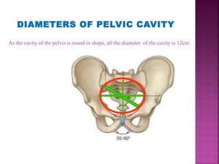
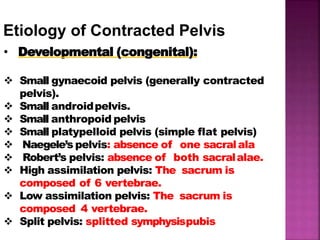
and
CONTRACTED PELVIS
FALSE PELVIS
The portion above the pelvic
brim.
Has no obstetrical significance
relevant to passage of fetus
through the pelvis
The bony canal that the fetus
needs to pass through during the
normal mechanism of childbirth
comprises brim, cavity & outlet
TRUE PELVIS
DIAMETERS OF PELVIC INLET
Antero -posterior diameters
•Anatomical conjugate (true
conjugate)= 12cm
•Obstetric conjugate = 11 cm
•Diagonal conjugate = 12 -13 cm
Oblique diameters
•Right oblique diameter =12 cm
• Left oblique diameter = 12 cm
Transverse diameters = 13cm
DIAMETERS OF PELVIC CAVITY
As the cavity of the pelvis is round in shape, all the diameter of the cavity is 12cm
Antero- posterior
diameters=13 cm
Oblique diameters = 12cm
Transverse diameters =10-
11cm
Oblique diameters = 12cm
DIAMETERS OF PELVIC
OUTLET
•Cephalo pelvic disproportion is the disparity in
relation between the head of baby and the mother’s
pelvis.
• It is a pelvis in which one or more of its diameter is
reduced below the normal by one or more
centimeter
CAUSES:-
1. Large baby due to:
• Hereditary factors
• Diabetes
• Postmaturity (still pregnant after due date
has passed)
• Multiparity (not the first pregnancy)
2. Abnormal fetal positions
3. contracted pelvis
4. Abnormally shaped pelvis
• Anatomical - It is a pelvis in which one or more of
its diameters is reduced below the normal by one or
more centimeters.
• Obstetric - It is a pelvis in which one or more of its
diameters is reduced so that it interferes with the
normal mechanism of labor.
1. Developmental factor: hereditaryor
congenital.
2. Racialfactor.
3. Nutritional factor: malnutrition resultsin
small pelvis.
4. Sexualfactor: asexcessiveandrogenmay
produce android pelvis.
5. Metabolic factor: asricketsandosteomalacia.
6. Trauma,diseasesor tumours of thebony
pelvis, legs orspines.
Factors influencing the size and
shape of the pelvis
Etiology of Contracted Pelvis
• Developmental (congenital):
 Small gynaecoid pelvis (generally contracted
pelvis).
 Small androidpelvis.
 Small anthropoid pelvis
 Small platypelloid pelvis (simple flat pelvis)
 Naegele’s pelvis: absence of one sacralala
 Robert’s pelvis: absence of both sacralalae.
 High assimilation pelvis: The sacrum is
composed of 6 vertebrae.
 Low assimilation pelvis: The sacrum is
composed 4 vertebrae.
 Split pelvis: splitted symphysispubis
• Metabolic:
Rickets.
Osteomalacia (triradiate pelvicbrim)
• Traumatic: as fractures.
• Neoplastic: as osteoma.
• Infection : TB
• Lumbar kyphosis
• Lumbar scoliosis
• Spondylolisthesis:
The 5thlumbar vertebra with the above vertebral column is pushed
forward while the promontory is pushed backwards and the tip of
the sacrum is pushed forwards leading to outlet contraction.
Causes in the lower limbs
• Dislocation of one or both femurs.
• Atrophy of one or both lowerlimbs.
N.B.oblique or asymmetric pelvis: one oblique
diameter is obviously shorter than the other. This can
be foundin:
• Diseases, fracture or tumors affecting one side.
• Otto’s pelvis – develop as result of
inflammatory process in the hip or knee
• Beaked (rostrate) pelvis – under
development of both sacral wings
• Spondylolithetic pelvis – formed due to
partial dislocation of last lumbar vertebra
in front of 1st sacral vertebra
• Osteomalacic pelvis
• Scoliotic pelvis – only the lumber region
cause deformity of the pelvis. The
acetabulum is pushed inwards on the
weight bearing side.
DIAGNOSIS OF CONTRACTED PELVIS
• History
• Rickets: is expected if there is a history
of delayed walking and dentition.
• Traumaor diseases: of the pelvis, spines
or lower limbs.
• Badobstetric history: e.g. prolonged
labour ended by:
 difficult forceps
 caesarean sectionor
 still birth.
•Examination
• Generalexamination:
 Gait:abnormal gait suggesting
abnormalities in the pelvis, spinesor lower
limbs.
 Height: women with lessthan 150cm
height usualy havecontractedpelvis.
 Spinesandlower limbs: mayhaveadisease
or lesion.( kyphosis,…)
Abdomen examination
• Pendulous abdomen in primigravida
• fetal head fails to enter a contracted
pelvis at the end of pregnancy and
floats high above inlet, failed growth
of uterus deviates upward and
anteriorly
• Non engagement in last 3-4 wks in
primigravida
• Acuminate (pointed)abdomen in
primigravida with a resilient abdominal
wall
• Pendulous abdomen in multiparous women
2 shapes of abdomen
DIAGNOSIS OF CPD AT BRIM
ABDOMINAL METHOD FOR CPD
• Patient is placed in dorsal position with
thigh flexes and separated.
• The head is grasped by the left hand.
• 2 fingers (index and middle) of theright hand
are placed above the symphysis pubis to
note the degree of overlapping. If when the
head is pushed downward and backward.
• The head can be pushed down in the pelvis without
overlapping of the parietal bone on the symphysis
pubis:- no disproportion
• Head can be pushed down a little but ther is
slightly overlapping of the parietal bone evidence
by touch on the under surface of finger overlapping
by 0.5cm:- moderate disproportion
• Head can not be pushed down and instead
the partial bone overhangs the symphysis
pubis displacing the finger – sever
disproportion
Some times the degree of disproportion is
difficult to found by this method because
of:-
• Deflexed head
• Thick abdominal wall
• Irritable uterus
• High floating head
ABDOMINAL-VAGINAL METHOD
• It is also called as MULLER – MUNRO KERR
• It is bimanual method.
Results :-
• the head can be pushed down up to the level
of ischial spines and there is no overlapping
of the parietal bone over the symphysis
pubis:- no disproportion
• The head can be pushed down a little but
not up to the level of ischial spine and ther
is slight overlapping of the parietal bone:-
slight or moderate disproportion
• The head can not be pushed down and
instead the parietal bone overhangs the
symphysis pubis displacing the thumb:-
sever disproportion.
• Pelvimetry :
It is assessment of the pelvic diameters andcapacity done at 38-39
weeks.It includes:
1. Clinicalpelvimetry:
 Internal pelvimetryfor:
 inlet
 cavity,and
 outlet.
 External pelvimetryfor:
 inlet and
 outlet.
2. Imagingpelvimetry:
 X-ray
.
 Computed tomography(CT).
 Magnetic resonanceimaging(MRI).
• N.B. CTandMRI are recent and accurate but expensiveandnot
alwaysavailablesotheyare not in commonuse.
CEPHALOMETRY
• Ultrasonography: is the safeaccurate and easy
method and candetect:
 The biparietal diameter(BPD)
 The occipito-frontaldiameter.
 Thecircumference of the head.
• Radiology (X-ray): is difficult to interpret.
DEGREES OF CONTRACTED PELVIS
1.Minor degree:Thetrue conjugateis9-10 cm. It
corresponds to minordisproportion.
2.Moderate degree:Thetrue conjugateis8-9 cm.
It corresponds to moderatedisproportion.
3.Severedegree:Thetrue conjugateis6-8 cm. It
corresponds to markeddisproportion.
4.Extremedegree:Thetrue conjugateislessthan 6 cm.
Vaginal delivery is impossible even after craniotomy asthe
bimastoid diameter (7.5 cm)is not crushed.
Management
Minor Moderate Sever
vaginal
delivery
trial labor, if
failed
caesarean
section.
caesarean
section
depends
mainly on the
degree of
disproportion
• Elective cesarean section at term is indicated in:-
Major degree of contraction
Major disproportion
Absolute contraction
Dead fetus
Patient not fit for trial labor
The operation is done in planned way any time
during last week of pregnancy.
• Emergency:-
when trial labor is failed
•Trial labor:-
• It is the conduction of spontaneous labor in a
moderate
institution
degree of disproportion,
under supervision with
in an
watchful
expectancy hoping for a vaginal delivery
or
Trial of labor is a test of labor allowing the patient
to enter into active labor putting all variable
( power, passage and passenger) into test and
determine whether vaginal delivery is possible or
not.
Careful fetal and maternal monitoring by
electronic fetal monitoring and non stress test
Oral feeding remain suspended and hydration is
maintained by intravenous drip
Adequate analgesic is administered
Augmentation of labor by pitocin
The progress of labor is mapped with
partograph:-
i) progressive descent of the head
ii) progressive dilatation of the cervix
After the membrane rupture, pelvic examination
is to be done:-
i) to exclude cord prolapse
ii) to note the color of liquor
iii) to assess the pelvis once or more
iv) to note the condition of the cervix
including pressure of the presenting part of the
cervix
in favorable cases, end spontaneously, low forcep and low
ventose.
In unfavorable cases, do caesarean section.
Successful trial:-
A trial is called successful, if a healthy baby is born
vaginally, spontaneous or by forcep or ventose with the
mother in good condition
Failure of trial labor:-
Delivery is by cesarean section or delivery of a dead
baby spontaneously or by craniotomy is called failure of trial
labor
• Lower incidence of cesarean section.
• A successful trial ensures the women a good
future obstetrics.
• May end before full cervix dilatation
• Increased fetal mortality and morbidity
• In failed trial operative risk increases.
• Check vitals every 4 hourly
• Monitor both contraction and fetus continuously
• Report immediately the sign of fetal distress
• Position the mother in ways to increase the pelvic
diameter such as sitting or squatting which increase
the outlet diameter and also aid in fetal descent
• Assess the fetus for hypoxia
• Provide support to the client and the family
members in coping with stress of a complicated
labor
COMPLICATION
•Maternal:
During pregnancy:
1. Incarcerated retroverted graviduterus.
2. Mal presentations.
3. Pendulous abdomen.
4. Non engagement.
5. Pyelonephritis especially in high assimilation
pelvis due to more compression of the ureters.
During labour:
1. Inertia, slow cervical dilatation and
prolonged labor.
2. Premature rupture of membranesand
cord prolapsed.
3. Obstructed labor and rupture uterus.
4. Necrotic genito-urinary fistula.
5. Injury topelvic joints or nerves from
difficult forceps delivery.
6. Postpartum hemorrhage.
• Fetal:
1. Intracranial
hemorrhage.
2. Asphyxia.
3. Fracture skull.
4. Nerve injuries.
5. Intra-amniotic infection.
BOOKS:-
•Beischer, N.A., Mackay, E.V. & Colditz, P.B. (1997). Obstetrics
and the New-born. (3rd ed.). London: W.B. Saunders.
•Dutta, D.C. (2004). Text book of obstetrics. (6th ed.).
Kolkata: New central book agency (P) ltd.
•Fraser, D.M. & Cooper,M.A. (2005). Myles Text book for
Midwives. (14th ed.). Edinburg: Churchill Livingstone.
•Littleton, L.V. & Engebeton, J.C. (2007). Maternity Nursing
care. (1st ed.). Australia: Thomson.
BIBLIOGRAPHY
THANK
YOU

CPD presentation

  • 1.
  • 3.
    FALSE PELVIS The portionabove the pelvic brim. Has no obstetrical significance relevant to passage of fetus through the pelvis The bony canal that the fetus needs to pass through during the normal mechanism of childbirth comprises brim, cavity & outlet TRUE PELVIS
  • 4.
    DIAMETERS OF PELVICINLET Antero -posterior diameters •Anatomical conjugate (true conjugate)= 12cm •Obstetric conjugate = 11 cm •Diagonal conjugate = 12 -13 cm Oblique diameters •Right oblique diameter =12 cm • Left oblique diameter = 12 cm Transverse diameters = 13cm
  • 5.
    DIAMETERS OF PELVICCAVITY As the cavity of the pelvis is round in shape, all the diameter of the cavity is 12cm
  • 6.
    Antero- posterior diameters=13 cm Obliquediameters = 12cm Transverse diameters =10- 11cm Oblique diameters = 12cm DIAMETERS OF PELVIC OUTLET
  • 11.
    •Cephalo pelvic disproportionis the disparity in relation between the head of baby and the mother’s pelvis. • It is a pelvis in which one or more of its diameter is reduced below the normal by one or more centimeter
  • 12.
    CAUSES:- 1. Large babydue to: • Hereditary factors • Diabetes • Postmaturity (still pregnant after due date has passed) • Multiparity (not the first pregnancy) 2. Abnormal fetal positions 3. contracted pelvis 4. Abnormally shaped pelvis
  • 14.
    • Anatomical -It is a pelvis in which one or more of its diameters is reduced below the normal by one or more centimeters. • Obstetric - It is a pelvis in which one or more of its diameters is reduced so that it interferes with the normal mechanism of labor.
  • 15.
    1. Developmental factor:hereditaryor congenital. 2. Racialfactor. 3. Nutritional factor: malnutrition resultsin small pelvis. 4. Sexualfactor: asexcessiveandrogenmay produce android pelvis. 5. Metabolic factor: asricketsandosteomalacia. 6. Trauma,diseasesor tumours of thebony pelvis, legs orspines. Factors influencing the size and shape of the pelvis
  • 16.
    Etiology of ContractedPelvis • Developmental (congenital):  Small gynaecoid pelvis (generally contracted pelvis).  Small androidpelvis.  Small anthropoid pelvis  Small platypelloid pelvis (simple flat pelvis)  Naegele’s pelvis: absence of one sacralala  Robert’s pelvis: absence of both sacralalae.  High assimilation pelvis: The sacrum is composed of 6 vertebrae.  Low assimilation pelvis: The sacrum is composed 4 vertebrae.  Split pelvis: splitted symphysispubis
  • 18.
    • Metabolic: Rickets. Osteomalacia (triradiatepelvicbrim) • Traumatic: as fractures. • Neoplastic: as osteoma. • Infection : TB • Lumbar kyphosis • Lumbar scoliosis • Spondylolisthesis: The 5thlumbar vertebra with the above vertebral column is pushed forward while the promontory is pushed backwards and the tip of the sacrum is pushed forwards leading to outlet contraction.
  • 19.
    Causes in thelower limbs • Dislocation of one or both femurs. • Atrophy of one or both lowerlimbs. N.B.oblique or asymmetric pelvis: one oblique diameter is obviously shorter than the other. This can be foundin: • Diseases, fracture or tumors affecting one side.
  • 20.
    • Otto’s pelvis– develop as result of inflammatory process in the hip or knee • Beaked (rostrate) pelvis – under development of both sacral wings • Spondylolithetic pelvis – formed due to partial dislocation of last lumbar vertebra in front of 1st sacral vertebra • Osteomalacic pelvis • Scoliotic pelvis – only the lumber region cause deformity of the pelvis. The acetabulum is pushed inwards on the weight bearing side.
  • 21.
    DIAGNOSIS OF CONTRACTEDPELVIS • History • Rickets: is expected if there is a history of delayed walking and dentition. • Traumaor diseases: of the pelvis, spines or lower limbs. • Badobstetric history: e.g. prolonged labour ended by:  difficult forceps  caesarean sectionor  still birth.
  • 22.
    •Examination • Generalexamination:  Gait:abnormalgait suggesting abnormalities in the pelvis, spinesor lower limbs.  Height: women with lessthan 150cm height usualy havecontractedpelvis.  Spinesandlower limbs: mayhaveadisease or lesion.( kyphosis,…)
  • 23.
    Abdomen examination • Pendulousabdomen in primigravida • fetal head fails to enter a contracted pelvis at the end of pregnancy and floats high above inlet, failed growth of uterus deviates upward and anteriorly • Non engagement in last 3-4 wks in primigravida
  • 24.
    • Acuminate (pointed)abdomenin primigravida with a resilient abdominal wall • Pendulous abdomen in multiparous women 2 shapes of abdomen
  • 25.
    DIAGNOSIS OF CPDAT BRIM ABDOMINAL METHOD FOR CPD • Patient is placed in dorsal position with thigh flexes and separated. • The head is grasped by the left hand. • 2 fingers (index and middle) of theright hand are placed above the symphysis pubis to note the degree of overlapping. If when the head is pushed downward and backward.
  • 26.
    • The headcan be pushed down in the pelvis without overlapping of the parietal bone on the symphysis pubis:- no disproportion • Head can be pushed down a little but ther is slightly overlapping of the parietal bone evidence by touch on the under surface of finger overlapping by 0.5cm:- moderate disproportion
  • 27.
    • Head cannot be pushed down and instead the partial bone overhangs the symphysis pubis displacing the finger – sever disproportion Some times the degree of disproportion is difficult to found by this method because of:- • Deflexed head • Thick abdominal wall • Irritable uterus • High floating head
  • 28.
    ABDOMINAL-VAGINAL METHOD • Itis also called as MULLER – MUNRO KERR • It is bimanual method.
  • 29.
    Results :- • thehead can be pushed down up to the level of ischial spines and there is no overlapping of the parietal bone over the symphysis pubis:- no disproportion • The head can be pushed down a little but not up to the level of ischial spine and ther is slight overlapping of the parietal bone:- slight or moderate disproportion • The head can not be pushed down and instead the parietal bone overhangs the symphysis pubis displacing the thumb:- sever disproportion.
  • 30.
    • Pelvimetry : Itis assessment of the pelvic diameters andcapacity done at 38-39 weeks.It includes: 1. Clinicalpelvimetry:  Internal pelvimetryfor:  inlet  cavity,and  outlet.  External pelvimetryfor:  inlet and  outlet. 2. Imagingpelvimetry:  X-ray .  Computed tomography(CT).  Magnetic resonanceimaging(MRI). • N.B. CTandMRI are recent and accurate but expensiveandnot alwaysavailablesotheyare not in commonuse.
  • 32.
    CEPHALOMETRY • Ultrasonography: isthe safeaccurate and easy method and candetect:  The biparietal diameter(BPD)  The occipito-frontaldiameter.  Thecircumference of the head. • Radiology (X-ray): is difficult to interpret.
  • 33.
    DEGREES OF CONTRACTEDPELVIS 1.Minor degree:Thetrue conjugateis9-10 cm. It corresponds to minordisproportion. 2.Moderate degree:Thetrue conjugateis8-9 cm. It corresponds to moderatedisproportion. 3.Severedegree:Thetrue conjugateis6-8 cm. It corresponds to markeddisproportion. 4.Extremedegree:Thetrue conjugateislessthan 6 cm. Vaginal delivery is impossible even after craniotomy asthe bimastoid diameter (7.5 cm)is not crushed.
  • 34.
    Management Minor Moderate Sever vaginal delivery triallabor, if failed caesarean section. caesarean section depends mainly on the degree of disproportion
  • 35.
    • Elective cesareansection at term is indicated in:- Major degree of contraction Major disproportion Absolute contraction Dead fetus Patient not fit for trial labor The operation is done in planned way any time during last week of pregnancy. • Emergency:- when trial labor is failed
  • 36.
    •Trial labor:- • Itis the conduction of spontaneous labor in a moderate institution degree of disproportion, under supervision with in an watchful expectancy hoping for a vaginal delivery or Trial of labor is a test of labor allowing the patient to enter into active labor putting all variable ( power, passage and passenger) into test and determine whether vaginal delivery is possible or not.
  • 37.
    Careful fetal andmaternal monitoring by electronic fetal monitoring and non stress test Oral feeding remain suspended and hydration is maintained by intravenous drip Adequate analgesic is administered Augmentation of labor by pitocin
  • 38.
    The progress oflabor is mapped with partograph:- i) progressive descent of the head ii) progressive dilatation of the cervix After the membrane rupture, pelvic examination is to be done:- i) to exclude cord prolapse ii) to note the color of liquor iii) to assess the pelvis once or more iv) to note the condition of the cervix including pressure of the presenting part of the cervix
  • 39.
    in favorable cases,end spontaneously, low forcep and low ventose. In unfavorable cases, do caesarean section. Successful trial:- A trial is called successful, if a healthy baby is born vaginally, spontaneous or by forcep or ventose with the mother in good condition Failure of trial labor:- Delivery is by cesarean section or delivery of a dead baby spontaneously or by craniotomy is called failure of trial labor
  • 40.
    • Lower incidenceof cesarean section. • A successful trial ensures the women a good future obstetrics.
  • 41.
    • May endbefore full cervix dilatation • Increased fetal mortality and morbidity • In failed trial operative risk increases.
  • 42.
    • Check vitalsevery 4 hourly • Monitor both contraction and fetus continuously • Report immediately the sign of fetal distress • Position the mother in ways to increase the pelvic diameter such as sitting or squatting which increase the outlet diameter and also aid in fetal descent • Assess the fetus for hypoxia • Provide support to the client and the family members in coping with stress of a complicated labor
  • 43.
    COMPLICATION •Maternal: During pregnancy: 1. Incarceratedretroverted graviduterus. 2. Mal presentations. 3. Pendulous abdomen. 4. Non engagement. 5. Pyelonephritis especially in high assimilation pelvis due to more compression of the ureters.
  • 44.
    During labour: 1. Inertia,slow cervical dilatation and prolonged labor. 2. Premature rupture of membranesand cord prolapsed. 3. Obstructed labor and rupture uterus. 4. Necrotic genito-urinary fistula. 5. Injury topelvic joints or nerves from difficult forceps delivery. 6. Postpartum hemorrhage.
  • 45.
    • Fetal: 1. Intracranial hemorrhage. 2.Asphyxia. 3. Fracture skull. 4. Nerve injuries. 5. Intra-amniotic infection.
  • 46.
    BOOKS:- •Beischer, N.A., Mackay,E.V. & Colditz, P.B. (1997). Obstetrics and the New-born. (3rd ed.). London: W.B. Saunders. •Dutta, D.C. (2004). Text book of obstetrics. (6th ed.). Kolkata: New central book agency (P) ltd. •Fraser, D.M. & Cooper,M.A. (2005). Myles Text book for Midwives. (14th ed.). Edinburg: Churchill Livingstone. •Littleton, L.V. & Engebeton, J.C. (2007). Maternity Nursing care. (1st ed.). Australia: Thomson. BIBLIOGRAPHY
  • 47.