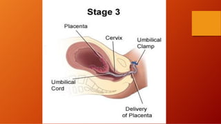
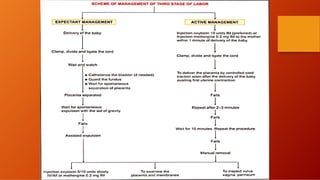
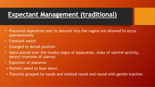
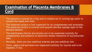
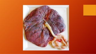
The third stage of labour involves the separation and expulsion of the placenta and membranes from the uterus. There are two main methods for managing the third stage - active management and expectant management. Active management involves providing oxytocin or methylergonovine within 1 minute of birth to help the uterus contract and quickly deliver the placenta, usually within 30 minutes. Expectant management allows the placenta to separate and deliver spontaneously on its own timeline, with close monitoring by a healthcare provider. After delivery of the placenta, the provider examines it and membranes for completeness and abnormalities before repairing any perineal trauma from birth.
The document summarizes information about the third stage of labor. It begins after the birth of the baby and ends with the delivery of the placenta and membranes. It describes the events that occur, including placental separation, descent of the placenta, and its eventual expulsion. It discusses management approaches like expectant management and active management, and interventions that may be needed like controlled cord traction or manual removal of the placenta. It provides details on examining the placenta and postpartum monitoring of the mother.
about the process of third stage of labor and management of post Partum Hemorrhage ,which is one of the major causes of blood loss in a pregnant women that needs active management.
The document provides information about the third stage of labor including its definition, duration, events, clinical course, and management. Some key points:
- The third stage lasts from birth of the baby until delivery of the placenta, usually 15-20 minutes. It involves placental separation, descent, and expulsion.
- Active management, using uterotonics and controlled cord traction, aims to shorten the third stage to around 5 minutes to reduce bleeding risk.
- Signs of placental separation include blood loss, lengthening cord, and changes in uterine shape and position.
- The placenta and membranes are examined for completeness after delivery. Complications of the third stage include postpart
NURSING MANAGEMENT OF THIRD AND FOURTH STAGE OF LABOUR.docx.pptxAyushi958023
In this ppt you will learn about Nursing management of third stage of labor(expected and active management) and Nursing management fourth stage of labor.
THIRD STAGE OF LABOUR AND ITS MANAGEMENT.pdfDolisha Warbi
definition, duration, events, (placenta separation, descend of placenta, expulsion of placenta , the Schultz mechanisms, Mathew Duncan mechanisms, signs of separation, expectant management, active management, complexion , examination of placenta and its membrane, complication.
The document summarizes key information about the human placenta, including its development, anatomy, separation and delivery during birth. It notes that the placenta develops from fetal and maternal tissues by 12 weeks and is discoid in shape. At term, the placenta is roughly 500g and occupies 30% of the uterine wall. It describes the fetal and maternal surfaces and explains that placental separation occurs due to uterine contraction and retraction over 3-5 minutes of the third stage of labor, after which the placenta is expelled. Active management with uterotonics and controlled cord traction is recommended to minimize bleeding.
Active management of the third stage of labor is preferred to prevent postpartum hemorrhage. It involves administering oxytocin within 1 minute of delivery to stimulate uterine contractions and facilitate rapid delivery of the placenta. This reduces blood loss and shortens the third stage. Expectant management allows spontaneous delivery but requires close monitoring in case interventions are needed. Both approaches require careful oversight during and after delivery of the placenta to manage any complications.
The third stage of labor involves the delivery of the placenta after childbirth. It is important to monitor for complications and allow the placenta to separate and deliver naturally without pulling on the umbilical cord. Signs of placental separation include lengthening of the cord and a gush of blood. Active management with controlled cord traction is commonly used to expedite delivery but physiological management without intervention is also appropriate for low risk births. Close monitoring during the third stage is important to detect any postpartum hemorrhage or retained placenta.
The document discusses the management of the third stage of labour, which begins with the birth of the baby and ends with delivery of the placenta. It describes the phases of placental separation, descent, and expulsion. It discusses expectant versus active management and the nursing care involved in each approach. The nursing diagnosis identifies risks for fluid deficit, lack of preparation for sensations, and energy expenditure from childbirth efforts. Nursing interventions include monitoring for signs of separation and bleeding, providing education and rest opportunities.
The document defines and discusses retained placenta, which occurs when the placenta is not expelled from the uterus within 30 minutes of delivery. There are several potential causes of retained placenta, including failure of the placenta to separate fully from the uterine wall due to issues like uterine atonicity. Management involves controlling bleeding if present and attempting controlled cord traction or manual removal of the placenta in the operating room if needed. Leaving the placenta retained poses risks like severe bleeding.
A retained placenta occurs when the placenta is not expelled from the uterus within 30 minutes of childbirth. Risk factors include previous retained placentas, uterine injuries or surgeries, preterm births, induced labor, and multiple pregnancies. Causes can include failure of the placenta to separate fully from the uterine wall or abnormal placenta attachments like placenta accreta. Treatment involves controlled cord traction, manual removal in the operating room if needed, or hysterectomy for deeply embedded placentas. Complications can be life-threatening bleeding, infections, or shock if not properly managed.
The document discusses the third stage of labor and postpartum hemorrhage. It describes the stages of labor including the third stage which involves placental separation and expulsion. The events of the third stage and mechanisms of placental separation are explained. Active management of the third stage is recommended to prevent postpartum hemorrhage. The etiology, diagnosis, and management of primary postpartum hemorrhage are outlined. Prevention focuses on risk assessment and active management of labor and delivery. True postpartum hemorrhage is managed through resuscitation, arresting bleeding, and involvement of senior staff.
This study evaluated risk factors and outcomes of manual removal of the placenta (MROP) at a tertiary hospital in Nigeria. The study compared 92 cases that underwent MROP to 91 matched controls. Risk factors for MROP included previous uterine scarring from procedures like dilation and curettage or cesarean section. Doctors performed MROP more often than midwives. On average, the third stage of labor was longer for those needing MROP. Establishing guidelines around diagnosing retained placenta could help reduce unnecessary MROP procedures.
Retained placenta is defined as failure to deliver the placenta within 30 minutes of childbirth. Risk factors include preterm delivery, induced labor, and uterine abnormalities. If the placenta is not expelled, manual removal may be needed to control bleeding and prevent complications like hemorrhage. Diagnosis involves assessing for signs of separation and feeling the placenta through the cervix. Treatment depends on the cause but may include controlled cord traction, manual removal in the operating room, or hysterectomy for deeply embedded placentas.
Pathophysiology of Normal Labor:
A series of events that take place in female genital organs to expel the product of conception that are fetus, placenta, membranes) out of womb through the vagina into the outer world. We further describe pathogenesis and features of different stages of labor
The document discusses various obstetrical operations including forceps delivery, vacuum delivery, c-section, and episiotomy. It describes the types of forceps used, indications for their use, prerequisites, and procedures. Complications of forceps delivery are also outlined. Similarly, the document discusses vacuum extractor procedures, indications, contraindications and complications. Cesarean section definitions, indications, preoperative preparation, intraoperative care, postoperative care and complications are summarized. Lastly, the document covers episiotomy including indications, advantages, disadvantages, procedures for performing and suturing an episiotomy, as well as post-care.
Pathophysiology of Normal Labour by Sunil Kumar Dahasunil kumar daha
Please find the power point on Management and pathophysiology of Normal Labour . I tried to present it on understandable way and all the contents are reviewed by experts and from very reliable references. Thank you
This document summarizes the stages of labor. It describes 4 stages: 1) preparatory stage with lightening and cervical changes, 2) active labor involving cervical dilation until fully dilated, 3) expulsion stage when the fetus is pushed through the birth canal, and 4) delivery of the placenta. Each stage is defined with details on duration, phenomena, cervical dilation curve, and management techniques to ensure delivery and control of hemorrhage.
This document discusses obstetric emergencies including prolapsed umbilical cord and uterine rupture. It defines a prolapsed cord as occurring when the umbilical cord precedes the presenting fetal part. Risk factors include premature rupture of membranes, multiparity, and malpresentation. Immediate management of a prolapsed cord with pulsation includes relieving pressure on the cord by holding the presenting part away from the cord with fingers in the vagina. Uterine rupture is defined as a full thickness tear through the uterus and can occur in scarred or unscarred uteruses. It is a medical emergency requiring prompt cesarean delivery and potential hysterectomy. Complications include hemorrhage, trauma to the fetus, and
Normal labour is defined as delivery of a single baby by vertex presentation through the vagina at term, with spontaneous onset and completion within 24 hours, leaving a healthy mother and baby. Labour is caused by hormonal and mechanical factors that lead to cervical dilation and descent and rotation of the fetal head through the birth canal in four stages. The first stage involves cervical dilation. The second stage is the birth of the baby. The third stage is delivery of the placenta, and the fourth involves recovery of the mother. A series of movements including engagement, descent, flexion, internal rotation, and extension help the fetal head navigate the birth canal.
The document discusses placental expulsion, which occurs after childbirth when the placenta exits the birth canal. It defines placental expulsion, discusses risk factors for retained placenta such as uterine atony or previous cesarean delivery. Methods of placental separation and expulsion discussed include the Schultz and Duncan methods, cord traction, and manual removal. Pharmacological, surgical, and nursing management of placental expulsion are also outlined.
Active management of the third stage of labour (AMTSL) involves administering a uterotonic drug, such as oxytocin, within one minute of delivery, controlled cord traction, and uterine massage after delivery of the placenta. This helps prevent postpartum hemorrhage, a leading cause of maternal mortality. AMTSL takes around 5-10 minutes and involves administering oxytocin to help the uterus contract followed by gentle traction on the umbilical cord to deliver the placenta once contractions occur. The uterus is then massaged to ensure it remains firm and bleeding is monitored.
Retained placenta occurs when the placenta is not delivered within 30 minutes of childbirth. Risk factors include previous retained placenta, uterine injuries or surgery, preterm delivery, and induced labor. Causes can be the placenta separating but not being expelled, simple adherent placenta, or morbidly adherent placenta which is attached deeply into the uterine wall. Management depends on the cause but may include controlled cord traction, manual removal of the placenta, or hysterectomy for deeply embedded placentas. Complications can include hemorrhage, infection, or reoccurrence with future pregnancies.
The document describes the stages of normal labor, including three stages:
1) First stage begins with uterine contractions and ends with full cervical dilation. It includes latent, active, and transition phases.
2) Second stage begins with full dilation and ends with baby's birth. It includes latent, active, and transition phases.
3) Third stage begins after birth and ends with placenta delivery. It includes separation and expulsion phases to deliver the placenta. Each stage follows physiological processes to progress labor and delivery successfully.
The document discusses the structure and function of the myometrium, the muscular layer of the uterine wall, during labor and delivery. It contains three layers of smooth muscle (longitudinal, circular, and oblique) that contract during labor due to hormones like oxytocin and prostaglandins. Calcium entry into uterine muscle cells allows the interaction of actin and myosin fibers to cause contractions. Synchronized contractions of the myometrium expel the fetus through the birth canal in three stages: cervical dilation and effacement in stage one; fetal expulsion in stage two; and placental separation and delivery in stage three.
Material management and inventory control 4th year.pptxNeharikaKumari5
This document discusses material management and inventory control in emergency and disaster situations. It begins by defining material management as the planning, organizing, and controlling of materials. Effective material management in emergencies ensures timely response, optimal resource use, logistical coordination, sustainability, risk reduction, and community support. Key aspects of inventory control in disasters include tracking resource availability and allocation, maintaining supply chain resilience, and facilitating coordination. The role of nurses is also summarized, focusing on prioritizing needs, conducting audits, communicating with others, and leveraging technology to manage medical supplies efficiently.
International health agencies provide aid to developing countries to improve healthcare. They include UNICEF, which focuses on child health, nutrition, and education. The Colombo Plan promotes economic and social development in Asia through technical cooperation. ILO works to establish social justice and decent working conditions globally. DANIDA, Denmark's development agency, prioritizes human rights, green growth, and stability in its assistance for health programs in India.
The document discusses Rh incompatibility, which occurs when a mother has Rh-negative blood and the fetus has Rh-positive blood. This can cause the mother's immune system to produce antibodies against the fetus's Rh-positive blood. The presentation covers the causes, symptoms, diagnostic tests, complications, treatments, and nursing management for Rh incompatibility. Nurses must ensure gentle removal of the placenta if needed, use of Rh-negative blood for transfusions, early umbilical cord clamping, and involvement of pediatricians in postpartum care including administration of IVIG if needed.
The document discusses India's national nutrition programs administered through various ministries. It outlines programs focused on vitamin A prophylaxis, anemia prevention, iodine deficiency control, special nutrition for vulnerable groups, balwadi pre-school meals, ICDS for children and mothers, and mid-day meals in schools. The goals are to reduce malnutrition, disease, and mortality while promoting child development, education, and maternal health. Nurse-led nutrition education teaches healthy diets, cooking, food handling and storage.
The Mini-Mental State Examination (MMSE) is a 30-point test used to screen for cognitive impairment. It tests several areas including orientation, registration, attention, recall, language, and visual-spatial skills. The test takes 10-15 minutes to administer and includes questions about orientation to time and place, registration of objects, attention and calculation, recall of objects, language skills, and the ability to follow commands. Scores are interpreted based on cut-offs, with scores below 24 indicating cognitive impairment. The MMSE provides a quick method to monitor changes over time but has limitations such as bias against those with low education.
This document defines and describes myasthenia gravis, an autoimmune disorder causing muscle weakness. It discusses the pathophysiology involving antibodies that impair signal transmission at the neuromuscular junction. Clinical manifestations usually involve the eyes and face, and can become generalized. Diagnosis involves tests like the Tensilon test and repetitive nerve stimulation. Treatment includes anticholinesterase medications to increase acetylcholine, immunosuppressants, plasmapheresis, and sometimes thymectomy. Management of exacerbations, called myasthenic crises, focuses on respiratory support and medication adjustments.
Delusion is a strong fixed unshakable belief irrespective of sociocultural background. The document defines delusion and lists 10 common types, including delusions of persecution, grandeur, and reference. It discusses managing delusions through psychological therapies, pharmacological treatments, strategies for working with delusional patients, and barriers to intervention. Nursing management focuses on addressing disturbed thought processes, impaired self-care and communication, ineffective health maintenance, and ineffective family coping.
The document discusses India's National Mental Health Programme (NMHP), which was launched in 1982 to address the country's high burden of mental illness. It defines mental health and mental illness, and outlines the aims, objectives, components, levels, and team involved in NMHP. These include providing mental healthcare access to all populations, integrating mental health into general health services, and improving quality of life. Nurses play a key role by identifying those with mental illness, providing first aid and education, assisting with therapies, and coordinating rehabilitation efforts in the community.
Physiological changes in puerperium normal puerperium.pptxNeharikaKumari5
The document summarizes the anatomical and physiological changes that occur during the postpartum period known as the puerperium. Key points include: the puerperium lasts around 6 weeks as the reproductive organs return to their non-pregnant state (involution); the uterus decreases rapidly in size from 1000g immediately after delivery to 50g by 6 weeks; lactation is initiated by withdrawal of progesterone and estrogen allowing prolactin to stimulate milk production; oxytocin triggers milk ejection in response to suckling.
The document discusses the Millennium Development Goals adopted by the UN in 2000 to improve global health and development. It outlines the 8 goals and their targets, which include eradicating extreme poverty and hunger, achieving universal primary education, promoting gender equality, reducing child and maternal mortality, combating diseases like HIV/AIDS and malaria, ensuring environmental sustainability, and developing a global partnership for development. It provides details on indicators to measure progress for each goal.
This document discusses common age-related physical and psychological changes, the responsibilities of nurses in caring for the elderly, and principles of geriatric care. Physically, aging is associated with decreases in skin elasticity, organ function, mobility, and sensory abilities. Psychologically, personality, memory, intelligence and sexuality can be impacted. Nurses should assess health, promote nutrition/exercise, provide preventive care and psychological support. Principles of care include considering individual needs, maintaining independence, comfort and hygiene, and ensuring annual physical exams. The goal of geriatric care is to maintain health, detect early disease, and prevent deterioration.
1. An eye examination assesses vision and eye health using tests like visual acuity, eye movement, pupil response, and ophthalmoscopy.
2. Examiners take a patient's ocular history and examine the eyes externally for issues like eyelid positioning and extraocular muscle function.
3. Diagnostic tests evaluate intraocular pressure, depth perception, color vision, and scan the retina, optic nerve and choroid using techniques like tonometry, Amsler grid, fundus photography, and angiography to detect disorders.
This slide is special for master students (MIBS & MIFB) in UUM. Also useful for readers who are interested in the topic of contemporary Islamic banking.
How to Add Chatter in the odoo 17 ERP ModuleCeline George
In Odoo, the chatter is like a chat tool that helps you work together on records. You can leave notes and track things, making it easier to talk with your team and partners. Inside chatter, all communication history, activity, and changes will be displayed.
Exploiting Artificial Intelligence for Empowering Researchers and Faculty, In...Dr. Vinod Kumar Kanvaria
Exploiting Artificial Intelligence for Empowering Researchers and Faculty,
International FDP on Fundamentals of Research in Social Sciences
at Integral University, Lucknow, 06.06.2024
By Dr. Vinod Kumar Kanvaria
Macroeconomics- Movie Location
This will be used as part of your Personal Professional Portfolio once graded.
Objective:
Prepare a presentation or a paper using research, basic comparative analysis, data organization and application of economic information. You will make an informed assessment of an economic climate outside of the United States to accomplish an entertainment industry objective.
A review of the growth of the Israel Genealogy Research Association Database Collection for the last 12 months. Our collection is now passed the 3 million mark and still growing. See which archives have contributed the most. See the different types of records we have, and which years have had records added. You can also see what we have for the future.
Physiology and chemistry of skin and pigmentation, hairs, scalp, lips and nail, Cleansing cream, Lotions, Face powders, Face packs, Lipsticks, Bath products, soaps and baby product,
Preparation and standardization of the following : Tonic, Bleaches, Dentifrices and Mouth washes & Tooth Pastes, Cosmetics for Nails.
This presentation was provided by Steph Pollock of The American Psychological Association’s Journals Program, and Damita Snow, of The American Society of Civil Engineers (ASCE), for the initial session of NISO's 2024 Training Series "DEIA in the Scholarly Landscape." Session One: 'Setting Expectations: a DEIA Primer,' was held June 6, 2024.
How to Build a Module in Odoo 17 Using the Scaffold MethodCeline George
Odoo provides an option for creating a module by using a single line command. By using this command the user can make a whole structure of a module. It is very easy for a beginner to make a module. There is no need to make each file manually. This slide will show how to create a module using the scaffold method.
it describes the bony anatomy including the femoral head , acetabulum, labrum . also discusses the capsule , ligaments . muscle that act on the hip joint and the range of motion are outlined. factors affecting hip joint stability and weight transmission through the joint are summarized.
Pride Month Slides 2024 David Douglas School District
Third stage of labour.pdf
1. OBSTETRICAL NURSING
Third stage of labour
Presented To :-
Resp. Sandhya Ma’am
Faculty
College of Nursing
VMMC & SJH
Presented by :-
Divyanshi
01250306619
Roll no. 08
B. Sc.(H) Nursing 4th
year
2. LABOUR
• Series of events that takes place in the genital organ in an
effort to expel the viable products of conception out of the
womb through the vagina into the outer world is called labour.
• It may occur prior to 37 completed weeks, when it is called
preterm labour.
• Delivery is the expulsion or extraction of viable fetus out of
the womb.
3.
4. STAGES OF LABOUR
First stage of labour
Second stage of labour
Third stage of labour
Fourth stage of labour
7. Events in Third Stage of Labour
1. Placental Separation
2. Separation of the Membranes
3. Expulsion of the Placenta with the membranes.
4. Mechanism of Control of Bleeding.
8. 1. Placental Separation
• Mechanism of separation: After delivery of the foetus, Marked
retraction reduces effectively the surface area at the placental
site to about its half. But as the placenta is inelastic, it cannot
keep pace with such an extent of diminution resulting in its
buckling. A shearing force is instituted between the placenta and
the placental site which brings about its ultimate separation.
9.
10. • There are two ways of separation of placenta.
(1) Central separation (Schultze): Detachment of placenta from
its uterine attachment starts at the center resulting in opening up
of few uterine sinuses and accumulation of blood behind the
placenta (retroplacental hematoma). With increasing contraction,
more and more detachment occurs facilitated by weight of the
placenta and retroplacental blood until whole of the placenta gets
detached.
(2) Marginal separation (Mathews-Duncan): Separation starts at
the margin as it is mostly unsupported. With progressive uterine
contraction, more and more areas of the placenta get separated.
Marginal separation is found more frequently.
11.
12. 2. Separation of Membranes
• The membranes, which are attached loosely in the active part,
are thrown into multiple folds. Those attached to the lower
segment are already separated during its stretching. The
separation is facilitated partly by uterine contraction and mostly
by weight of the placenta as it descends down from the active
part. The membranes so separated carry with them remnants of
decidua vera giving the outer surface of the chorion its
characteristic roughness.
13. 3. Expulsion of Placenta
• After complete separation of the placenta, it is forced down into
the flabby lower uterine segment or upper part of the vagina by
effective contraction and retraction of the uterus. Thereafter, it
is expelled out either by voluntary contraction of abdominal
muscles (bearing down efforts) or by manual procedure.
14. 4. Mechanism of Control of Bleeding
• After placental separation, innumerable torn sinuses which have free
circulation of blood from uterine and ovarian vessels have to be obliterated.
The occlusion is affected by com plete retraction whereby the arterioles, as
they pass tortuously through the interlacing intermediate layer of the
myometrium, are literally clamped. It (living ligature) is the principal
mechanism of hemostasis. However, thrombosis occurs to occlude the torn
sinuses, a phenomenon, which is facilitated by hypercoagulable state of
pregnancy. Apposition of the walls of the uterus following expulsion of the
placenta (myotamponade) also contributes to minimize blood loss.
15.
16. Clinical Course of Third Stage of Labour
• Third stage includes separation, descent and expulsion
of the placenta with its membranes.
• PAIN: For a short time, the patient experiences no pain.
However, intermittent discomfort in the lower abdomen
reappears, corresponding with the uterine contractions.
17. Before Separation
• Per abdomen uterus becomes discoid in shape, firm in feel and
non-ballottable. Fundal height reaches slightly below the
umbilicus.
• Per vaginam: There may be slight trickling of blood.Length of
the umbilical cord as visible from outside remains static.
18. After Separation
• It takes about 5 minutes in conventional management for the
placenta to separate.
• Per abdomen:
. Uterus becomes globular, firm, and ballottable.
. The fundal height is slightly raised as the separated placenta
comes down in the lower segment and the contracted uterus
rests on top of it.
. Slight bulging in the suprapubic region due to distension of the
lower segment by the separated placenta.
19. • Per vaginam:
4. Slight gush of vaginal bleeding.
5. Permanent lengthening of the cord is established.
This can be elicited by pushing down the fundus when a
length of cord comes outside the vulva, which remains
permanent even after the pressure is released.
Alternatively, on suprapubic pressure upward by the
fingers, there is no indrawing of the cord and the same
lies unchanged outside the vulva.
20. Expulsion of Placenta & Membranes
• The expulsion is achieved either by voluntary bearing-down
efforts or more commonly aided by manipulative procedure. The
afterbirth delivery is soon followed by slight to moderate bleeding
amounting to 100-250 mL.
Maternal Signs
• There may be chills and occasional shivering. Slight transient
hypotension is not unusual.
22. Steps of Management
• Third stage is the most crucial stage of labor. Previously uneventful
first and second stage can become abnormal within a minute with
disastrous consequences.
• The principles underlying the management of third stage are to
ensure strict vigilance and to follow the management guidelines
strictly in practice so as to prevent the complications, the important
one being postpartum hemorrhage.
• STEPS OF MANAGEMENT: Two methods of management are currently
in practice.
1. Active Management
2. Expectant Management
23.
24. Nursing Diagnosis
• Risk for deficient fluid volume related to :
- Blood loss occurring after placental separation and expulsion.
- Inadequate contraction of the uterus.
• Anxiety related to:
-Lack of knowledge regarding separation and expulsion of the placenta.
-Occurrence of perineal trauma and the need for repair.
• Fatigue related to :
-energy expenditure associated with childbirth and the bearing-down efforts of
the second stage.
25. Active Management
• The underlying principle In active management is to excite
powerful uterine contractions within 1 minute of delivery of
the baby (WHO) by giving parenteral oxytocic.
• The advantages are-(a) to minimize blood loss in third stage
approximately to one-fifth and (b) to shorten the duration of
third stage to half; and (c) Reduces maternal anemia.
• The only disadvantage is slight increased incidence of retained
placenta (1-2%) and consequent increased incidence of manual
removal.
26. Procedures :-
• Injection oxytocin 10 units IM (pre ferred) or methergine 0.2 mg
IM is given within 1 minute of delivery of the baby (WHO). The
placenta is expected to be delivered soon following delivery of the
baby. If the placenta is not delivered thereafter, it should be
delivered forthwith by controlled cord traction (Brandt -Andrews)
technique after clamping the cord while the uterus still remains
contracted. If the first attempt fails, another attempt is made after
2-3 minutes failing which another attempt is made at 10 minutes. If
this still fails, manual removal is to be done.
27. Assisted Expulsion
• Brandt-andrews Method (Controlled Cord Traction)
The palmar surface of the fingers of the left hand is placed (above the
symphysis pubis) approximately at the junction of upper and lower uterine
segment. The body of the uterus is pushed upward and backward, toward the
umbilicus while by the right hand steady tension (but not too strong traction) is
given in downward and backward direction holding the clamp until the placenta
comes outside the introitus. It is thus more a uterine elevation which facilitates
expulsion of the placenta. The procedure is to be adopted only when the uterus
ishard and contracted.
28.
29. • Fundal Pressure
The fundus is pushed down ward and backward after placing four
fingers behind the fundus and the thumb in front using the uterus as
sort of piston. Pressure must be given only when the uterus becomes
hard. If it is not, then make it hard by gentle rubbing. The pressure is
to be withdrawn as soon as the placenta passes through the introitus.
If the baby is macerated or premature, this method is preferable to
cord traction as the tensile strength of the cord is much reduced in
both the instances.The cord may be accidentally torn which is not
likely to cause any problem. The sterile gloved hand should be
introduced, and the placenta is to be grasped and extracted.
30.
31. Expectant Management (traditional)
• Placental separation and its descent into the vagina are allowed to occur
spontaneously
• Constant watch
• Changed to dorsal position
• Hand placed over the fundus (signs of separation, state of uterine activity,
detect inversion of uterus)
• Expulsion of placenta
• Patient asked to bear down
• Placenta grasped by hands and twisted round and round with gentle traction
32. Examination of Placenta Membranes &
Cord
• The placenta is placed on a tray and is washed out in running tap water to
remove the blood and clots.
• The maternal surface is first inspected for its completeness and anomalies.
The maternal surface is covered with grayish decidua (spongy layer of the
decidua basalis).
• The membranes-chorion and amnion are to be examined carefully for
completeness and presence of abnormal vessels indicative of succenturiate
lobe.
• Normally, there are two umbilical arteries and one umbilical vein.
• Vulva, vagina and perineum are inspected carefully for injuries and to be
repaired, if any.