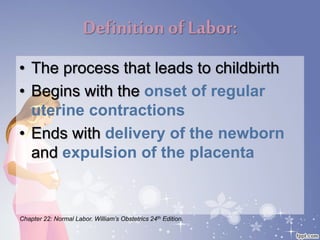
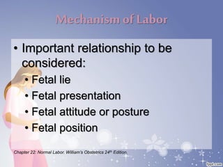
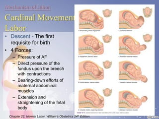
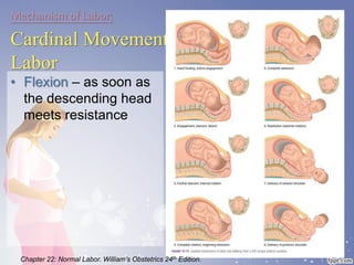
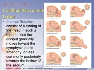
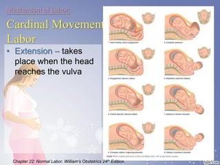
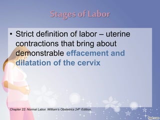
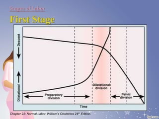
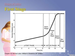
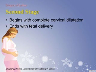
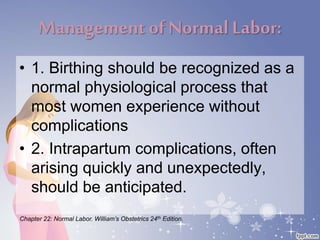
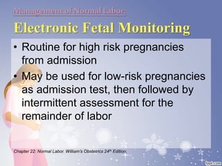
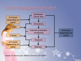
This document summarizes the normal labor and delivery process. It defines labor as beginning with regular uterine contractions and ending with childbirth. Labor involves three stages - first stage is cervical dilation from 0-10cm, second stage is birth of the baby, and third is delivery of the placenta. Key aspects of managing normal labor are admitting women in early labor, monitoring the fetus, allowing freedom of movement, and active management including amniotomy and oxytocin to shorten stages of labor. The goal is a safe birth for both mother and child with minimal medical intervention.
Caesarean section is a surgical procedure used to deliver babies through incisions in the abdominal wall and uterus. It is performed when vaginal delivery would put the mother or baby's health at risk. The document discusses the history of c-sections and various techniques for performing them, including types of incisions, anesthesia methods, and post-operative care recommendations. Key aspects are a lower transverse uterine incision, controlled cord traction to deliver the placenta, a two-layer closure of the uterine incision, and facilitating early skin-to-skin contact between mother and baby.
Learn what is "fetal attitude", "fetal lie", different types of fetal presentation (cephalic, breech, compound, shoulder), how to determine fetal position, and how to do Leopold's maneuvers
1) Vacuum and forceps deliveries are indicated for maternal exhaustion, prolonged second stage of labor, or fetal distress.
2) Proper technique using mnemonics like "ABCDEFG" are important to safely perform instrumental deliveries and minimize complications.
3) Potential maternal complications include vaginal lacerations and trauma, while fetal risks include scalp injuries, cephalohematomas, and rarely intracranial hemorrhage. Forceps carry higher risks than vacuum extraction.
MANAGEMENT OF ACUTE UTERINE INVERSION BY DR SHASHWAT JANIDR SHASHWAT JANI
1) Acute uterine inversion occurs within 24 hours of delivery when the uterus turns inside out, often due to fundal placenta insertion and uterine atony.
2) Immediate manual replacement of the inverted uterus without anesthesia has the highest chance of success and should be attempted promptly.
3) If manual replacement fails or the patient presents later, general anesthesia and tocolytic agents like nitroglycerin are administered to relax the uterus before again attempting manual replacement while controlling hemorrhage. Prompt treatment is critical to prevent complications like shock.
The document discusses the partograph, a graphical record used to monitor labor. It was developed by the WHO to allow early detection of abnormal labor progress.
The partograph includes sections to monitor the fetal condition, labor progress, and maternal condition over time. It graphs cervical dilation against time to identify normal vs prolonged labor. Lines are included to indicate when extra vigilance or interventions are needed.
The partograph aims to prevent prolonged labor and its complications by allowing early decision making about transfers, augmentations, or terminations. Its use requires adequate training but it can effectively reduce problems from prolonged labor for both mother and baby when properly implemented.
This document discusses breech presentation, which occurs when a baby is positioned bottom or feet first in the uterus instead of head first. It defines the different types of breech positions and discusses risk factors, diagnosis, management options, and complications associated with vaginal breech delivery and cesarean section for breech babies. Management options include external cephalic version, vaginal delivery, or cesarean section depending on the specific situation. Risks and procedures for both vaginal delivery and cesarean section are outlined.
Shoulder dystocia is when the fetal shoulders become lodged at the maternal pelvis after delivery of the head, occurring in 0.2-2% of births. Risk factors include maternal diabetes, obesity, macrosomia, and prior shoulder dystocia. Management involves calling for help, applying suprapubic pressure and the McRoberts maneuver to widen the pelvis, and rotating the shoulders using maneuvers like Woods screw or Rubin. If unsuccessful, procedures include delivering the posterior arm or rarely symphysiotomy. Fetal risks are brachial plexus injury, fractures, and hypoxic brain injury. Maternal risks include perineal tears and postpartum hemorrhage. Prevention focuses
The document discusses various topics related to fetal lie, presentation, position, and labor including:
- The fetal lie can be longitudinal, transverse, or oblique relative to the mother's long axis.
- Cephalic presentation is most common, with other possibilities including breech, face, brow, and transverse lie.
- Fetal position describes the relationship of parts of the presenting fetal head to the mother's right or left side.
- Leopold's maneuvers are used to determine fetal position and presentation during vaginal exams.
- The cardinal movements of labor are engagement, descent, flexion, internal rotation, extension, and external rotation.
The document discusses several topics related to labour and delivery:
- The physiological mechanisms that initiate labour, including hormonal and anatomical changes in the mother and fetus.
- How uterine contractions progress cervical dilation and effacement in the first stage of labour.
- The second stage where contractions expel the fetus through the birth canal.
- The third stage where the placenta is delivered.
- Methods for assessing and monitoring labour including physical exams, cardiotocography to monitor the fetal heart rate, and use of the partogram to track labour progress.
This document provides information on operative vaginal delivery using forceps. It describes the types of forceps including long curved forceps, short curved forceps, and Kielland's forceps. It details the parts of the forceps including the blades, shanks, locks, handles, and screws. It explains how to identify and apply the forceps blades for low forceps delivery. The steps taken are identification and application of the blades, locking the blades, applying traction, and removing the blades. Precautions and techniques are outlined to ensure a safe operative vaginal delivery using forceps.
This document discusses unstable lie and version in pregnancy. It defines unstable lie as a condition where the fetal presentation constantly changes beyond 36 weeks of pregnancy when it should have stabilized. Causes include factors that prevent the presenting part from remaining fixed in the lower uterus. Complications include cord entanglement and increased risk of perinatal death. External cephalic version can be attempted to correct the malpresentation if there are no contraindications. Hospitalization is recommended at 37 weeks to monitor for premature rupture of membranes or cord prolapse. Elective c-section is often required, especially if complicating factors are present.
Prelabour Rupture of Membrane (PROM) by Sunil Kumar Dahasunil kumar daha
Please find the power point on Prelabour Rupture of Membrane (PROM). I tried to present it on understandable way and all the contents are reviewed by experts and from very reliable references. Thank you
The document discusses the physiology of labor and pain pathways. It describes the theories behind the onset of labor, including progesterone withdrawal and estrogen stimulation. It outlines the stages of labor and differences between true and false labor. The passage of the fetus through the birth canal involves changes in position called cardinal movements. Factors that can affect labor include the passenger (fetus), passageway (maternal pelvis), and powers (uterine contractions and maternal efforts). Pain in labor is transmitted via neural pathways and can stimulate various physiological responses.
The pelvis is made up of four bones - two innominate bones, the sacrum, and coccyx. It forms the inlet, cavity, and outlet of the true pelvis through which the fetus must pass during childbirth. The key diameters of the inlet are the anteroposterior (11cm), obstetric conjugate (10cm), and transverse (13cm). The cavity is roughly spherical with diameters of 12cm. The outlet's diameters are the anteroposterior (13cm), oblique (12cm), and transverse between ischial spines (11cm), which is the smallest diameter requiring baby to rotate.
This document outlines the mechanism of normal labor, including the criteria, onset, and three stages. The first stage involves cervical effacement and dilation. The second stage involves descent, flexion, internal rotation, crowning, extension, restitution, and delivery of the shoulders and body. Key mechanisms in the second stage include synclitism, engagement, and internal and external rotation. The third stage involves delivery of the placenta by either the Mathews-Duncan or Schultz mechanism. Management of the second stage includes controlling delivery of the head, performing episiotomy if needed, and repairing any tears or episiotomy.
Caesarean section is a surgical procedure to deliver a baby through abdominal and uterine incisions after 28 weeks of pregnancy. The incidence of C-sections has increased due to abandoning difficult procedures and increased use for breech births. Indications include maternal conditions like pelvic abnormalities and previous C-sections, as well as fetal indications like distress. A lower segment transverse incision is most common. Complications can include hemorrhage, injury, infection and rupture of the uterine scar in subsequent pregnancies.
This document provides an overview of shock in obstetrics, including definitions, classifications, pathophysiology, diagnosis, and management. It discusses the main types of shock seen in obstetrics such as hypovolemic (hemorrhagic), septic, cardiogenic, distributive, and anaphylactic shock. For each type, it outlines the causes, clinical features, and specific management approaches. Initial management focuses on maintaining airway, breathing, and circulation while treating the underlying cause.
The document summarizes information about the third stage of labor. It begins after the birth of the baby and ends with the delivery of the placenta and membranes. It describes the events that occur, including placental separation, descent of the placenta, and its eventual expulsion. It discusses management approaches like expectant management and active management, and interventions that may be needed like controlled cord traction or manual removal of the placenta. It provides details on examining the placenta and postpartum monitoring of the mother.
This document defines and classifies uterine abnormalities, or mullerian duct anomalies. It describes 7 classes of anomalies resulting from abnormal development of the mullerian ducts during embryogenesis. Class 1 involves complete or partial mullerian agenesis. Classes 2 through 5 involve various degrees of failure of the mullerian ducts to fully fuse, resulting in anomalies like a unicornuate, didelphys, bicornuate, or septate uterus. Class 6 is an arcuate uterus and Class 7 involves DES exposure in utero causing a T-shaped uterus. Symptoms may include pain or infertility. Diagnosis involves imaging tests. Treatment depends on symptoms but may involve surgery to correct the anomaly
The document describes the normal mechanism of labor, including the three stages of labor and the fetus' seven passive movements that enable it to navigate the birth canal. The first stage involves cervical dilation. The second stage is when the fetus is delivered. The third stage involves delivery of the placenta. Key movements include engagement, descent, flexion, internal rotation, extension, restitution/external rotation, and expulsion. Close monitoring of the fetus and mother is important throughout labor.
Uterine rupture is a life-threatening condition where the wall of the uterus tears, potentially exposing the fetus and placenta to the mother's abdominal cavity. It most commonly occurs in women with a previous cesarean section scar. Signs include acute abdominal pain, fetal distress, and hemorrhage. Diagnosis is often made using ultrasound or MRI to detect tears in the uterine wall. Immediate exploratory laparotomy and cesarean delivery is usually required for treatment. Conservative uterine repair may be attempted for some cases but hysterectomy is often necessary due to severe hemorrhage. Prevention focuses on careful management of trial of labor for women with previous scars.
- Palpates fetal back anteriorly.
- Applies counter pressure over fetal back to prevent version.
Obstetrician:
- Applies pressure over fetal presenting part (breech) to flex it.
- Applies pressure over fetal back to extend it.
- Applies pressure over fetal head to flex it.
- Rotates fetal presenting part out of pelvis.
- Rotates fetal head into pelvis.
• Version is complete when head engages.
Osama Warda 34
BREECH PRESENTATION- MANAGEMENT
BREECH
PRESENTATION-
MANAGEMENT
Osama
Ward
Vaginal delivery
DescriptionA vaginal delivery is the giving birth to offspring in mammals through the vagina. It is the natural method of birth for all mammals except monotremes, which lay eggs into the external environment.
This document discusses the management of normal labor and delivery. It defines labor as regular painful uterine contractions associated with cervical changes. The stages of labor are outlined as first stage being cervical dilation, second stage being fetal expulsion, and third stage being placental delivery. Fetal head shape and size changes during labor through moulding and position changes to negotiate the maternal pelvis. Monitoring of maternal well-being, fetal well-being, and labor progress is important for managing normal labor.
This document summarizes the normal process of labor and delivery in 3 stages:
1) The first stage of labor begins with the onset of contractions and ends with full cervical dilation. It is divided into latent and active phases.
2) The second stage begins at full dilation and ends with delivery of the baby. It has passive and active phases.
3) The third stage begins with delivery of the baby and ends with delivery of the placenta, usually within 10-20 minutes.
This document provides an outline on normal labor and the mechanism of labor. It defines labor as the physiological process of expelling the fetus, placenta, and membranes through the birth canal after 24 weeks of pregnancy. Normal labor fulfills criteria of spontaneous onset at term, vertex presentation, no prolongation, and natural termination with minimal aids. The document describes the three stages of labor as well as the cervical changes, fetal positioning, and mechanisms involved in labor including engagement, descent, flexion, rotations, and expulsion of the fetus through the birth canal. It emphasizes the importance of classifying pregnancies as high or low risk and addressing any medical issues to ensure a safe delivery.
This chapter provides an overview of obstetrics and discusses pregnancy, signs and symptoms of pregnancy, complications of pregnancy, labor signs and symptoms, and distinguishing false labor from true labor. Key points include pregnancy being divided into trimesters, common physiological changes during pregnancy like weight gain and skin changes, signs of labor including bloody show and lightening, and characteristics of true labor contractions being regular, longer, and more intense versus irregular and shorter false labor pains.
Normal Labour/ Stages of Labour/ Mechanism of LabourWasim Ak
Normal labor is also termed spontaneous labor, defined as the natural physiological process through which the fetus, placenta, and membranes are expelled from the uterus through the birth canal at term (37 to 42 weeks
1) The document summarizes the management of normal labor and the use of the partograph to monitor labor. It describes the stages of labor, mechanisms of labor, and complications that can occur.
2) The partograph is a graphic record that aids in early detection of problems in the mother and fetus. It includes monitoring of cervical dilation, fetal descent, uterine contractions, and fetal and maternal conditions.
3) Key principles of the WHO partograph include commencing the active phase at 3cm dilation, the latent phase not exceeding 8 hours, and cervical dilation slowing to less than 1cm/hr requiring intervention.
This document provides information about physical changes in pregnancy, the stages of labor including dilation, expulsion, and placental delivery, sample questions for taking an obstetric history, definitions of gravidity and parity, potential obstetric emergencies like preeclampsia and placental abnormalities, procedures for childbirth and newborn care, and the resuscitation triangle for depressed newborns. It includes diagrams of fetal circulation, the placenta, stages of labor, and techniques for situations like breech births and shoulder dystocia.
This document provides information on the stages of labor and management of the first stage of labor. It discusses the normal progression through the latent, active, and transition phases of the first stage. It also covers monitoring during labor including vital signs, contractions, and fetal heart rate. Active management of labor is described which includes interventions like amniotomy and oxytocin if progress is unsatisfactory. The nurse's role in caring for the woman in the first stage is also summarized.
This document provides an overview of obstetrics and pregnancy. It discusses topics such as the definition of obstetrics, the stages of pregnancy, fetal development, physiological changes during pregnancy, signs and symptoms of pregnancy, calculating the due date, discomforts of pregnancy, complications of pregnancy, signs and symptoms of labor, diagnosing true labor versus false labor, and common diagnostic techniques, treatments and procedures in obstetrics such as AFP screening, amniocentesis, cesarean section, and contraction stress tests.
The document describes the stages of labor:
1) The first stage begins with onset of true labor pain and ends with full dilation of the cervix. It includes the latent and active phases.
2) The second stage begins with full dilation and ends with delivery of the fetus.
3) The third stage begins with delivery of the fetus and ends with delivery of the placenta.
4) The fourth stage is a 1 hour observation period after delivery of the placenta.
Clinical methods to assess cephalopelvic disproportion include the abdominal method, Ian Donald method, and the Munro Kerr-Muller method involving pelvic measurements.
Early pregnancy bleeding can be caused by issues related to the pregnancy itself like miscarriage or ectopic pregnancy, or issues associated with pregnancy like cervical lesions. Examination of a woman with bleeding includes general exam to check for signs of heavy bleeding, abdominal exam to check for masses, and pelvic exam including speculum and bimanual exams to examine the cervix and uterus. Common causes of early pregnancy bleeding are threatened abortion where bleeding has started but pregnancy is still viable, inevitable abortion where continuation is impossible, complete abortion where all pregnancy tissue is expelled, incomplete abortion where tissue remains inside, and missed abortion where the fetus has died but remains in utero. Treatment depends on the situation and may include monitoring, uterine evacuation, or cure
NORMAL LABOR.. (EUTOCIA) ABNORMAL LABOR ALSO EXPLAINED. Series of events that take place in the genital organs in an effort to expel the viable products of conception out of the womb through the vagina into the outer world is called LABOR.
what is labor and what is the normal?
what are the signs of labor?
what are the stages of labor?
what are the mechanism of labor?
what are the factors that affect the labor?
Management of normal labour Final yr.pptxIram Chaudhry
This document provides an outline and overview of the management of normal labor. It defines the three stages of labor as follows:
1) First stage (cervical dilation from 0-10cm): divided into latent phase and active phase
2) Second stage (fetal descent and expulsion): from full dilation until delivery of the baby
3) Third stage (placental expulsion): from delivery of the baby until delivery of the placenta
It describes the assessment, monitoring and care provided during each stage, including vaginal exams, partograms to monitor progress, fetal monitoring, positioning, pain management, and active management of the third stage to prevent postpartum hemorrhage. The
The document discusses normal labor and delivery, including:
1. It describes the phases (latent and active) and stages (first, second, third) of labor, as well as the cardinal movements that the fetus undergoes during delivery.
2. The variables that influence normal labor are discussed, including the powers of uterine contractions, characteristics of the fetus or passenger, and the maternal pelvis or passageway.
3. Management of normal labor is outlined, including monitoring labor progress, fetal wellbeing, maternal nutrition and positioning, and criteria for prolonged stages of labor.
This document discusses labor and delivery. It begins by defining labor and its normal stages. The first stage of labor is divided into latent and active phases. Diagnosis of labor requires painful contractions accompanied by cervical changes. Upon admission, management includes IV placement, monitoring, and determining labor progress through cervical exams and fetal heart rate monitoring. Active management is described as controlling labor progress through early interventions like amniotomy and oxytocin if needed. Monitoring protocols recommend frequent maternal and fetal assessments including intermittent fetal heart rate checks and recording labor on a partogram.
Normal labour is defined as the spontaneous expulsion of a single, mature fetus through the birth canal within 18 hours without complications. It involves 3 stages: 1) cervical dilation, 2) delivery of the baby, 3) delivery of the placenta. The document discusses evaluating and monitoring labour, managing each stage, relieving pain, and recording labour progress on a partogram. Fetal well-being and maternal vital signs are closely watched throughout. [END SUMMARY]
Here is the updated list of Top Best Ayurvedic medicine for Gas and Indigestion and those are Gas-O-Go Syp for Dyspepsia | Lavizyme Syrup for Acidity | Yumzyme Hepatoprotective Capsules etc
Local Advanced Lung Cancer: Artificial Intelligence, Synergetics, Complex Sys...Oleg Kshivets
Overall life span (LS) was 1671.7±1721.6 days and cumulative 5YS reached 62.4%, 10 years – 50.4%, 20 years – 44.6%. 94 LCP lived more than 5 years without cancer (LS=2958.6±1723.6 days), 22 – more than 10 years (LS=5571±1841.8 days). 67 LCP died because of LC (LS=471.9±344 days). AT significantly improved 5YS (68% vs. 53.7%) (P=0.028 by log-rank test). Cox modeling displayed that 5YS of LCP significantly depended on: N0-N12, T3-4, blood cell circuit, cell ratio factors (ratio between cancer cells-CC and blood cells subpopulations), LC cell dynamics, recalcification time, heparin tolerance, prothrombin index, protein, AT, procedure type (P=0.000-0.031). Neural networks, genetic algorithm selection and bootstrap simulation revealed relationships between 5YS and N0-12 (rank=1), thrombocytes/CC (rank=2), segmented neutrophils/CC (3), eosinophils/CC (4), erythrocytes/CC (5), healthy cells/CC (6), lymphocytes/CC (7), stick neutrophils/CC (8), leucocytes/CC (9), monocytes/CC (10). Correct prediction of 5YS was 100% by neural networks computing (error=0.000; area under ROC curve=1.0).
Adhd Medication Shortage Uk - trinexpharmacy.comreignlana06
The UK is currently facing a Adhd Medication Shortage Uk, which has left many patients and their families grappling with uncertainty and frustration. ADHD, or Attention Deficit Hyperactivity Disorder, is a chronic condition that requires consistent medication to manage effectively. This shortage has highlighted the critical role these medications play in the daily lives of those affected by ADHD. Contact : +1 (747) 209 – 3649 E-mail : sales@trinexpharmacy.com
Histololgy of Female Reproductive System.pptxAyeshaZaid1
Dive into an in-depth exploration of the histological structure of female reproductive system with this comprehensive lecture. Presented by Dr. Ayesha Irfan, Assistant Professor of Anatomy, this presentation covers the Gross anatomy and functional histology of the female reproductive organs. Ideal for students, educators, and anyone interested in medical science, this lecture provides clear explanations, detailed diagrams, and valuable insights into female reproductive system. Enhance your knowledge and understanding of this essential aspect of human biology.
These lecture slides, by Dr Sidra Arshad, offer a quick overview of the physiological basis of a normal electrocardiogram.
Learning objectives:
1. Define an electrocardiogram (ECG) and electrocardiography
2. Describe how dipoles generated by the heart produce the waveforms of the ECG
3. Describe the components of a normal electrocardiogram of a typical bipolar lead (limb II)
4. Differentiate between intervals and segments
5. Enlist some common indications for obtaining an ECG
6. Describe the flow of current around the heart during the cardiac cycle
7. Discuss the placement and polarity of the leads of electrocardiograph
8. Describe the normal electrocardiograms recorded from the limb leads and explain the physiological basis of the different records that are obtained
9. Define mean electrical vector (axis) of the heart and give the normal range
10. Define the mean QRS vector
11. Describe the axes of leads (hexagonal reference system)
12. Comprehend the vectorial analysis of the normal ECG
13. Determine the mean electrical axis of the ventricular QRS and appreciate the mean axis deviation
14. Explain the concepts of current of injury, J point, and their significance
Study Resources:
1. Chapter 11, Guyton and Hall Textbook of Medical Physiology, 14th edition
2. Chapter 9, Human Physiology - From Cells to Systems, Lauralee Sherwood, 9th edition
3. Chapter 29, Ganong’s Review of Medical Physiology, 26th edition
4. Electrocardiogram, StatPearls - https://www.ncbi.nlm.nih.gov/books/NBK549803/
5. ECG in Medical Practice by ABM Abdullah, 4th edition
6. Chapter 3, Cardiology Explained, https://www.ncbi.nlm.nih.gov/books/NBK2214/
7. ECG Basics, http://www.nataliescasebook.com/tag/e-c-g-basics
- Video recording of this lecture in English language: https://youtu.be/kqbnxVAZs-0
- Video recording of this lecture in Arabic language: https://youtu.be/SINlygW1Mpc
- Link to download the book free: https://nephrotube.blogspot.com/p/nephrotube-nephrology-books.html
- Link to NephroTube website: www.NephroTube.com
- Link to NephroTube social media accounts: https://nephrotube.blogspot.com/p/join-nephrotube-on-social-media.html
TEST BANK For An Introduction to Brain and Behavior, 7th Edition by Bryan Kol...rightmanforbloodline
TEST BANK For An Introduction to Brain and Behavior, 7th Edition by Bryan Kolb, Ian Q. Whishaw, Verified Chapters 1 - 16, Complete Newest Versio
TEST BANK For An Introduction to Brain and Behavior, 7th Edition by Bryan Kolb, Ian Q. Whishaw, Verified Chapters 1 - 16, Complete Newest Version
TEST BANK For Community Health Nursing A Canadian Perspective, 5th Edition by...Donc Test
TEST BANK For Community Health Nursing A Canadian Perspective, 5th Edition by Stamler, Verified Chapters 1 - 33, Complete Newest Version Community Health Nursing A Canadian Perspective, 5th Edition by Stamler, Verified Chapters 1 - 33, Complete Newest Version Community Health Nursing A Canadian Perspective, 5th Edition by Stamler Community Health Nursing A Canadian Perspective, 5th Edition TEST BANK by Stamler Test Bank For Community Health Nursing A Canadian Perspective, 5th Edition Pdf Chapters Download Test Bank For Community Health Nursing A Canadian Perspective, 5th Edition Pdf Download Stuvia Test Bank For Community Health Nursing A Canadian Perspective, 5th Edition Study Guide Test Bank For Community Health Nursing A Canadian Perspective, 5th Edition Ebook Download Stuvia Test Bank For Community Health Nursing A Canadian Perspective, 5th Edition Questions and Answers Quizlet Test Bank For Community Health Nursing A Canadian Perspective, 5th Edition Studocu Test Bank For Community Health Nursing A Canadian Perspective, 5th Edition Quizlet Test Bank For Community Health Nursing A Canadian Perspective, 5th Edition Stuvia Community Health Nursing A Canadian Perspective, 5th Edition Pdf Chapters Download Community Health Nursing A Canadian Perspective, 5th Edition Pdf Download Course Hero Community Health Nursing A Canadian Perspective, 5th Edition Answers Quizlet Community Health Nursing A Canadian Perspective, 5th Edition Ebook Download Course hero Community Health Nursing A Canadian Perspective, 5th Edition Questions and Answers Community Health Nursing A Canadian Perspective, 5th Edition Studocu Community Health Nursing A Canadian Perspective, 5th Edition Quizlet Community Health Nursing A Canadian Perspective, 5th Edition Stuvia Community Health Nursing A Canadian Perspective, 5th Edition Test Bank Pdf Chapters Download Community Health Nursing A Canadian Perspective, 5th Edition Test Bank Pdf Download Stuvia Community Health Nursing A Canadian Perspective, 5th Edition Test Bank Study Guide Questions and Answers Community Health Nursing A Canadian Perspective, 5th Edition Test Bank Ebook Download Stuvia Community Health Nursing A Canadian Perspective, 5th Edition Test Bank Questions Quizlet Community Health Nursing A Canadian Perspective, 5th Edition Test Bank Studocu Community Health Nursing A Canadian Perspective, 5th Edition Test Bank Quizlet Community Health Nursing A Canadian Perspective, 5th Edition Test Bank Stuvia
TEST BANK For Community Health Nursing A Canadian Perspective, 5th Edition by...
Normal labor and delivery
1. Normal Labor and Delivery
Resident Lecturer: A. Polintan, MD
Moderator: V. Espallardo, MD, FPOGS
2. Outline:
• Definition of Labor
• Mechanism of Labor
• Stages of Labor
• Management of Normal Labor
3. Definition ofLabor:
• The process that leads to childbirth
• Begins with the onset of regular
uterine contractions
• Ends with delivery of the newborn
and expulsion of the placenta
Chapter 22: Normal Labor. William’s Obstetrics 24th Edition.
4. Outline:
• Definition of Labor
• Mechanism of Labor
• Stages of Labor
• Management of Normal Labor
5. Mechanism of Labor
• Important relationship to be
considered:
• Fetal lie
• Fetal presentation
• Fetal attitude or posture
• Fetal position
Chapter 22: Normal Labor. William’s Obstetrics 24th Edition.
6. Mechanismof Labor:
Fetal Lie
• Definition: The relation of the fetal
long axis to that of the mother.
• Longitudinal
• Transverse
• Oblique
Chapter 22: Normal Labor. William’s Obstetrics 24th Edition.
7. Mechanismof Labor:
Fetal Presentation
• Definition: The presenting part is that
portion of the fetal body that is either
foremost within the birth canal or in
closest proximity to it.
Chapter 22: Normal Labor. William’s Obstetrics 24th Edition.
11. Mechanismof Labor:
Fetal Attitude or Posture
• Definition: A characteristic posture
that the fetus assumes in the later
months of pregnancy
• Usually convex
• May rarely be concave
Chapter 22: Normal Labor. William’s Obstetrics 24th Edition.
12. Mechanismof Labor:
Fetal Position
• Definition: Refers to the relationship
of an arbitrary chosen portion of the
fetal presenting part to the right or left
side of the birth canal.
• May be directed anteriorly,
transversely or posteriorly
Chapter 22: Normal Labor. William’s Obstetrics 24th Edition.
14. Mechanismof Labor:
Abdominal Palpation – Leopolds
Maneuver
• LM 1: Identifies which fetal
pole occupies the fundus
• LM 2: Performed to
determine the fetal lie
• LM3: Determined if the
presenting part is engaged
• LM4: Readily differentiates
the anterior shoulder
Chapter 22: Normal Labor. William’s Obstetrics 24th Edition.
15. Mechanism of Labor:
Cardinal Movements of
Labor
• Engagement – the
mechanism by
which the biparietal
diameter passes
through the pelvic
inlet
Chapter 22: Normal Labor. William’s Obstetrics 24th Edition.
16. Mechanism of Labor:
Cardinal Movements of
Labor
• Descent - The first
requisite for birth
• 4 Forces:
– Pressure of AF
– Direct pressure of the
fundus upon the breech
with contractions
– Bearing-down efforts of
maternal abdominal
muscles
– Extension and
straightening of the fetal
body
Chapter 22: Normal Labor. William’s Obstetrics 24th Edition.
17. Mechanism of Labor:
Cardinal Movements of
Labor
• Flexion – as soon as
the descending head
meets resistance
Chapter 22: Normal Labor. William’s Obstetrics 24th Edition.
18. Mechanism of Labor:
Cardinal Movements of
Labor
• Internal Rotation -
consist of a turning of
the head in such a
manner that the
occiput gradually
moves toward the
symphysis pubis
anteriorly, or less
commonly posteriorly
towards the hollow of
the sacrum.
Chapter 22: Normal Labor. William’s Obstetrics 24th Edition.
19. Mechanism of Labor:
Cardinal Movements of
Labor
• Extension – takes
place when the head
reaches the vulva
Chapter 22: Normal Labor. William’s Obstetrics 24th Edition.
20. Mechanism of Labor:
Cardinal Movements of
Labor
• External Rotation
(Restitution) –
Corresponds to the
rotation of the fetal
body and serves to
bring its bisacromial
diameter into the
relation with the AP
diameter of the pelvic
outlet
Chapter 22: Normal Labor. William’s Obstetrics 24th Edition.
21. Mechanism of Labor:
Cardinal Movements of
Labor
• Expulsion
Chapter 22: Normal Labor. William’s Obstetrics 24th Edition.
22. Outline:
• Definition of Labor
• Mechanism of Labor
• Stages of Labor
• Management of Normal Labor
23. Stages of Labor
• Strict definition of labor – uterine
contractions that bring about
demonstrable effacement and
dilatation of the cervix
Chapter 22: Normal Labor. William’s Obstetrics 24th Edition.
24. Stages of Labor
First Stage
Chapter 22: Normal Labor. William’s Obstetrics 24th Edition.
25. Stages of Labor
First Stage
Chapter 22: Normal Labor. William’s Obstetrics 24th Edition.
26. Stages of Labor
Second Stage
Chapter 22: Normal Labor. William’s Obstetrics 24th Edition.
• Begins with complete cervical dilatation
• Ends with fetal delivery
27. Outline:
• Definition of Labor
• Mechanism of Labor
• Stages of Labor
• Management of Normal Labor
28. Management of NormalLabor:
• 1. Birthing should be recognized as a
normal physiological process that
most women experience without
complications
• 2. Intrapartum complications, often
arising quickly and unexpectedly,
should be anticipated.
Chapter 22: Normal Labor. William’s Obstetrics 24th Edition.
30. Managementof NormalLabor:
Identification of Labor
• Uterine contractions 5 minutes apart
for 1 hour
• Cervical dilatation ≥4cm
Chapter 22: Normal Labor. William’s Obstetrics 24th Edition.
31. Managementof NormalLabor:
Electronic Fetal Monitoring
• Routine for high risk pregnancies
from admission
• May be used for low-risk pregnancies
as admission test, then followed by
intermittent assessment for the
remainder of labor
Chapter 22: Normal Labor. William’s Obstetrics 24th Edition.
35. Managementof NormalLabor:
First Stage of Labor
• Intrapartum Fetal Monitoring
– OB Normal
• FHT monitoring at least every 30 minutes during
the 1st stage
• FHT monitoing at least every 15 minutes during
the 2nd stage
– FHT
• Immediately after a contraction at least every 30
minutes and then every 15 minutes during the 2nd
stage
Chapter 22: Normal Labor. William’s Obstetrics 24th Edition.
36. Managementof NormalLabor:
First Stage of Labor
• Intrapartum Fetal Monitoring
– High Risk Pregnancy
• FHT monitoring every 15 minutes and every 5
minutes during the 2nd stage of labor
Chapter 22: Normal Labor. William’s Obstetrics 24th Edition.
37. Managementof NormalLabor:
First Stage of Labor
• Cervical Examination
– 2-3 hours intervals during the 1st stage of
labor
• Oral Intake
– Food should be witheld during active labor
and delivery
Chapter 22: Normal Labor. William’s Obstetrics 24th Edition.
38. Managementof NormalLabor:
First Stage of Labor
• Intravenous Fluid
– No actual need unless analgesia has been
given
– 60 -120mL/hour
• Maternal Position
– Let the woman assume the position most
comfortable to her
Chapter 22: Normal Labor. William’s Obstetrics 24th Edition.
39. Managementof NormalLabor:
Second Stage of Labor
• Bearing-down efforts
• Active Management of Labor
– Amniotomy
– Oxytocin
Chapter 22: Normal Labor. William’s Obstetrics 24th Edition.
Intro: At the onset of labor, the position of the fetus with respect to the birth canal is critical to the route of delivery and thus should be determined in early labor.
It may either be any of the 3:
Longitudinal lie is seen in 99% of pregnancies
Predisposition to transverse lie include multiparity, placenta previa, hydamios and uterine animalies
Oblique lie is when the fetal and maternal axes may cross at a 45degree angle, it is unstable and becomes either longitudinal or transverse during labor
The cephalic presentation is classified according to the relationship between the head and body of the fetus.
Vertex or Occiput Presentation – Head is flexed sharply so that the chin is in contact with the thorax. The posterior (occipital) fontanel is the presenting part.
Face Presentation- Fetal neck is sharply extended to that the occiput and back come in contact.
Sinciput (Military) Presentation- midway between vertex and face, the anterior (bregma) fontanel is the presenting part.
Brow Presentation – the head is partially extended.
If the fetus is breech in presentation, the fetus often changes polarity to make us of the roomier fundus for its bulkier and more mobile podalic pole (breech).
Incidence of breech presentation decreases with gestational age:
25% at 28weeks
17% at 30weeks
11% at 32 weeks
3% at term
Complete Breech – the lower extremities are flexed at the hip, and wither one of both knees are flexed
Frank Breech – the lower extremities are flexed at the hips and extended at the knees
Footling Presentation – a presentation wherein one of both feet are below the breech
Convex meaning the fetus is folded or bent on itself, in such that the shin is touching the chest and the thighs are flexed over the abdomen, and the umbilical cord lies in the space between them.
Concave occurs as the fetal head becomes progressively more extended from the vertex to the face presentation.
The fetal occiput, chin, and sacrum are the determining points in vertex, face and breech presentations.
LO, RO, LM, RM, LS and RS
A, T, P
Approximately 2/3 of vertex presentation are in the left occiput position, an done third in the right.
In shoulder presentation, the acromion is the portion of the fetus arbitrary chosen for orientation with the maternal pelvis. The acromion may be directed anteriorly or posteriorly and superiorly or inferiorly.
It is impossible to differentiate exactly the several varieties of shoulder presentation by clinical examination and serves no practical purpose.
More practically it may be written as transverse lie or shoulder presentation with back up or back down which is clinically important when deciding the incision type for cesarean delivery.
LM1:
Breech – sensation of a large, nodular mass
Head – hard and round, mobile and ballotable
LM2:
Hard resistant structure
Numerous small, irregular, mobile parts
LM3:
Not engaged – movable mass
Engaged
Asynclitism – lateral deflection to a more anterior or posterior position in the pelvis
Anterior asynclitism – the sagittal suture approaches the sacral promotory, more of the anterior parietal bone presents itself to the examining fingers.
Posterior asynclitism – the sagittal suture lies close to the symphysis, more of the posterior parietal bone will present
If severe, the condition, is a common reason for CPD even with an otherwise normal sized pelvis.
Nulliparas- engagement may take place before the onset of labor, and further descent may not follow until the onset of the 2nd stage
Multiparas – descent usually begins with engagement
Functional Labor Division:
Preparatory Division
cervix dilates littles, connective tissue components change considerably
Sedation and conduction of analgesia are capable of arresting this labor division
Dilatation Division
dilatation proceeds at its most rapid rate
Unaffected by sedation
Pelvic Division
a. Commence with the deceleration phase of cervical dilatation
Two phases of cervical dilatation:
Latent Phase – corresponds to the preparatory division
Point at which the mother perceives regular contractions
3-5cm dilatation
Active Phase – subdivided into:
Acceleration Phase
Phase of Maximum Slope
Decceleration Phase
Median duration is 50 minutes for nulliparas and 20 minutes for multiparas
Rupture of Membranes is significant for 3 reasons:
If the presenting part is not fixed in the pelvis, the possibility of umbilical cord prolapse and compression is greatly increased
Labor is likely to begin soon if the pregnancy is at or near term
If delivery is delayed after membrane rupture, intrauterine infection is more likely as the time interval increases
Cervical Assessment includes:
Degree of cervical effacement – expressed in terms of the length of the cervical canal compared to that of an uneffaced cervix
Cervical dilatation – determined by estimating the average diameter of the cervical opening by sweeping the examining finger from the margin of the cervical opening.
Position – detremined by the relationship of the cervical os to the fetal head (posterior, mid-posterior or anterior)
Consistency is detremined to be soft, firm or intermediate
Level or Station – described in relationship to the ischial spine, and is designated to be station 0, negative stations above station 0 and plus stations below station 0 at an increment of 1cm
Wome with no prior prenatal consultations should be considered to be high risk for syphilis, hepatits and HIV
Women with no prior prenatal consultations should be considered to be high risk for syphilis, hepatits and HIV