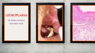
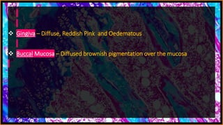
DR. ISHITA SINGHAL
MDS FIRST YEAR
PERSONAL
DATA
CASE NO. 2
OPD NO. 559656
NAME PREM SINGH
AGE 73 YEARS
SEX MALE
ADDRESS HARYANA
RELIGION HINDU
CHIEF
COMPLAINT
Patient complained of pain in lower left back
tooth region since 2-3 months.
Patient also complained of sensitivity to hot
and cold food since 3 months.
ONSET
Since 3 months.
ASSOCIATED
SYMPTOMS
 Pain and sensitivity in the associated area
 Burning sensation was present
 Past Medical History – No relevant history
 Family History – No relevant family history
 Past Dental History – Prosthesis: 31, 32, 41 and 42
 General Physical Gait – Normal Built, Co-operative with Normal Gait
PERSONAL
HISTORY
 Married/ Unmarried –Married
HABITS
 Patient cleaned his teeth with toothpaste
once daily for 2 minutes
 He smoked 5 bidis daily since 50 years
 He had hookah occasionally
EXTRA ORAL
EXAMINATION
 Patient had Wheatish complexion
 Lips were Pinkish Brown in Colour
 TMJ Movements – Normal
 Lymph Nodes – Non-palpable
INTRA ORAL
EXAMINATION
 Number Of Teeth Present - 28
 Missing Teeth – 4
 Supernumerary Teeth – NIL
 Mobility – NIL
 Caries – NIL
 Cervical Abrasion – 34 and 35
 Erosion – 11 and 21
 Plaque & Calculus – Generalised Moderate
Stains and Calculus Present
 Generalised Attrition
 Generalised Gingival Recession
 Gingiva – Diffuse, Reddish Pink and Oedematous
 Buccal Mucosa – Diffused brownish pigmentation over the mucosa
PATCH
 Number – 1
 Location And Extent – On the left retro-
commissural area of lip
 Non-Scrapable
 Non-Tender
 Colour – Mixed: Reddish-white
 Size – 2 cm x 1 cm
 Shape – Irregular and slightly elevated
Contoso
S u i t e s
SINGLE REDDISH WHITE PATCH
IRREGULAR SHAPE AND SLIGHTLY ELEVATED
LEFT RETRO COMMISSURAL AREA OF THE LIP
PROVISIONAL
DIAGNOSIS
On the basis of clinical presentation of the
patch, the provisional diagnosis of Speckled
Leukoplakia i.r.t. Left Retro-Commissural area
of lip was made
DIFFERENTIAL
DIAGNOSIS
1. Leukoplakia
2. Chronic Hyperplastic Candidiasis
3. Frictional Keratosis
INVESTIGATIO
N
 HEMOGRAM –
1. HB - 12.8 gm%
2. BT - 1 minute 50 seconds
3. CT - 5 minutes 45 seconds
4. RBS - 108 mg/dl
BIOPSY
Punch biopsy was done in the Department of
Oral and Maxillofacial Surgery and the tissue
was sent to the Department of Oral Pathology
for histopathological examination
MACROSCOPIC
DETAILS
 Received 1 small soft tissue specimens
measuring
 1.5 cm x 1 cm
 Firm in consistency
 Yellowish-white in colour
MICROSCOPIC
EXAMINATION
The H & E stained tissue section revealed
stratified squamous epithelium exhibiting
basal cell hyperplasia, loss of basal polarity,
acanthosis. Underlying connective tissue
shows inflammatory cells, collagen fibers and
blood vessels. In one area, dense inflammatory
cells are masking the epithelial connective
tissue junction.
Contoso
S u i t e s
OVERLYING EPITHELIUM
UNDERLYING CONNECTIVE TISSUE
Contoso
S u i t e s
PARAKERATINIZED STRATIFIED
SQUAMOUS EPITHELIUM
INFLAMMATORY CELLS, COLLAGEN
BUNDLES AND BLOOD VESSELS
ACANTOSIS
BASAL CELL HYPERPLASIA
BROAD RETE RIDGES
FINAL
DIAGNOSIS
The Histopathological features were suggestive
of Mild Dysplasia
DEFINITION
Leukoplakia can be defined as a “white patch” or “plaque” in the oral
cavity, which cannot be scrapped off and which cannot be
characterized clinically or pathologically as any other disease.
This excludes lesions such as lichen planus, candidiasis, leukoedema,
white spongy nevus, and obvious frictional keratosis.
The term leukoplakia should be used to recognise white plaques of
questionable risk having excluded (other) known diseases or
disorders that carry no risk for cancer-WHO 2005.
Carrard VC, van der Waal I. A clinical diagnosis of oral leukoplakia; A guide for dentists. Med Oral Patol Oral Cir Bucal. 2018;23(1):e59–e64.
The clinical color results from a thickened surface keratin layer, which
appears white when wet, or a thickened spinous layer, which masks
the normal vascularity (redness) of the underlying connective tissue.
NEVILLE’S TEXTBOOK OF ORAL AND MAXILLOFACIAL PATHOLOGY 3RD EDITION
ETIOLOGY
ETIOLOGICAL FACTORS OF LEUKOPLAKIA
Tobacco Candidiasis
Alcohol Viral infections
Ultraviolet radiation Chronic irritation
Sanguinaria
Odell, E. W., and R. A. Cawson. Cawson's Essentials of Oral Pathology and Oral Medicine. 2017.
Leukoplakia is idiopathic – no cause is known because the patch
cannot be characterised as any other definable lesion.
Tobacco:
During smoking a significantly large amount of tobacco end products
are produced in the oral cavity, these products in association with the
heat, cause severe irritation to the oral mucous membrane and can
cause leukoplakia.
There are higher rate of occurrence of leukoplakia among the “reverse
smokers”.
The risk of development of leukoplakia in a person depends upon the
frequency and duration of the tobacco habits, and the age and sex of
the person concerned.
The leukoplakic lesions regress when the tobacco habits are
discontinued or reduced.
SHAFER’S TEXTBOOK OF ORAL PATHOLOGY 7TH EDITION
Alcohol:
Alcohol, seems to have a strong synergistic effect with tobacco relative
to oral cancer production, but has not been associated with
leukoplakia.
People who excessively use mouth rinses with an alcohol content
greater than 25% may have greyish buccal mucosal plaques, but these
are not considered true leukoplakia.
Ultraviolet radiation:
It is accepted as a causative factor for leukoplakia of the lower lip
vermilion. This is usually associated with actinic cheilosis.
Immunocompromised persons, especially transplant patients, are
especially prone to the development of leukoplakia and squamous cell
carcinoma of the lower lip vermilion.
NEVILLE’S TEXTBOOK OF ORAL AND MAXILLOFACIAL PATHOLOGY 3RD EDITION
Sanguinaria:
Persons who use toothpaste or mouth rinses containing the herbal
extract, sanguinaria, may develop a true leukoplakia.
It is usually located in the maxillary vestibule or on the alveolar
mucosa of the maxilla.
The affected epithelium may demonstrate dysplasia identical to that
seen in other leukoplakias, although the potential for the
development of cancer is uncertain.
NEVILLE’S TEXTBOOK OF ORAL AND MAXILLOFACIAL PATHOLOGY 3RD EDITION
Candidiasis:
Chronic Candidal infections are often associated with leukoplakia,
however, it is not very clear whether the fungi are directly responsible
for the initiation of the disease or they are only producing secondary
infections in a pre-existing leukoplakia.
It has been observed that the Candida associated leukoplakias develop
more epithelial dysplasia than the non-candidal lesions.
Viral Infections:
Experimental studies indicate that, oral mucosal infections caused by
HPV may have some role in the development of leukoplakia.
SHAFER’S TEXTBOOK OF ORAL PATHOLOGY 7TH EDITION
NEVILLE’S TEXTBOOK OF ORAL AND MAXILLOFACIAL PATHOLOGY 3RD EDITION
Chronic Irritation:
Several keratotic lesions, which until recently had been viewed as
variants of leukoplakia, are now considered not to be precancers.
Nicotine stomatitis is a generalized white palatal alteration that seems
to be a hyperkeratotic response to the heat generated by tobacco
smoking, rather than a response to the carcinogens within the smoke.
Its malignant transformation potential is low.
Chronic mechanical irritation can produce a white lesion with a
roughened keratotic surface, termed frictional keratosis. Although the
resulting lesion is clinically similar to true leukoplakia, such a lesion is
now thought to be no more than a normal hyperplastic response.
NEVILLE’S TEXTBOOK OF ORAL AND MAXILLOFACIAL PATHOLOGY 3RD EDITION
Keratoses of this type are readily reversible after elimination of the
trauma, and obviously traumatic lesions such as linea alba,
morsicatio, and toothbrush gingival “abrasion” have not been
documented to have transformed into malignancy.
In addition, the presence of dentures or broken and missing teeth has
not been shown to increase the cancer risk.
Alveolar ridge keratoses—involving the retromolar pad or crest of an
edentulous alveolar ridge—represent another form of frictional
keratosis caused by masticatory function or denture trauma.
Frictional keratosis should be differentiated from the group of oral
precancers.
NEVILLE’S TEXTBOOK OF ORAL AND MAXILLOFACIAL PATHOLOGY 3RD EDITION
Regezi, Joseph A, James J. Sciubba, and Richard C. K. Jordan. Oral Pathology: Clinical Pathologic Correlations.
CLINICAL FEATURES
Regezi, Joseph A, James J. Sciubba, and Richard C. K. Jordan. Oral Pathology: Clinical Pathologic Correlations.
CLINICAL PRESENTATION
Early and mild lesions appear as slightly elevated grey or grey-white
plaques, which may appear somewhat translucent, fissured, or
wrinkled and are typically soft and flat. They usually have sharply
demarcated borders but occasionally blend gradually into normal
mucosa. Mild or thin leukoplakia, which seldom shows dysplasia on
biopsy, may disappear or continue unchanged.
NEVILLE’S TEXTBOOK OF ORAL AND MAXILLOFACIAL PATHOLOGY 3RD EDITION
For tobacco smokers who do not reduce their habit, as many as two
thirds of such lesions slowly extend laterally, become thicker, and
acquire a distinctly white appearance. The affected mucosa may
become leathery to palpation, and fissures may deepen and become
more numerous. The lesion is often called a homogeneous or thick
leukoplakia.
Most thick, smooth lesions remain indefinitely at this stage. Some,
perhaps as many as one third, regress or disappear; a few become even
more severe, develop increased surface irregularities, and are then
called granular or nodular leukoplakia.
NEVILLE’S TEXTBOOK OF ORAL AND MAXILLOFACIAL PATHOLOGY 3RD EDITION
Some lesions demonstrate sharp or blunt projections and have been
called verrucous leukoplakia.
NEVILLE’S TEXTBOOK OF ORAL AND MAXILLOFACIAL PATHOLOGY 3RD EDITION
A special high-risk form of leukoplakia, proliferative verrucous
leukoplakia (PVL), is characterized by the development of multiple
keratotic plaques with roughened surface projections.
The multiple PVL plaques tend to slowly spread and involve
additional oral mucosal sites.
NEVILLE’S TEXTBOOK OF ORAL AND MAXILLOFACIAL PATHOLOGY 3RD EDITION
The gingiva is frequently involved, although other sites may be
affected as well.
The lesions typically begin as simple, flat hyperkeratosis that are
indistinguishable from ordinary leukoplakic lesions, PVL exhibits
persistent growth, eventually becoming exophytic and verrucous in
nature.
As the lesions progress, they may go through a stage indistinguishable
from verrucous carcinoma, but they later usually develop dysplastic
changes and transform into full-fledged squamous cell carcinoma.
These lesions rarely regress despite therapy.
PVL is unusual among the leukoplakia variants in having a strong
female predilection (1:4 male-to-female ratio) and minimal
association with tobacco use.
NEVILLE’S TEXTBOOK OF ORAL AND MAXILLOFACIAL PATHOLOGY 3RD EDITION
Leukoplakia may become dysplastic, even invasive, with no change in
its clinical appearance.
However, some lesions eventually demonstrate scattered patches of
redness, called erythroplakia.
Such areas usually represent sites in which epithelial cells are so
immature or atrophic that they can no longer produce keratin.
This intermixed red-and-white lesion, called erythroleukoplakia or
speckled leukoplakia, represents a pattern of leukoplakia that
frequently reveals advanced dysplasia on biopsy.
NEVILLE’S TEXTBOOK OF ORAL AND MAXILLOFACIAL PATHOLOGY 3RD EDITION
DIFFERENTIAL DIAGNOSIS
The first step in developing a differential
diagnosis for a white patch on the oral mucosa
is to determine whether the lesion can be
removed with a gauze square or a tongue blade.
If the lesion can be removed, it may represent a
pseudomembrane, a fungus colony, or debris.
If chronic trauma or tobacco use is elicited in the patient’s
history, frictional or tobacco-associated hyperkeratosis,
respectively, should be considered.
If the lesion in question is not removable and is not clinically
diagnostic, it should be considered an idiopathic leukoplakia and a
biopsy should be performed.
For extensive lesions, multiple biopsies may be necessary to avoid
sample error. The clinically most suspicious areas (red, ulcerated, or
indurated areas) should be included in the area to be biopsied.
Regezi, Joseph A, James J. Sciubba, and Richard C. K. Jordan. Oral Pathology: Clinical Pathologic Correlations.
Mortazavi H, Safi Y, Baharvand M, Jafari S, Anbari F, Rahmani S. Oral White Lesions: An Updated Clinical Diagnostic Decision Tree. Dent J (Basel).
2019;7(1):15. Published 2019 Feb 7.
Odell, E. W., and R. A. Cawson. Cawson's Essentials of Oral Pathology and Oral Medicine. 2017.
HISTOLOGICFEATURES
Histologic changes range from hyperkeratosis, dysplasia, and
carcinoma in situ to invasive squamous cell carcinoma.
Regezi, Joseph A, James J. Sciubba, and Richard C. K. Jordan. Oral Pathology: Clinical Pathologic Correlations.
Under microscope leukoplakia generally presents hyper-
orthokeratinization or hyper-parakeratinization or both, with or
without the presence of epithelial dysplasia.
Normal epithelium shows a variety of histologic changes in terms of
the following factors:
1. Changes in keratinization pattern
2. Changes in the cellular layers
3. Thickness of the epithelium
4. Alterations in the underlying connective tissue stroma
1. Changes in keratinization pattern:
Hyper-keratinization of the epithelium refers to formation of
keratinized layers in the epithelium which are normally
nonkeratinized or abnormal increase in the thickness of the existing
keratin layer in the epithelium, which are normally keratinized.
Hyper-orthokeratinization-
 Orthokeratin is a homogenous layer of keratin present on the
superficial part of the epithelium, which does not contain any nuclear
remnants.
 In case of leukoplakia, an abnormal increase in the thickness of
orthokeratin layer is seen in the areas of epithelium which are usually
keratinized.
 Besides this, some degree of orthokeratinization may also be seen in
the areas of epithelium, which are usually nonkeratinized.
Hyper-parakeratinization-
 When there is an increase in the thickness of parakeratin layer
(keratin that contains some nuclear remnants) on the epithelium, it is
called hyper-parakeratinization.
 An important histologic criteria of leukoplakia is the presence of
hyper-parakeratinization of the normally keratinized epithelium, or
some amount of parakeratin deposition in the areas of epithelium
which are usually non-keratinized.
 Epithelial dysplasia is more frequently associated with hyper-
parakeratinized lesions.
 In few lesions of leukoplakia, both hyper-orthokeratinization and
hyper-parakeratinization may be seen.
NEVILLE’S TEXTBOOK OF ORAL AND MAXILLOFACIAL PATHOLOGY 3RD EDITION
2. Changes in cellular level:
When the precancerous changes in a lesion develop only at the
cellular level, it is known as ‘cellular atypia’.
At this situation, the overall alterations in the tissue in the direction of
precancerous changes are not fully expressed.
When the precancerous changes in a lesion worsen further and the
changes (both physical and morphological) begin to express
themselves in the overall tissue levels, it is called ‘epithelial dysplasia’.
Dysplasia (dys: abnormal and plasia: formation) –
 Epithelial dysplasia is the hallmark in the histological changes seen in
the epithelium, in case of leukoplakia and its presence is an important
indicator of the precancerous nature of the disease.
Odell, E. W., and R. A. Cawson. Cawson's Essentials of Oral Pathology and Oral Medicine. 2017.
Types of Dysplasia–
 Depending upon the degree and the extent to which the dysplastic
changes have developed in a lesion of leukoplakia; epithelial dysplasia
may be divided into three categories namely the:
1. Mild epithelial dysplasia
2. Moderate epithelial dysplasia
3. Severe epithelial dysplasia
 The degree of dysplasia is of immense value in predicting the
malignant potential of a precancerous lesion.
 However, the dysplastic changes in a lesion are reversible and if the
predisposing factors are removed, the dysplastic cells can turn back
towards normal.
NEVILLE’S TEXTBOOK OF ORAL AND MAXILLOFACIAL PATHOLOGY 3RD EDITION
When the basal cells show slight
disorganisation, there is increased
proliferation with several layers of
basaloid cells with large nuclei, and
among them are scattered cells with
very abnormal shape or size.
It has more layers of basaloid cells and
usually increased intercellular spaces
as a result of loss of cohesion. There is
a disorganised higgledy-piggledy
appearance in the basal layers. Mitotic
figures and very abnormal cells are
seen in the basal cells but in the
middle of the epithelium where basal
cells proliferate upward at the expense
of prickle cells.
It contains cells with the most marked
abnormalities and loss of the normal
layered structure of the epithelium.
Most of the thickness is occupied by
basal type cells. There are few or no
prickle cells or organised keratin layer,
but individual cells may keratinise at
any level. Loss of cohesion is usually
marked.
Odell, E. W., and R. A. Cawson. Cawson's Essentials of Oral Pathology and Oral Medicine. 2017.
NEVILLE’S TEXTBOOK OF ORAL AND MAXILLOFACIAL PATHOLOGY 3RD EDITION
3. Thickness of the epithelium:
 In leukoplakia, the thickness of the epithelium is often altered and it
occurs in the form of epithelial atrophy or epithelial hyperplasia or
acanthosis.
 Acanthosis: An abnormal increase in the thickness of stratum
spinosum of the epithelium is called acanthosis. In case of
leukoplakia, acanthosis is commonly observed in multiple areas of the
epithelium, which often causes elongation, thickening and blunting
of the rete pegs.
4. Alterations in the underlying connective tissue stroma:
 In leukoplakia, there are often variable degrees of destruction of the
collagen fibers and moreover chronic inflammatory cell infiltration is
also present in the underlying connective tissue stroma.
TREATMENT
Stoppage of all oral habits
Surgical excision /Electrocautery/Cryosurgery/Laser surgery
Long-term follow-up after removal is extremely important because
recurrences are frequent and additional leukoplakias may develop.
Leukoplakia not exhibiting dysplasia often is not excised, but clinical
evaluation every 6 months is recommended because of the possibility
of progression toward epithelial dysplasia. Additional biopsies are
recommended if smoking continues or if the clinical changes increase
in severity.
Although dysplasia may be present in any leukoplakia, each clinical
appearance or phase of leukoplakia has a different malignant
transformation potential.
NEVILLE’S TEXTBOOK OF ORAL AND MAXILLOFACIAL PATHOLOGY 3RD EDITION
1. Thin Leukoplakia seldom becomes malignant without
demonstrating a clinical change.
2. Homogeneous, Thick Leukoplakia - 1% to 7%
3. Granular or Verruciform Leukoplakia - 4% to 15%
4. Erythroleukoplakia - 18% to 47%
5. Moderate Dysplasia - 4% to 11%
6. Severe Dysplasia - 20% to 35%
7. Cancers from dysplastic lesions usually develop within 3 years of the
dysplasia diagnosis, but they can occur much later. Additionally, one
in three dysplasia will recur after complete removal.
8. Floor of the Mouth and Ventral Surface of the Tongue- 16% to 39%
9. Leukoplakia in Females - 47%
NEVILLE’S TEXTBOOK OF ORAL AND MAXILLOFACIAL PATHOLOGY 3RD EDITION
Isotretinoin, a form of vitamin A—alone or in combination with beta-
carotene—has been reported to reduce or eliminate some leukoplakic
lesions in short-term studies.
Agents such as bleomycin, lycopene, and COX-2 inhibitors have been
investigated as potential chemo preventive agents as well.
However, to date there is insufficient evidence from well-designed
clinical trials to support the effectiveness of such medical therapies in
treating oral dysplasia or preventing the progression of oral dysplasia
to squamous cell carcinoma.
NEVILLE’S TEXTBOOK OF ORAL AND MAXILLOFACIAL PATHOLOGY 3RD EDITION
Regezi, Joseph A, James J. Sciubba, and Richard C. K. Jordan. Oral Pathology: Clinical Pathologic Correlations.
REFERENCES
 SHAFER’S TEXTBOOK OF ORAL PATHOLOGY 7TH EDITION
 NEVILLE’S TEXTBOOK OF ORAL AND MAXILLOFACIAL PATHOLOGY 3RD
EDITION
 Regezi, Joseph A, James J. Sciubba, and Richard C. K. Jordan. Oral Pathology:
Clinical Pathologic Correlations.
 Odell, E. W., and R. A. Cawson. Cawson's Essentials of Oral Pathology and Oral
Medicine. 2017.
 Carrard VC, van der Waal I. A clinical diagnosis of oral leukoplakia; A guide for
dentists. Med Oral Patol Oral Cir Bucal. 2018;23(1):e59–e64.
 Mortazavi H, Safi Y, Baharvand M, Jafari S, Anbari F, Rahmani S. Oral White
Lesions: An Updated Clinical Diagnostic Decision Tree. Dent J (Basel).
2019;7(1):15. Published 2019 Feb 7.
Leukoplakia case presentation

Leukoplakia case presentation

  • 1.
  • 2.
    PERSONAL DATA CASE NO. 2 OPDNO. 559656 NAME PREM SINGH AGE 73 YEARS SEX MALE ADDRESS HARYANA RELIGION HINDU
  • 3.
    CHIEF COMPLAINT Patient complained ofpain in lower left back tooth region since 2-3 months. Patient also complained of sensitivity to hot and cold food since 3 months.
  • 4.
    ONSET Since 3 months. ASSOCIATED SYMPTOMS Pain and sensitivity in the associated area  Burning sensation was present
  • 5.
     Past MedicalHistory – No relevant history  Family History – No relevant family history  Past Dental History – Prosthesis: 31, 32, 41 and 42  General Physical Gait – Normal Built, Co-operative with Normal Gait
  • 6.
    PERSONAL HISTORY  Married/ Unmarried–Married HABITS  Patient cleaned his teeth with toothpaste once daily for 2 minutes  He smoked 5 bidis daily since 50 years  He had hookah occasionally
  • 7.
    EXTRA ORAL EXAMINATION  Patienthad Wheatish complexion  Lips were Pinkish Brown in Colour  TMJ Movements – Normal  Lymph Nodes – Non-palpable
  • 8.
    INTRA ORAL EXAMINATION  NumberOf Teeth Present - 28  Missing Teeth – 4  Supernumerary Teeth – NIL  Mobility – NIL  Caries – NIL  Cervical Abrasion – 34 and 35  Erosion – 11 and 21  Plaque & Calculus – Generalised Moderate Stains and Calculus Present  Generalised Attrition  Generalised Gingival Recession
  • 9.
     Gingiva –Diffuse, Reddish Pink and Oedematous  Buccal Mucosa – Diffused brownish pigmentation over the mucosa
  • 10.
    PATCH  Number –1  Location And Extent – On the left retro- commissural area of lip  Non-Scrapable  Non-Tender  Colour – Mixed: Reddish-white  Size – 2 cm x 1 cm  Shape – Irregular and slightly elevated
  • 11.
    Contoso S u it e s SINGLE REDDISH WHITE PATCH IRREGULAR SHAPE AND SLIGHTLY ELEVATED LEFT RETRO COMMISSURAL AREA OF THE LIP
  • 12.
    PROVISIONAL DIAGNOSIS On the basisof clinical presentation of the patch, the provisional diagnosis of Speckled Leukoplakia i.r.t. Left Retro-Commissural area of lip was made
  • 13.
    DIFFERENTIAL DIAGNOSIS 1. Leukoplakia 2. ChronicHyperplastic Candidiasis 3. Frictional Keratosis
  • 14.
    INVESTIGATIO N  HEMOGRAM – 1.HB - 12.8 gm% 2. BT - 1 minute 50 seconds 3. CT - 5 minutes 45 seconds 4. RBS - 108 mg/dl
  • 15.
    BIOPSY Punch biopsy wasdone in the Department of Oral and Maxillofacial Surgery and the tissue was sent to the Department of Oral Pathology for histopathological examination
  • 16.
    MACROSCOPIC DETAILS  Received 1small soft tissue specimens measuring  1.5 cm x 1 cm  Firm in consistency  Yellowish-white in colour
  • 17.
    MICROSCOPIC EXAMINATION The H &E stained tissue section revealed stratified squamous epithelium exhibiting basal cell hyperplasia, loss of basal polarity, acanthosis. Underlying connective tissue shows inflammatory cells, collagen fibers and blood vessels. In one area, dense inflammatory cells are masking the epithelial connective tissue junction.
  • 18.
    Contoso S u it e s OVERLYING EPITHELIUM UNDERLYING CONNECTIVE TISSUE
  • 19.
    Contoso S u it e s PARAKERATINIZED STRATIFIED SQUAMOUS EPITHELIUM INFLAMMATORY CELLS, COLLAGEN BUNDLES AND BLOOD VESSELS ACANTOSIS BASAL CELL HYPERPLASIA BROAD RETE RIDGES
  • 20.
    FINAL DIAGNOSIS The Histopathological featureswere suggestive of Mild Dysplasia
  • 22.
    DEFINITION Leukoplakia can bedefined as a “white patch” or “plaque” in the oral cavity, which cannot be scrapped off and which cannot be characterized clinically or pathologically as any other disease. This excludes lesions such as lichen planus, candidiasis, leukoedema, white spongy nevus, and obvious frictional keratosis. The term leukoplakia should be used to recognise white plaques of questionable risk having excluded (other) known diseases or disorders that carry no risk for cancer-WHO 2005. Carrard VC, van der Waal I. A clinical diagnosis of oral leukoplakia; A guide for dentists. Med Oral Patol Oral Cir Bucal. 2018;23(1):e59–e64.
  • 23.
    The clinical colorresults from a thickened surface keratin layer, which appears white when wet, or a thickened spinous layer, which masks the normal vascularity (redness) of the underlying connective tissue. NEVILLE’S TEXTBOOK OF ORAL AND MAXILLOFACIAL PATHOLOGY 3RD EDITION
  • 24.
    ETIOLOGY ETIOLOGICAL FACTORS OFLEUKOPLAKIA Tobacco Candidiasis Alcohol Viral infections Ultraviolet radiation Chronic irritation Sanguinaria Odell, E. W., and R. A. Cawson. Cawson's Essentials of Oral Pathology and Oral Medicine. 2017. Leukoplakia is idiopathic – no cause is known because the patch cannot be characterised as any other definable lesion.
  • 25.
    Tobacco: During smoking asignificantly large amount of tobacco end products are produced in the oral cavity, these products in association with the heat, cause severe irritation to the oral mucous membrane and can cause leukoplakia. There are higher rate of occurrence of leukoplakia among the “reverse smokers”. The risk of development of leukoplakia in a person depends upon the frequency and duration of the tobacco habits, and the age and sex of the person concerned. The leukoplakic lesions regress when the tobacco habits are discontinued or reduced. SHAFER’S TEXTBOOK OF ORAL PATHOLOGY 7TH EDITION
  • 26.
    Alcohol: Alcohol, seems tohave a strong synergistic effect with tobacco relative to oral cancer production, but has not been associated with leukoplakia. People who excessively use mouth rinses with an alcohol content greater than 25% may have greyish buccal mucosal plaques, but these are not considered true leukoplakia. Ultraviolet radiation: It is accepted as a causative factor for leukoplakia of the lower lip vermilion. This is usually associated with actinic cheilosis. Immunocompromised persons, especially transplant patients, are especially prone to the development of leukoplakia and squamous cell carcinoma of the lower lip vermilion. NEVILLE’S TEXTBOOK OF ORAL AND MAXILLOFACIAL PATHOLOGY 3RD EDITION
  • 27.
    Sanguinaria: Persons who usetoothpaste or mouth rinses containing the herbal extract, sanguinaria, may develop a true leukoplakia. It is usually located in the maxillary vestibule or on the alveolar mucosa of the maxilla. The affected epithelium may demonstrate dysplasia identical to that seen in other leukoplakias, although the potential for the development of cancer is uncertain. NEVILLE’S TEXTBOOK OF ORAL AND MAXILLOFACIAL PATHOLOGY 3RD EDITION
  • 28.
    Candidiasis: Chronic Candidal infectionsare often associated with leukoplakia, however, it is not very clear whether the fungi are directly responsible for the initiation of the disease or they are only producing secondary infections in a pre-existing leukoplakia. It has been observed that the Candida associated leukoplakias develop more epithelial dysplasia than the non-candidal lesions. Viral Infections: Experimental studies indicate that, oral mucosal infections caused by HPV may have some role in the development of leukoplakia. SHAFER’S TEXTBOOK OF ORAL PATHOLOGY 7TH EDITION
  • 29.
    NEVILLE’S TEXTBOOK OFORAL AND MAXILLOFACIAL PATHOLOGY 3RD EDITION
  • 30.
    Chronic Irritation: Several keratoticlesions, which until recently had been viewed as variants of leukoplakia, are now considered not to be precancers. Nicotine stomatitis is a generalized white palatal alteration that seems to be a hyperkeratotic response to the heat generated by tobacco smoking, rather than a response to the carcinogens within the smoke. Its malignant transformation potential is low. Chronic mechanical irritation can produce a white lesion with a roughened keratotic surface, termed frictional keratosis. Although the resulting lesion is clinically similar to true leukoplakia, such a lesion is now thought to be no more than a normal hyperplastic response. NEVILLE’S TEXTBOOK OF ORAL AND MAXILLOFACIAL PATHOLOGY 3RD EDITION
  • 31.
    NEVILLE’S TEXTBOOK OFORAL AND MAXILLOFACIAL PATHOLOGY 3RD EDITION
  • 32.
    Keratoses of thistype are readily reversible after elimination of the trauma, and obviously traumatic lesions such as linea alba, morsicatio, and toothbrush gingival “abrasion” have not been documented to have transformed into malignancy. In addition, the presence of dentures or broken and missing teeth has not been shown to increase the cancer risk. Alveolar ridge keratoses—involving the retromolar pad or crest of an edentulous alveolar ridge—represent another form of frictional keratosis caused by masticatory function or denture trauma. Frictional keratosis should be differentiated from the group of oral precancers. NEVILLE’S TEXTBOOK OF ORAL AND MAXILLOFACIAL PATHOLOGY 3RD EDITION
  • 33.
    Regezi, Joseph A,James J. Sciubba, and Richard C. K. Jordan. Oral Pathology: Clinical Pathologic Correlations. CLINICAL FEATURES
  • 34.
    Regezi, Joseph A,James J. Sciubba, and Richard C. K. Jordan. Oral Pathology: Clinical Pathologic Correlations.
  • 35.
    CLINICAL PRESENTATION Early andmild lesions appear as slightly elevated grey or grey-white plaques, which may appear somewhat translucent, fissured, or wrinkled and are typically soft and flat. They usually have sharply demarcated borders but occasionally blend gradually into normal mucosa. Mild or thin leukoplakia, which seldom shows dysplasia on biopsy, may disappear or continue unchanged. NEVILLE’S TEXTBOOK OF ORAL AND MAXILLOFACIAL PATHOLOGY 3RD EDITION
  • 36.
    For tobacco smokerswho do not reduce their habit, as many as two thirds of such lesions slowly extend laterally, become thicker, and acquire a distinctly white appearance. The affected mucosa may become leathery to palpation, and fissures may deepen and become more numerous. The lesion is often called a homogeneous or thick leukoplakia. Most thick, smooth lesions remain indefinitely at this stage. Some, perhaps as many as one third, regress or disappear; a few become even more severe, develop increased surface irregularities, and are then called granular or nodular leukoplakia. NEVILLE’S TEXTBOOK OF ORAL AND MAXILLOFACIAL PATHOLOGY 3RD EDITION
  • 37.
    NEVILLE’S TEXTBOOK OFORAL AND MAXILLOFACIAL PATHOLOGY 3RD EDITION
  • 38.
    Some lesions demonstratesharp or blunt projections and have been called verrucous leukoplakia. NEVILLE’S TEXTBOOK OF ORAL AND MAXILLOFACIAL PATHOLOGY 3RD EDITION
  • 39.
    A special high-riskform of leukoplakia, proliferative verrucous leukoplakia (PVL), is characterized by the development of multiple keratotic plaques with roughened surface projections. The multiple PVL plaques tend to slowly spread and involve additional oral mucosal sites. NEVILLE’S TEXTBOOK OF ORAL AND MAXILLOFACIAL PATHOLOGY 3RD EDITION
  • 40.
    The gingiva isfrequently involved, although other sites may be affected as well. The lesions typically begin as simple, flat hyperkeratosis that are indistinguishable from ordinary leukoplakic lesions, PVL exhibits persistent growth, eventually becoming exophytic and verrucous in nature. As the lesions progress, they may go through a stage indistinguishable from verrucous carcinoma, but they later usually develop dysplastic changes and transform into full-fledged squamous cell carcinoma. These lesions rarely regress despite therapy. PVL is unusual among the leukoplakia variants in having a strong female predilection (1:4 male-to-female ratio) and minimal association with tobacco use. NEVILLE’S TEXTBOOK OF ORAL AND MAXILLOFACIAL PATHOLOGY 3RD EDITION
  • 41.
    Leukoplakia may becomedysplastic, even invasive, with no change in its clinical appearance. However, some lesions eventually demonstrate scattered patches of redness, called erythroplakia. Such areas usually represent sites in which epithelial cells are so immature or atrophic that they can no longer produce keratin. This intermixed red-and-white lesion, called erythroleukoplakia or speckled leukoplakia, represents a pattern of leukoplakia that frequently reveals advanced dysplasia on biopsy. NEVILLE’S TEXTBOOK OF ORAL AND MAXILLOFACIAL PATHOLOGY 3RD EDITION
  • 42.
    NEVILLE’S TEXTBOOK OFORAL AND MAXILLOFACIAL PATHOLOGY 3RD EDITION
  • 43.
    DIFFERENTIAL DIAGNOSIS The firststep in developing a differential diagnosis for a white patch on the oral mucosa is to determine whether the lesion can be removed with a gauze square or a tongue blade. If the lesion can be removed, it may represent a pseudomembrane, a fungus colony, or debris.
  • 44.
    If chronic traumaor tobacco use is elicited in the patient’s history, frictional or tobacco-associated hyperkeratosis, respectively, should be considered.
  • 49.
    If the lesionin question is not removable and is not clinically diagnostic, it should be considered an idiopathic leukoplakia and a biopsy should be performed. For extensive lesions, multiple biopsies may be necessary to avoid sample error. The clinically most suspicious areas (red, ulcerated, or indurated areas) should be included in the area to be biopsied. Regezi, Joseph A, James J. Sciubba, and Richard C. K. Jordan. Oral Pathology: Clinical Pathologic Correlations.
  • 50.
    Mortazavi H, SafiY, Baharvand M, Jafari S, Anbari F, Rahmani S. Oral White Lesions: An Updated Clinical Diagnostic Decision Tree. Dent J (Basel). 2019;7(1):15. Published 2019 Feb 7.
  • 51.
    Odell, E. W.,and R. A. Cawson. Cawson's Essentials of Oral Pathology and Oral Medicine. 2017.
  • 52.
    HISTOLOGICFEATURES Histologic changes rangefrom hyperkeratosis, dysplasia, and carcinoma in situ to invasive squamous cell carcinoma. Regezi, Joseph A, James J. Sciubba, and Richard C. K. Jordan. Oral Pathology: Clinical Pathologic Correlations.
  • 53.
    Under microscope leukoplakiagenerally presents hyper- orthokeratinization or hyper-parakeratinization or both, with or without the presence of epithelial dysplasia. Normal epithelium shows a variety of histologic changes in terms of the following factors: 1. Changes in keratinization pattern 2. Changes in the cellular layers 3. Thickness of the epithelium 4. Alterations in the underlying connective tissue stroma
  • 54.
    1. Changes inkeratinization pattern: Hyper-keratinization of the epithelium refers to formation of keratinized layers in the epithelium which are normally nonkeratinized or abnormal increase in the thickness of the existing keratin layer in the epithelium, which are normally keratinized. Hyper-orthokeratinization-  Orthokeratin is a homogenous layer of keratin present on the superficial part of the epithelium, which does not contain any nuclear remnants.  In case of leukoplakia, an abnormal increase in the thickness of orthokeratin layer is seen in the areas of epithelium which are usually keratinized.  Besides this, some degree of orthokeratinization may also be seen in the areas of epithelium, which are usually nonkeratinized.
  • 55.
    Hyper-parakeratinization-  When thereis an increase in the thickness of parakeratin layer (keratin that contains some nuclear remnants) on the epithelium, it is called hyper-parakeratinization.  An important histologic criteria of leukoplakia is the presence of hyper-parakeratinization of the normally keratinized epithelium, or some amount of parakeratin deposition in the areas of epithelium which are usually non-keratinized.  Epithelial dysplasia is more frequently associated with hyper- parakeratinized lesions.  In few lesions of leukoplakia, both hyper-orthokeratinization and hyper-parakeratinization may be seen.
  • 56.
    NEVILLE’S TEXTBOOK OFORAL AND MAXILLOFACIAL PATHOLOGY 3RD EDITION
  • 57.
    2. Changes incellular level: When the precancerous changes in a lesion develop only at the cellular level, it is known as ‘cellular atypia’. At this situation, the overall alterations in the tissue in the direction of precancerous changes are not fully expressed. When the precancerous changes in a lesion worsen further and the changes (both physical and morphological) begin to express themselves in the overall tissue levels, it is called ‘epithelial dysplasia’. Dysplasia (dys: abnormal and plasia: formation) –  Epithelial dysplasia is the hallmark in the histological changes seen in the epithelium, in case of leukoplakia and its presence is an important indicator of the precancerous nature of the disease.
  • 58.
    Odell, E. W.,and R. A. Cawson. Cawson's Essentials of Oral Pathology and Oral Medicine. 2017.
  • 59.
    Types of Dysplasia– Depending upon the degree and the extent to which the dysplastic changes have developed in a lesion of leukoplakia; epithelial dysplasia may be divided into three categories namely the: 1. Mild epithelial dysplasia 2. Moderate epithelial dysplasia 3. Severe epithelial dysplasia  The degree of dysplasia is of immense value in predicting the malignant potential of a precancerous lesion.  However, the dysplastic changes in a lesion are reversible and if the predisposing factors are removed, the dysplastic cells can turn back towards normal.
  • 61.
    NEVILLE’S TEXTBOOK OFORAL AND MAXILLOFACIAL PATHOLOGY 3RD EDITION When the basal cells show slight disorganisation, there is increased proliferation with several layers of basaloid cells with large nuclei, and among them are scattered cells with very abnormal shape or size. It has more layers of basaloid cells and usually increased intercellular spaces as a result of loss of cohesion. There is a disorganised higgledy-piggledy appearance in the basal layers. Mitotic figures and very abnormal cells are seen in the basal cells but in the middle of the epithelium where basal cells proliferate upward at the expense of prickle cells. It contains cells with the most marked abnormalities and loss of the normal layered structure of the epithelium. Most of the thickness is occupied by basal type cells. There are few or no prickle cells or organised keratin layer, but individual cells may keratinise at any level. Loss of cohesion is usually marked. Odell, E. W., and R. A. Cawson. Cawson's Essentials of Oral Pathology and Oral Medicine. 2017.
  • 62.
    NEVILLE’S TEXTBOOK OFORAL AND MAXILLOFACIAL PATHOLOGY 3RD EDITION
  • 63.
    3. Thickness ofthe epithelium:  In leukoplakia, the thickness of the epithelium is often altered and it occurs in the form of epithelial atrophy or epithelial hyperplasia or acanthosis.  Acanthosis: An abnormal increase in the thickness of stratum spinosum of the epithelium is called acanthosis. In case of leukoplakia, acanthosis is commonly observed in multiple areas of the epithelium, which often causes elongation, thickening and blunting of the rete pegs. 4. Alterations in the underlying connective tissue stroma:  In leukoplakia, there are often variable degrees of destruction of the collagen fibers and moreover chronic inflammatory cell infiltration is also present in the underlying connective tissue stroma.
  • 64.
    TREATMENT Stoppage of alloral habits Surgical excision /Electrocautery/Cryosurgery/Laser surgery Long-term follow-up after removal is extremely important because recurrences are frequent and additional leukoplakias may develop. Leukoplakia not exhibiting dysplasia often is not excised, but clinical evaluation every 6 months is recommended because of the possibility of progression toward epithelial dysplasia. Additional biopsies are recommended if smoking continues or if the clinical changes increase in severity. Although dysplasia may be present in any leukoplakia, each clinical appearance or phase of leukoplakia has a different malignant transformation potential. NEVILLE’S TEXTBOOK OF ORAL AND MAXILLOFACIAL PATHOLOGY 3RD EDITION
  • 65.
    1. Thin Leukoplakiaseldom becomes malignant without demonstrating a clinical change. 2. Homogeneous, Thick Leukoplakia - 1% to 7% 3. Granular or Verruciform Leukoplakia - 4% to 15% 4. Erythroleukoplakia - 18% to 47% 5. Moderate Dysplasia - 4% to 11% 6. Severe Dysplasia - 20% to 35% 7. Cancers from dysplastic lesions usually develop within 3 years of the dysplasia diagnosis, but they can occur much later. Additionally, one in three dysplasia will recur after complete removal. 8. Floor of the Mouth and Ventral Surface of the Tongue- 16% to 39% 9. Leukoplakia in Females - 47% NEVILLE’S TEXTBOOK OF ORAL AND MAXILLOFACIAL PATHOLOGY 3RD EDITION
  • 66.
    Isotretinoin, a formof vitamin A—alone or in combination with beta- carotene—has been reported to reduce or eliminate some leukoplakic lesions in short-term studies. Agents such as bleomycin, lycopene, and COX-2 inhibitors have been investigated as potential chemo preventive agents as well. However, to date there is insufficient evidence from well-designed clinical trials to support the effectiveness of such medical therapies in treating oral dysplasia or preventing the progression of oral dysplasia to squamous cell carcinoma. NEVILLE’S TEXTBOOK OF ORAL AND MAXILLOFACIAL PATHOLOGY 3RD EDITION
  • 67.
    Regezi, Joseph A,James J. Sciubba, and Richard C. K. Jordan. Oral Pathology: Clinical Pathologic Correlations.
  • 69.
    REFERENCES  SHAFER’S TEXTBOOKOF ORAL PATHOLOGY 7TH EDITION  NEVILLE’S TEXTBOOK OF ORAL AND MAXILLOFACIAL PATHOLOGY 3RD EDITION  Regezi, Joseph A, James J. Sciubba, and Richard C. K. Jordan. Oral Pathology: Clinical Pathologic Correlations.  Odell, E. W., and R. A. Cawson. Cawson's Essentials of Oral Pathology and Oral Medicine. 2017.  Carrard VC, van der Waal I. A clinical diagnosis of oral leukoplakia; A guide for dentists. Med Oral Patol Oral Cir Bucal. 2018;23(1):e59–e64.  Mortazavi H, Safi Y, Baharvand M, Jafari S, Anbari F, Rahmani S. Oral White Lesions: An Updated Clinical Diagnostic Decision Tree. Dent J (Basel). 2019;7(1):15. Published 2019 Feb 7.