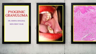
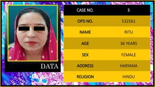
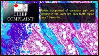
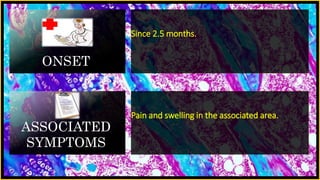
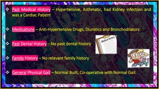
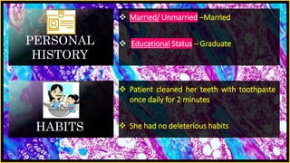
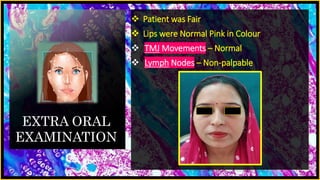
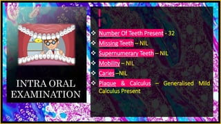
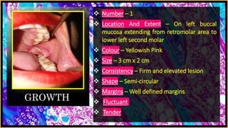
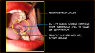
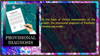
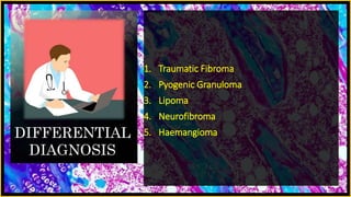
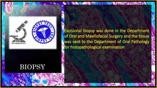
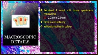
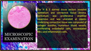
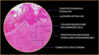
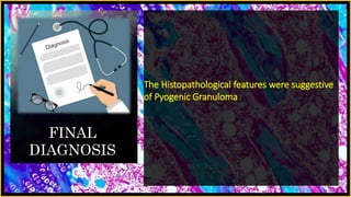
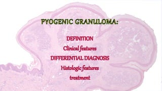
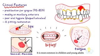
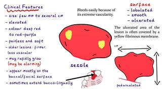
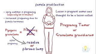
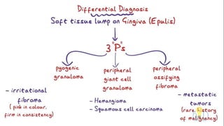
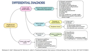
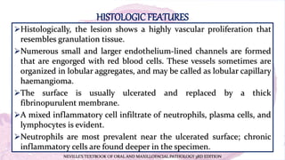
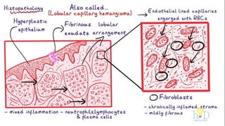
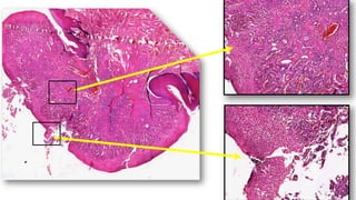
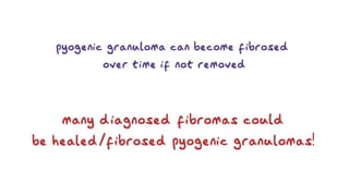
DR. ISHITA SINGHAL
MDS FIRST YEAR
PERSONAL
DATA
CASE NO. 3
OPD NO. 532561
NAME RITU
AGE 36 YEARS
SEX FEMALE
ADDRESS HARYANA
RELIGION HINDU
CHIEF
COMPLAINT
Patient complained of occasional pain and
swelling in the lower left back tooth region
since 2.5 months.
ONSET
Since 2.5 months.
ASSOCIATED
SYMPTOMS
Pain and swelling in the associated area.
 Past Medical History – Hypertensive, Asthmatic, had Kidney Infection and
was a Cardiac Patient
 Medications – Anti-Hypertensive Drugs, Diuretics and Bronchodilators
 Past Dental History – No past dental history
 Family History – No relevant family history
 General Physical Gait – Normal Built, Co-operative with Normal Gait
PERSONAL
HISTORY
 Married/ Unmarried –Married
 Educational Status – Graduate
HABITS
 Patient cleaned her teeth with toothpaste
once daily for 2 minutes
 She had no deleterious habits
EXTRA ORAL
EXAMINATION
 Patient was Fair
 Lips were Normal Pink in Colour
 TMJ Movements – Normal
 Lymph Nodes – Non-palpable
INTRA ORAL
EXAMINATION
 Number Of Teeth Present - 32
 Missing Teeth – NIL
 Supernumerary Teeth – NIL
 Mobility – NIL
 Caries –NIL
 Plaque & Calculus – Generalised Mild
Calculus Present
GROWTH
 Number – 1
 Location And Extent – On left buccal
mucosa extending from retromolar area to
lower left second molar
 Colour – Yellowish Pink
 Size – 3 cm x 2 cm
 Consistency – Firm and elevated lesion
 Shape – Semi-circular
 Margins – Well defined margins
 Fluctuant
 Tender
Contoso
S u i t e s
YELLOWISH PINK IN COLOUR
SEMI-CIRCULAR SHAPE WITH WELL
DEFINED MARGINS
ON LEFT BUCCAL MUCOSA EXTENDING
FROM RETROMOLAR AREA TO LOWER
LEFT SECOND MOLAR
PROVISIONAL
DIAGNOSIS
On the basis of Clinical presentation of the
growth, the provisional diagnosis of Traumatic
Fibroma was made
DIFFERENTIAL
DIAGNOSIS
1. Traumatic Fibroma
2. Pyogenic Granuloma
3. Lipoma
4. Neurofibroma
5. Haemangioma
INVESTIGATIO
N
 HEMOGRAM –
1. HB - 11.7 gm%
2. BT - 1 minute 40 seconds
3. CT - 6 minutes 15 seconds
4. RBS – 108 mg/dl
BIOPSY
Excisional biopsy was done in the Department
of Oral and Maxillofacial Surgery and the tissue
was sent to the Department of Oral Pathology
for histopathological examination
MACROSCOPIC
DETAILS
 Received 1 small soft tissue specimens
measuring
 2.2 cm x 2.0 cm
 Firm in consistency
 Yellowish-white in colour
MICROSCOPIC
EXAMINATION
The H & E stained tissue section revealed
epithelium and connective tissue stroma.
Epithelium was proliferative stratified
squamous and was ulcerated at places.
Underlying connective tissue was composed of
collagen bundles, numerous blood vessels,
proliferating endothelial cells, extravasated
RBCs and inflammatory cells.
Contoso
S u i t e s
OVERLYING EPITHELIUM
UNDERLYING CONNECTIVE TISSUE
WITH MINOR SALIVARY GLANDS
Contoso
S u i t e s
STRATIFIED SQUAMOUS
EPITHELIUM
CONNECTIVE TISSUE STROMA
COLLAGEN BUNDLES AND
INFLAMMATORY CELLS
ULCERATED EPITHELIUM
ENDOTHELIUM LINED BLOOD
VESSELS AND EXTRAVASATED RBCs
FINAL
DIAGNOSIS
The Histopathological features were suggestive
of Pyogenic Granuloma
DEFINITION
It is most common in children and young adults
The ulcerated area of the
lesion is often covered by a
yellow fibrinous membrane.
Bleeds easily because of
its extreme vascularity.
DIFFERENTIAL DIAGNOSIS
Mortazavi H, Safi Y, Baharvand M, Rahmani S, Jafari S. Peripheral Exophytic Oral Lesions: A Clinical Decision Tree. Int J Dent. 2017;2017:9193831.
HISTOLOGICFEATURES
Histologically, the lesion shows a highly vascular proliferation that
resembles granulation tissue.
Numerous small and larger endothelium-lined channels are formed
that are engorged with red blood cells. These vessels sometimes are
organized in lobular aggregates, and may be called as lobular capillary
haemangioma.
The surface is usually ulcerated and replaced by a thick
fibrinopurulent membrane.
A mixed inflammatory cell infiltrate of neutrophils, plasma cells, and
lymphocytes is evident.
Neutrophils are most prevalent near the ulcerated surface; chronic
inflammatory cells are found deeper in the specimen.
NEVILLE’S TEXTBOOK OF ORAL AND MAXILLOFACIAL PATHOLOGY 3RD EDITION
TREATMENT
Pyogenic granuloma is treated by surgical excision.
Excision and biopsy of the lesion is the recommended line of
treatment unless it would produce a marked deformity and in such a
case incisional biopsy is recommended. Conservative surgical excision
of the lesion with the removal of irritants such as plaque, calculus,
and foreign materials is recommended for small painless nonbleeding
lesions. Excision of the gingival lesions up to the periosteum with
through scaling and root planning of adjacent teeth to remove all
visible sources of irritation is recommended.
Newadkar UR, Khairnar S, Dodamani A. Pyogenic granuloma: A clinicopathological analysis of fifty cases. J Oral Res
Rev 2018;10:7-10
REFERENCES
SHAFER’S TEXTBOOK OF ORAL PATHOLOGY 7TH EDITION
NEVILLE’S TEXTBOOK OF ORAL AND MAXILLOFACIAL
PATHOLOGY 3RD EDITION
Newadkar UR, Khairnar S, Dodamani A. Pyogenic granuloma: A
clinicopathological analysis of fifty cases. J Oral Res Rev 2018;10:7-10
Mortazavi H, Safi Y, Baharvand M, Rahmani S, Jafari S. Peripheral
Exophytic Oral Lesions: A Clinical Decision Tree. Int J Dent.
2017;2017:9193831.
Pyogenic granuloma

Pyogenic granuloma

  • 1.
  • 2.
    PERSONAL DATA CASE NO. 3 OPDNO. 532561 NAME RITU AGE 36 YEARS SEX FEMALE ADDRESS HARYANA RELIGION HINDU
  • 3.
    CHIEF COMPLAINT Patient complained ofoccasional pain and swelling in the lower left back tooth region since 2.5 months.
  • 4.
    ONSET Since 2.5 months. ASSOCIATED SYMPTOMS Painand swelling in the associated area.
  • 5.
     Past MedicalHistory – Hypertensive, Asthmatic, had Kidney Infection and was a Cardiac Patient  Medications – Anti-Hypertensive Drugs, Diuretics and Bronchodilators  Past Dental History – No past dental history  Family History – No relevant family history  General Physical Gait – Normal Built, Co-operative with Normal Gait
  • 6.
    PERSONAL HISTORY  Married/ Unmarried–Married  Educational Status – Graduate HABITS  Patient cleaned her teeth with toothpaste once daily for 2 minutes  She had no deleterious habits
  • 7.
    EXTRA ORAL EXAMINATION  Patientwas Fair  Lips were Normal Pink in Colour  TMJ Movements – Normal  Lymph Nodes – Non-palpable
  • 8.
    INTRA ORAL EXAMINATION  NumberOf Teeth Present - 32  Missing Teeth – NIL  Supernumerary Teeth – NIL  Mobility – NIL  Caries –NIL  Plaque & Calculus – Generalised Mild Calculus Present
  • 9.
    GROWTH  Number –1  Location And Extent – On left buccal mucosa extending from retromolar area to lower left second molar  Colour – Yellowish Pink  Size – 3 cm x 2 cm  Consistency – Firm and elevated lesion  Shape – Semi-circular  Margins – Well defined margins  Fluctuant  Tender
  • 10.
    Contoso S u it e s YELLOWISH PINK IN COLOUR SEMI-CIRCULAR SHAPE WITH WELL DEFINED MARGINS ON LEFT BUCCAL MUCOSA EXTENDING FROM RETROMOLAR AREA TO LOWER LEFT SECOND MOLAR
  • 11.
    PROVISIONAL DIAGNOSIS On the basisof Clinical presentation of the growth, the provisional diagnosis of Traumatic Fibroma was made
  • 12.
    DIFFERENTIAL DIAGNOSIS 1. Traumatic Fibroma 2.Pyogenic Granuloma 3. Lipoma 4. Neurofibroma 5. Haemangioma
  • 13.
    INVESTIGATIO N  HEMOGRAM – 1.HB - 11.7 gm% 2. BT - 1 minute 40 seconds 3. CT - 6 minutes 15 seconds 4. RBS – 108 mg/dl
  • 14.
    BIOPSY Excisional biopsy wasdone in the Department of Oral and Maxillofacial Surgery and the tissue was sent to the Department of Oral Pathology for histopathological examination
  • 15.
    MACROSCOPIC DETAILS  Received 1small soft tissue specimens measuring  2.2 cm x 2.0 cm  Firm in consistency  Yellowish-white in colour
  • 16.
    MICROSCOPIC EXAMINATION The H &E stained tissue section revealed epithelium and connective tissue stroma. Epithelium was proliferative stratified squamous and was ulcerated at places. Underlying connective tissue was composed of collagen bundles, numerous blood vessels, proliferating endothelial cells, extravasated RBCs and inflammatory cells.
  • 17.
    Contoso S u it e s OVERLYING EPITHELIUM UNDERLYING CONNECTIVE TISSUE WITH MINOR SALIVARY GLANDS
  • 18.
    Contoso S u it e s STRATIFIED SQUAMOUS EPITHELIUM CONNECTIVE TISSUE STROMA COLLAGEN BUNDLES AND INFLAMMATORY CELLS ULCERATED EPITHELIUM ENDOTHELIUM LINED BLOOD VESSELS AND EXTRAVASATED RBCs
  • 19.
    FINAL DIAGNOSIS The Histopathological featureswere suggestive of Pyogenic Granuloma
  • 21.
  • 22.
    It is mostcommon in children and young adults
  • 23.
    The ulcerated areaof the lesion is often covered by a yellow fibrinous membrane. Bleeds easily because of its extreme vascularity.
  • 26.
    DIFFERENTIAL DIAGNOSIS Mortazavi H,Safi Y, Baharvand M, Rahmani S, Jafari S. Peripheral Exophytic Oral Lesions: A Clinical Decision Tree. Int J Dent. 2017;2017:9193831.
  • 27.
    HISTOLOGICFEATURES Histologically, the lesionshows a highly vascular proliferation that resembles granulation tissue. Numerous small and larger endothelium-lined channels are formed that are engorged with red blood cells. These vessels sometimes are organized in lobular aggregates, and may be called as lobular capillary haemangioma. The surface is usually ulcerated and replaced by a thick fibrinopurulent membrane. A mixed inflammatory cell infiltrate of neutrophils, plasma cells, and lymphocytes is evident. Neutrophils are most prevalent near the ulcerated surface; chronic inflammatory cells are found deeper in the specimen. NEVILLE’S TEXTBOOK OF ORAL AND MAXILLOFACIAL PATHOLOGY 3RD EDITION
  • 31.
    TREATMENT Pyogenic granuloma istreated by surgical excision. Excision and biopsy of the lesion is the recommended line of treatment unless it would produce a marked deformity and in such a case incisional biopsy is recommended. Conservative surgical excision of the lesion with the removal of irritants such as plaque, calculus, and foreign materials is recommended for small painless nonbleeding lesions. Excision of the gingival lesions up to the periosteum with through scaling and root planning of adjacent teeth to remove all visible sources of irritation is recommended. Newadkar UR, Khairnar S, Dodamani A. Pyogenic granuloma: A clinicopathological analysis of fifty cases. J Oral Res Rev 2018;10:7-10
  • 32.
    REFERENCES SHAFER’S TEXTBOOK OFORAL PATHOLOGY 7TH EDITION NEVILLE’S TEXTBOOK OF ORAL AND MAXILLOFACIAL PATHOLOGY 3RD EDITION Newadkar UR, Khairnar S, Dodamani A. Pyogenic granuloma: A clinicopathological analysis of fifty cases. J Oral Res Rev 2018;10:7-10 Mortazavi H, Safi Y, Baharvand M, Rahmani S, Jafari S. Peripheral Exophytic Oral Lesions: A Clinical Decision Tree. Int J Dent. 2017;2017:9193831.