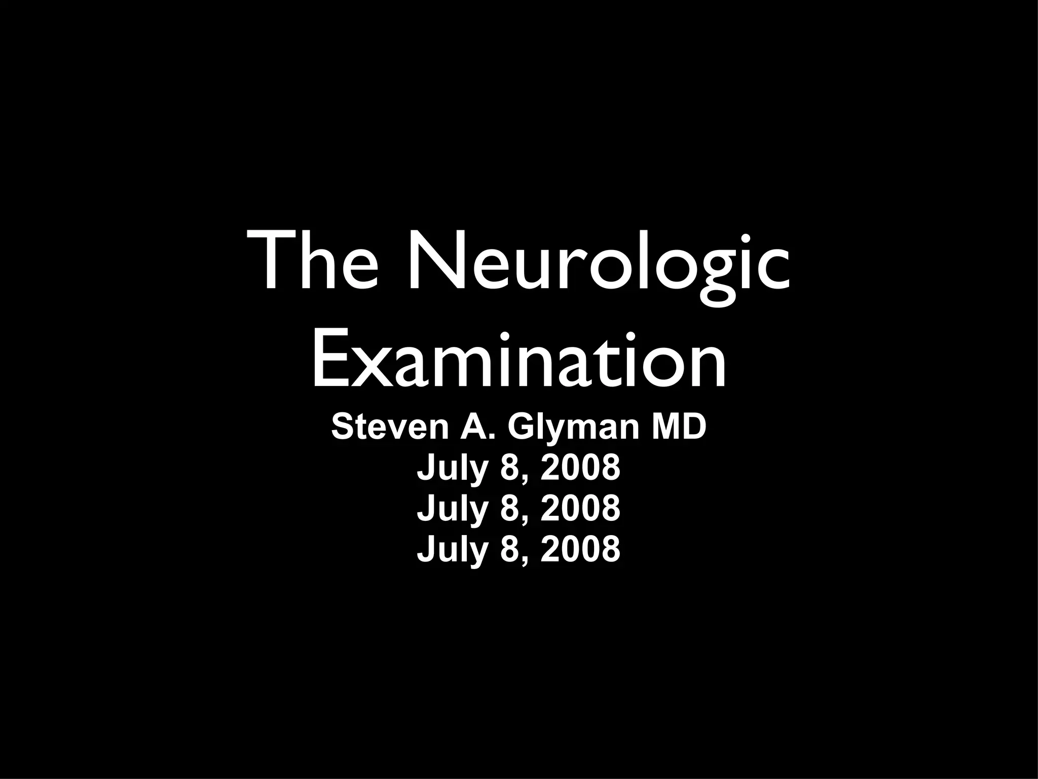
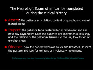
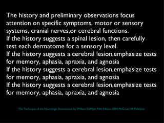
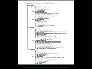
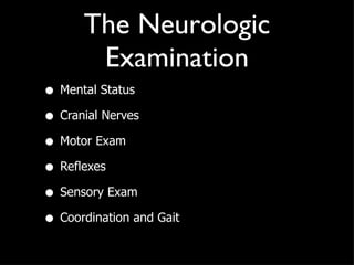
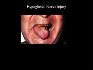
The Neurologic Examination Steven A. Glyman MD July 8, 2008 July 8, 2008 July 8, 2008
Resources Neuroanatomy through Clinical Cases by Hal Blumenfeld  http://www.neuroexam.com / http://www.utoronto.ca/neuronotes/NeuroExam/main.htm The Technique of the Neurologic Examination by William DeMyer DeJong’s Neurologic Examination by William W. Campbell
 
A proper neurologic examination begins with a careful clinical history.
http://www.cbu.edu/~mcondren/IRM/Stop-Look-Listen-sign-IRM-7-7-07.jpg
You must do a minimum basic examination on every patient but you don’t need to do every test
A mechanic does not need to use every tool on every project
The history guides the neurologic examination
"You can see a lot just by observing." Yogi Berra
The Neurologic Exam often can be completed during the clinical history Assess :  the patient’s articulation, content of speech, and overall mental status Inspect : the patient’s facial features,facial movement,and and note any asymmetry. Note the patient’s eye movements, blinking, and the relation of the palpebral fissures to the iris, look for en or exophthalmos.  Observe :  how the patient swallows saliva and breathes. Inspect the posture and look for tremors or involuntary movements The Technique of the Neurologic Examination by William DeMyer Fifth Edition 2004 McGraw Hill Publisher
The history and preliminary observations focus attention on specific symptoms, motor or sensory systems, cranial nerves,or cerebral functions.  If the history suggests a spinal lesion, then carefully test each dermatome for a sensory level.  If the history suggests a cerebral lesion,emphasize tests for memory, aphasia, apraxia, and agnosia If the history suggests a cerebral lesion,emphasize tests for memory, aphasia, apraxia, and agnosia If the history suggests a cerebral lesion,emphasize tests for memory, aphasia, apraxia, and agnosia The Technique of the Neurologic Examination by William DeMyer Fifth Edition 2004 McGraw Hill Publisher
The Technique of the Neurologic Examination by William DeMyer  McGraw Hill  2004
Tools of the trade
http://www.ptup.org/files/2007-SJ-Monterey-Tour/TheWholeEnchilada.jpg
The Neurologic Examination Mental Status Cranial Nerves Motor Exam Reflexes Sensory Exam Coordination and Gait
Mental Status Exam 1. Level of alertness,attention and cooperation 2. Orientation for time,place,and person 3. Speech and language 4. Memory for immediate,recent,and remote events 5. Calculations and right/left orientation,finger agnosia,and agraphia 6. Apraxia 7. Constructions and neglect 8. Logic and abstractions 9. Sequencing tasks and frontal release signs 10. Delusions and Hallucinations 11. Mood
Mental Status Exam  Folstein Mini-mental status exam This is a screening tool used to follow the cognitive decline associated with dementia. It has been in wide use since 1975 and takes 5-10 minutes to administer. It is a limited test instrument. This examination is not suitable for making a diagnosis but can be used to indicate the presence of cognitive impairment, such as when  dementia  or  head injury  are suspected. People from different cultural groups or low intelligence or education may score poorly on this examination in the absence of cognitive impairment and well educated people may score well despite having cognitive impairment
 
Mental Status Exam 1. Level of alertness,attention and cooperation We can test attention by seeing if the patient can remain focused on a simple task, such as spelling a short word forward and backward (W-O-R-L-D / D-L-R-O-W is a standard), repeating a string of integers forward and backward (digit span), or naming the months forward and then backward. Normal digit span is 6 or more forward, and 4 or more backward, depending slightly on age and education. Degree of cooperation should be noted, especially if it is abnormal, since this will influence many aspects of the exam. http://www.neuroexam.com /
Mental Status Exam 2. Orientation for time,place, and person Ask for the patient's full name, the location, and the date, and note the exact response.  http://www.neuroexam.com /
Mental Status Exam 3. Speech and language Spontaneous speech:  Note the patient's fluency, including phrase length, rate, and abundance of spontaneous speech. Also note tonal modulation and whether paraphasic errors (inappropriately substituted words or syllables), neologisms (nonexistent words), or errors in grammar are present. Comprehension:  Can the patient understand simple questions and commands? Comprehension of grammatical structure should be tested as well Naming:  Ask the patient to name some easy (pen, watch, tie, etc.) and some more difficult (fingernail, belt buckle, stethoscope, etc.) objects Repetition:  Can the patient repeat single words and sentences (a standard is "no ifs ands or buts")? Reading:  Ask the patient to read single words, a brief passage, and the front page of the newspaper aloud and test for comprehension.  Writing:  Ask the patient to write their name and write a sentence. http://www.neuroexam.com /
Mental Status Exam 4. Memory for recent and remote events Recent memory:  Ask the patient to recall three items or a brief story after a delay of 3 to 5 minutes. Be sure the information has been registered by asking the patient to repeat it immediately before initiating the delay. Provide distracters during the delay to prevent the patient from rehearsing the items repeatedly. Remote memory:  Ask the patient about historical or verifiable personal events. Memory can be impaired on many different timescales. Impaired ability to register and recall something within a few seconds after it was said is an abnormality that blends into the category of impaired attention. If immediate recall is intact, then difficulty with recall after about 1 to 5 minutes usually signifies damage to the limbic memory structures located in the medial temporal lobes and medial diencephalon http://www.neuroexam.com /
Mental Status Exam 5. Calculations and right/left orientation,finger agnosia,and agraphia Impairment of all four of these functions in an otherwise intact patient is referred to as  Gerstmann's syndrome . Since Gerstmann's syndrome is caused by lesions in the dominant parietal lobe, aphasia is often (but not always) present as well, which can make the diagnosis difficult or impossible. Each of the individual components of Gerstmann's syndrome is poorly localizing on its own, but they are worth documenting as part of the assessment of overall cognitive function http://www.neuroexam.com /
Mental Status Exam 6. Apraxia The term  apraxia will be used here to mean inability to follow a motor command that is not due to a primary motor deficit or a language impairment.  It is apparently caused by a deficit in higher-order planning or conceptualization of the motor task. You can test for apraxia by asking the patient to do complex tasks, using commands such as  "Pretend to comb your hair" or "Pretend to strike a match and blow it out"  and so on. Patients with apraxia perform awkward movements that only minimally resemble those requested, despite having intact comprehension and an otherwise normal motor exam. This kind of apraxia is sometimes called  ideomotor apraxia . In some patients, rather than affecting the distal extremities, apraxia can involve primarily the mouth and face, or movements of the whole body, such as walking or turning around. http://www.neuroexam.com /
Mental Status Exam 7. Constructions and neglect Hemineglect is an abnormality in attention to one side of the universe that is not due to a primary sensory or motor disturbance . In sensory neglect, patients ignore visual, somatosensory, or auditory stimuli on the affected side, despite intact primary sensation. This can often be demonstrated by testing for extinction on double simultaneous stimulation. Thus, patients may detect a stimulus on the affected side when presented alone, but when stimuli are presented simultaneously on both sides, only the stimulus on the unaffected side may be detected. In motor neglect, normal strength may be present, however, the patient often does not move the affected limb unless attention is strongly directed toward it. Sensory and motor neglect are usually tested as part of the visual, auditory, somatosensory, and motor exams. During the reading and writing portions of the language exam, patients may be noted to neglect one side of the page. http://www.neuroexam.com /
http://www.mrc-cbu.cam.ac.uk/research/rehabilitation/research.html
Mental Status Exam 8. Logic and abstractions Can the patients solve simple problems such as the following:  "If Mary is taller than Jane, and Jane is taller than Ann, who's the tallest?"   How do they interpret proverbs such as  "Don't cry over spilled milk?”   How well can they comprehend similarities such as  "How are a car and an airplane alike?”  Educational background must always be taken into account in interpretations of these tests. http://www.neuroexam.com /
Mental Status Exam 9. Sequencing tasks and frontal release signs Frontal lobe lesions in adults can cause the reemergence of certain primitive reflexes that are normally present in infants. These so-called frontal release signs include the grasp, snout, root, and suck reflexes. Of these reflexes, the  grasp reflex  is the most useful in evaluating frontal lobe dysfunction. Patients with frontal lobe dysfunction may have particular difficulty in changing from one action to the next when asked to perform a repeated sequence of actions.This phenomenon is called  perseveration http://www.neuroexam.com /
Perseveration
Mental Status Exam 10. Delusions and Hallucinations Does the patient have any delusional thought processes? Does he have auditory or visual hallucinations? Ask questions such as, "Do you ever hear things that other people don't hear or see things that other people don't see?" "Do you feel that someone is watching you or trying to hurt you?" "Do you have any special abilities or powers?" These abnormalities can be seen in toxic or metabolic abnormalities and other causes of diffuse brain dysfunction, and in primary psychiatric disorders. In addition, abnormal sensory phenomena can be caused by focal lesions or seizures in visual, somatosensory, or auditory cortex, and thought disorders can be caused by lesions in the association cortex and limbic system http://www.neuroexam.com /
Mental Status Exam 11. Mood Signs of major depression include depressed mood, changes in eating and sleeping patterns, loss of energy and initiative, low self-esteem, poor concentration, lack of enjoyment of previously pleasurable activities, and self-destructive or suicidal thoughts and behavior. Anxiety disorders are characterized by preoccupation with worrisome thoughts. Mania causes patients to be abnormally active and cognitively disorganized.  http://www.neuroexam.com /
The Cranial Nerves
Cranial Nerve1 Olfaction Olfaction Not tested much unless a frontal lobe tumor is suspected May be damaged in patients with closed head injuries,nasal obstruction,viral infections, and can be abnormal in Parkinsons disease, Alzheimer’s,and Multiple Sclerosis  Test by asking if patients can smell,coffee,vanilla, or cinnamon in each nostril.  Avoid noxious odors ( ie NH3) http://www.neuroexam.com /
http://commons.wikimedia.org/wiki/Image:Head_olfactory_nerve.jpg http://www.braininjury.com/images/cranialnerveinjury.jpg
Cranial Nerve 2 Optic Nerve Optic Nerve Visual Acuity (test with hand card) Color Vision (loss of color vision especially  red  is an important symptom of optic neuritis) Visual Fields (can be tested at the bedside by counting fingers in each quadrant) Visual Extinction (to detect visual neglect) Funduscopic Examination http://www.neuroexam.com /
http://www.merck.com/media/mmhe2/figures/MMHE_20_235_01_eps.gif http://arapaho.nsuok.edu/~fulk/Images/Img0013.JPG
Cranial Nerve 2 and 3 Pupillary responses Pupillary responses The  size and shape  of the pupil should be recorded at rest. Under normal conditions, the pupil constricts in response to light. Note the  direct response , meaning constriction of the illuminated pupil, as well as the  consensual response , meaning constriction of the opposite pupil. Test the pupillary response to  accommodation . Normally, the pupils constrict while fixating on an object being moved from far away to near the eyes. Direct response (pupil illuminated).  The direct response is impaired in lesions of the ipsilateral optic nerve, the pretectal area, the ipsilateral parasympathetics traveling in CN III, or the pupillary constrictor muscle of the iris. Consensual response (contralateral pupil illuminated).  The consensual response is impaired in lesions of the contralateral optic nerve, the pretectal area, the ipsilateral parasympathetics traveling in CN III, or the pupillary constrictor muscle. Accommodation (response to looking at something moving toward the eye).  Accommodation is impaired in lesions of the ipsilateral optic nerve, the ipsilateral parasympathetics traveling in CN III, or the pupillary constrictor muscle, or in bilateral lesions of the pathways from the optic tracts to the visual cortex . Accommodation is spared in lesions of the pretectal area. http://www.neuroexam.com /
Pupillary Size is determined by the light input, sympathetic and parasympathetic tone Text http://www.psych.ndsu.nodak.edu/mccourt/Psy460/Central%20visual%20pathways/pupillary%20light%20reflex.gif http://www.abdn.ac.uk/~opt065/Web%20pages%20only/drh-g085.gif
Cranial Nerve 3,4,6 Extraocular Movements Extraocular Movements Observe the eyes at rest  to see if there are any abnormalities such as spontaneous nystagmus (see below)or dysconjugate gaze (eyes not both fixated on the same point) resulting in diplopia (double vision) Test smooth pursuit  by having the patient follow an object moved across their full range of horizontal and vertical eye movements.  Test convergence  movements by having the patient fixate on an object as it is moved slowly towards a point right between the patient's eyes In comatose or severely lethargic patients , the vestibulo-ocular reflex can be used to test whether brainstem eye movement pathways are intact. The oculocephalic reflex, a form of the vestibulo-ocular reflex, is tested by holding the eyes open and rotating the head from side to side or up and down http://www.neuroexam.com /
Cranial Nerve 5 Facial Sensation and Muscles of Mastication Facial Sensation and Muscles of Mastication Test facial sensation  using a cotton wisp and a sharp object. Also test for tactile extinction using double simultaneous stimulation. The corneal reflex , which involves both CN 5 and CN 7, is tested by touching each cornea gently with a cotton wisp and observing any asymmetries in the blink response. Feel the masseter muscles  during jaw clench. Test for a jaw jerk reflex by gently tapping on the jaw with the mouth slightly open. http://www.neuroexam.com /
http://www.healthatoz.com/healthatoz/Atoz/images/ency/00044039.jpg http://www.dalcross.com/staging/_images/trigeminal.JPG
Cranial Nerve 7 Muscles of Facial Expression and Taste  Muscles of Facial Expression and Taste  Look for asymmetry in facial shape  or in depth of furrows such as the nasolabial fold. Also look for asymmetries in spontaneous facial expressions and blinking. Ask patient to smile, puff out their cheeks, clench their eyes tight, wrinkle their brow, and so on. Old photographs of the patient can often aid your recognition of subtle changes Check taste  with sugar, salt, or lemon juice on cotton swabs applied to the lateral aspect of each side of the tongue. Like olfaction, taste is often tested only when specific pathology is suspected, such as in lesions of the facial nerve, or in lesions of the gustatory nucleus The upper motor neurons for the upper face project to the facial nuclei bilaterally. Therefore, upper motor neuron lesions, such as a stroke, cause contralateral face weakness sparing the forehead, while lower motor neuron lesions, such as a facial nerve injury, typically cause weakness involving the whole ipsilateral face. http://www.neuroexam.com /
 
Cranial Nerve 8  Hearing and Balance Hearing and Balance Test to see can the patient hear fingers rubbed together or words whispered  just outside of the auditory canal and identify which ear hears the sound? A tuning fork can be used to perform the Weber and Rinne test to evaluate sensorineural and conductive hearing loss respectively Hearing loss can be caused by lesions in the acoustic and mechanical elements of the ear, the neural elements of the cochlea, or the acoustic nerve (CN VIII). After the hearing pathways enter the brainstem, they cross over at multiple levels and ascend bilaterally to the thalamus and auditory cortex. Therefore, clinically significant unilateral hearing loss is invariably caused by peripheral neural or mechanical lesions. Vestibular testing is not done routinely. http://www.neuroexam.com /
Cranial Nerve 9 and 10 Palatal Elevation and Gag Reflex Palatal Elevation and Gag Reflex Does the palate elevate symmetrically when the patient says, "Aah"? Does the patient gag when the posterior pharynx is brushed?  The gag reflex needs to be tested only in patients with suspected brainstem pathology, impaired consciousness, or impaired swallowing. Palate elevation and the gag reflex are impaired in lesions involving CN 9, CN 10, the neuromuscular junction, or the pharyngeal muscles. http://www.neuroexam.com /
Cranial Nerves 5,7,9,10,12 Muscles of Articulation  Muscles of Articulation  Is the patient's speech hoarse, slurred, quiet, breathy, nasal, low or high pitched, and so on?  Note that dysarthria, or abnormal pronunciation of speech, is not the same as aphasia, which is an abnormality in language production or comprehension. Abnormal articulation of speech can occur in lesions involving the muscles of articulation, the neuromuscular junction, or the peripheral or central portions of CN 5,7,9,10,and 12. Furthermore, speech production can be abnormal as a result of lesions in the motor cortex, cerebellum, basal ganglia, or descending pathways to the brainstem. http://www.neuroexam.com /
Cranial Nerve11 Sternocleidomastoid and Trapezius Muscles Sternocleidomastoid and Trapezius Muscles Ask the patient to shrug their shoulders, turn their head in both directions, and raise their head from the bed, flexing forward against the force of your hands . Weakness in the sternocleidomastoid or trapezius muscles can be caused by lesions in the muscles, neuromuscular junction, or lower motor neurons of the accessory spinal nerve (CN XI). Unilateral upper motor neuron lesions in the cortex or descending pathways cause contralateral weakness of the trapezius, with relative sparing of sternocleidomastoid strength http://www.neuroexam.com /
Cranial Nerve12 Note any atrophy or fasciculations (spontaneous quivering movements caused by firing of muscle motor units) of the tongue while it is resting on the floor of the mouth. Ask the patient to stick their tongue straight out and note whether it curves to one side or the other. Ask the patient to move their tongue from side to side and push it forcefully against the inside of each cheek Fasciculations and atrophy are signs of lower motor neuron lesions. Unilateral tongue weakness causes the tongue to deviate toward the weak side. Tongue weakness can result from lesions of the tongue muscles, the neuromuscular junction, the lower motor neurons of the hypoglossal nerve (CN XII), or the upper motor neurons originating in the motor cortex. Lesions of the motor cortex cause contralateral tongue weakness. http://www.neuroexam.com /
Hypoglossal Nerve Injury
Motor Examination
Motor Examination Observe:  Look for any twitches, tremors, abnormal movements or postures. Look carefully for hypokinesia,decreased eye blinking or staring which could be indicative or an extrapyramidal disorder such as Parkinson’s disease In suspected lower motor neuron disorders,look for muscle wasting or fasiculations Palpate  muscles in cases of suspected myopathy to check for muscle tenderness Passively move  each limb to check muscle tone. Ask the patient to relax before beginning http://www.neuroexam.com /
Motor Examination Test for subtle weakness first by checking pronator drift, finger tapping, pronation/supination movements and toe tapping.  Then check individual muscles for strength using the MRC scale to rate strength http://www.neuroexam.com /
0  No contraction 1  Flicker or trace contraction 2  Active movement with gravity eliminated 3  Active movement against gravity 4  Active movement against gravity and resistance 5  Normal power MRC Scale  Aids to the Examination of the Peripheral Nervous System Fourth Edition WB Saunders
Note the parkinsonian stare,masked facies,flexed posture,hypokinesia typical of extrapyramidal disorders  http://www.ananova.com/news/story/sm_1417224.html
Muscle Stretch Reflexes 0  : absent reflex   1 +  : trace, or seen only with reinforcement 2 +  : normal 3 +  : brisk 4 +  : nonsustained clonus (i.e., repetitive vibratory movements) 5 +  : sustained clonus
Reflex Arcs Jaw Jerk  Trigeminal, Facial Pectoral  C5 Biceps  C5,C6 Triceps  C6,C7 Brachioradialis  C7 Patellar  L3,L4 Achilles Tendon  S1
Plantar Response Test the plantar response by scraping an object across the sole of the foot beginning from the heel, moving forward toward the small toe, and then arcing medially toward the big toe. The normal response is downward contraction of the toes. The abnormal response, called  Babinski's sign , is characterized by an upgoing big toe and fanning outward of the other toes.The presence of Babinski's sign is always abnormal in adults, but it is often present in infants, up to the age of about 1 year. http://www.neuroexam.com /
http://commons.wikimedia.org/wiki/Image:Babinski's_sign01.jpg
Coordination Normal performance of these motor tasks depends on the integrated functioning of multiple sensory and motor subsystems. These include position sense pathways, lower motor neurons, upper motor neurons, the basal ganglia, and the cerebellum . Thus, in order to convincingly demonstrate that abnormalities are due to a cerebellar lesion, one must first test for normal joint position sense, strength, and reflexes and confirm the absence of involuntary movements caused by basal ganglia lesions. As already mentioned, limb ataxia is usually caused by lesions of the cerebellar hemispheres and associated pathways, while truncal ataxia is often caused by damage to the midline cerebellar vermis and associated pathway Tests for Limb Ataxia include: Rapid alternating movements, Finger Nose Finger test, and Heel Knee Shin test http://www.neuroexam.com /
Romberg test With the eyes open, three sensory systems provide input to the cerebellum to maintain truncal stability.  These are vision, proprioception, and vestibular sense . If there is a mild lesion in the vestibular or proprioception systems, the patient is usually able to compensate with the eyes open. When the patient closes their eyes, however, visual input is removed and instability can be brought out. If there is a more severe proprioceptive or vestibular lesion, or if there is a midline cerebellar lesion causing truncal instability, the patient will be unable to maintain this position even with their eyes open http://www.neuroexam.com /
Gait Gait involves multiple sensory and motor systems . These include vision, proprioception, lower motor neurons, upper motor neurons, basal ganglia, the cerebellum, and higher-order motor planning systems in the association cortex Observe: Stance, how far apart are the feet, posture, stability, how high the feet are raised off the floor, trajectory of leg swing and whether there is circumduction (an arced trajectory in the medial to lateral direction), leg stiffness and degree of knee bending, arm swing, tendency to fall or swerve in any particular direction, rate and speed, difficulty initiating or stopping gait, and any involuntary movements that are brought out by walking. Turns should also be observed closely. The patient's ability to rise from a chair with or without assistance should also be recorded. To bring out abnormalities in gait and balance, ask the patient to do more difficult maneuvers : ie Tandem Gait http://www.neuroexam.com /
Sensory Exam The sensory exam relies to a large extent on the ability or willingness of the patient to report what he is feeling. It can therefore often be the most difficult and unreliable part of the neurologic exam
Primary Sensation Light Touch Pinprick Vibration Joint Position Temperature Two point discrimination The pattern of sensory loss can provide important information that helps localize lesions to particular nerves, nerve roots, and regions of the spinal cord, brainstem, thalamus, or cortex
Cortical sensation Graphesthesia Sterognosis Double Simultaneous Stimulation Intact primary sensation with deficits in cortical sensation such as agraphesthesia or astereognosis suggests a lesion in the contralateral sensory cortex . Note, however, that severe cortical lesions can cause deficits in primary sensation as well. Extinction with intact primary sensation is a form of hemineglect that is most commonly associated with lesions of the right parietal lobe. Extinction can also be seen in right frontal or subcortical lesions, or sometimes in left hemisphere lesions causing mild right hemineglect http://www.neuroexam.com /
 

The neurologicexamination

  • 1.
    The Neurologic ExaminationSteven A. Glyman MD July 8, 2008 July 8, 2008 July 8, 2008
  • 2.
    Resources Neuroanatomy throughClinical Cases by Hal Blumenfeld http://www.neuroexam.com / http://www.utoronto.ca/neuronotes/NeuroExam/main.htm The Technique of the Neurologic Examination by William DeMyer DeJong’s Neurologic Examination by William W. Campbell
  • 3.
  • 4.
    A proper neurologicexamination begins with a careful clinical history.
  • 5.
  • 6.
    You must doa minimum basic examination on every patient but you don’t need to do every test
  • 7.
    A mechanic doesnot need to use every tool on every project
  • 8.
    The history guidesthe neurologic examination
  • 9.
    "You can seea lot just by observing." Yogi Berra
  • 10.
    The Neurologic Examoften can be completed during the clinical history Assess : the patient’s articulation, content of speech, and overall mental status Inspect : the patient’s facial features,facial movement,and and note any asymmetry. Note the patient’s eye movements, blinking, and the relation of the palpebral fissures to the iris, look for en or exophthalmos. Observe : how the patient swallows saliva and breathes. Inspect the posture and look for tremors or involuntary movements The Technique of the Neurologic Examination by William DeMyer Fifth Edition 2004 McGraw Hill Publisher
  • 11.
    The history andpreliminary observations focus attention on specific symptoms, motor or sensory systems, cranial nerves,or cerebral functions. If the history suggests a spinal lesion, then carefully test each dermatome for a sensory level. If the history suggests a cerebral lesion,emphasize tests for memory, aphasia, apraxia, and agnosia If the history suggests a cerebral lesion,emphasize tests for memory, aphasia, apraxia, and agnosia If the history suggests a cerebral lesion,emphasize tests for memory, aphasia, apraxia, and agnosia The Technique of the Neurologic Examination by William DeMyer Fifth Edition 2004 McGraw Hill Publisher
  • 12.
    The Technique ofthe Neurologic Examination by William DeMyer McGraw Hill 2004
  • 13.
  • 14.
  • 15.
    The Neurologic ExaminationMental Status Cranial Nerves Motor Exam Reflexes Sensory Exam Coordination and Gait
  • 16.
    Mental Status Exam1. Level of alertness,attention and cooperation 2. Orientation for time,place,and person 3. Speech and language 4. Memory for immediate,recent,and remote events 5. Calculations and right/left orientation,finger agnosia,and agraphia 6. Apraxia 7. Constructions and neglect 8. Logic and abstractions 9. Sequencing tasks and frontal release signs 10. Delusions and Hallucinations 11. Mood
  • 17.
    Mental Status Exam Folstein Mini-mental status exam This is a screening tool used to follow the cognitive decline associated with dementia. It has been in wide use since 1975 and takes 5-10 minutes to administer. It is a limited test instrument. This examination is not suitable for making a diagnosis but can be used to indicate the presence of cognitive impairment, such as when dementia or head injury are suspected. People from different cultural groups or low intelligence or education may score poorly on this examination in the absence of cognitive impairment and well educated people may score well despite having cognitive impairment
  • 18.
  • 19.
    Mental Status Exam1. Level of alertness,attention and cooperation We can test attention by seeing if the patient can remain focused on a simple task, such as spelling a short word forward and backward (W-O-R-L-D / D-L-R-O-W is a standard), repeating a string of integers forward and backward (digit span), or naming the months forward and then backward. Normal digit span is 6 or more forward, and 4 or more backward, depending slightly on age and education. Degree of cooperation should be noted, especially if it is abnormal, since this will influence many aspects of the exam. http://www.neuroexam.com /
  • 20.
    Mental Status Exam2. Orientation for time,place, and person Ask for the patient's full name, the location, and the date, and note the exact response. http://www.neuroexam.com /
  • 21.
    Mental Status Exam3. Speech and language Spontaneous speech: Note the patient's fluency, including phrase length, rate, and abundance of spontaneous speech. Also note tonal modulation and whether paraphasic errors (inappropriately substituted words or syllables), neologisms (nonexistent words), or errors in grammar are present. Comprehension: Can the patient understand simple questions and commands? Comprehension of grammatical structure should be tested as well Naming: Ask the patient to name some easy (pen, watch, tie, etc.) and some more difficult (fingernail, belt buckle, stethoscope, etc.) objects Repetition: Can the patient repeat single words and sentences (a standard is "no ifs ands or buts")? Reading: Ask the patient to read single words, a brief passage, and the front page of the newspaper aloud and test for comprehension. Writing: Ask the patient to write their name and write a sentence. http://www.neuroexam.com /
  • 22.
    Mental Status Exam4. Memory for recent and remote events Recent memory: Ask the patient to recall three items or a brief story after a delay of 3 to 5 minutes. Be sure the information has been registered by asking the patient to repeat it immediately before initiating the delay. Provide distracters during the delay to prevent the patient from rehearsing the items repeatedly. Remote memory: Ask the patient about historical or verifiable personal events. Memory can be impaired on many different timescales. Impaired ability to register and recall something within a few seconds after it was said is an abnormality that blends into the category of impaired attention. If immediate recall is intact, then difficulty with recall after about 1 to 5 minutes usually signifies damage to the limbic memory structures located in the medial temporal lobes and medial diencephalon http://www.neuroexam.com /
  • 23.
    Mental Status Exam5. Calculations and right/left orientation,finger agnosia,and agraphia Impairment of all four of these functions in an otherwise intact patient is referred to as Gerstmann's syndrome . Since Gerstmann's syndrome is caused by lesions in the dominant parietal lobe, aphasia is often (but not always) present as well, which can make the diagnosis difficult or impossible. Each of the individual components of Gerstmann's syndrome is poorly localizing on its own, but they are worth documenting as part of the assessment of overall cognitive function http://www.neuroexam.com /
  • 24.
    Mental Status Exam6. Apraxia The term apraxia will be used here to mean inability to follow a motor command that is not due to a primary motor deficit or a language impairment. It is apparently caused by a deficit in higher-order planning or conceptualization of the motor task. You can test for apraxia by asking the patient to do complex tasks, using commands such as "Pretend to comb your hair" or "Pretend to strike a match and blow it out" and so on. Patients with apraxia perform awkward movements that only minimally resemble those requested, despite having intact comprehension and an otherwise normal motor exam. This kind of apraxia is sometimes called ideomotor apraxia . In some patients, rather than affecting the distal extremities, apraxia can involve primarily the mouth and face, or movements of the whole body, such as walking or turning around. http://www.neuroexam.com /
  • 25.
    Mental Status Exam7. Constructions and neglect Hemineglect is an abnormality in attention to one side of the universe that is not due to a primary sensory or motor disturbance . In sensory neglect, patients ignore visual, somatosensory, or auditory stimuli on the affected side, despite intact primary sensation. This can often be demonstrated by testing for extinction on double simultaneous stimulation. Thus, patients may detect a stimulus on the affected side when presented alone, but when stimuli are presented simultaneously on both sides, only the stimulus on the unaffected side may be detected. In motor neglect, normal strength may be present, however, the patient often does not move the affected limb unless attention is strongly directed toward it. Sensory and motor neglect are usually tested as part of the visual, auditory, somatosensory, and motor exams. During the reading and writing portions of the language exam, patients may be noted to neglect one side of the page. http://www.neuroexam.com /
  • 26.
  • 27.
    Mental Status Exam8. Logic and abstractions Can the patients solve simple problems such as the following: "If Mary is taller than Jane, and Jane is taller than Ann, who's the tallest?" How do they interpret proverbs such as "Don't cry over spilled milk?” How well can they comprehend similarities such as "How are a car and an airplane alike?” Educational background must always be taken into account in interpretations of these tests. http://www.neuroexam.com /
  • 28.
    Mental Status Exam9. Sequencing tasks and frontal release signs Frontal lobe lesions in adults can cause the reemergence of certain primitive reflexes that are normally present in infants. These so-called frontal release signs include the grasp, snout, root, and suck reflexes. Of these reflexes, the grasp reflex is the most useful in evaluating frontal lobe dysfunction. Patients with frontal lobe dysfunction may have particular difficulty in changing from one action to the next when asked to perform a repeated sequence of actions.This phenomenon is called perseveration http://www.neuroexam.com /
  • 29.
  • 30.
    Mental Status Exam10. Delusions and Hallucinations Does the patient have any delusional thought processes? Does he have auditory or visual hallucinations? Ask questions such as, "Do you ever hear things that other people don't hear or see things that other people don't see?" "Do you feel that someone is watching you or trying to hurt you?" "Do you have any special abilities or powers?" These abnormalities can be seen in toxic or metabolic abnormalities and other causes of diffuse brain dysfunction, and in primary psychiatric disorders. In addition, abnormal sensory phenomena can be caused by focal lesions or seizures in visual, somatosensory, or auditory cortex, and thought disorders can be caused by lesions in the association cortex and limbic system http://www.neuroexam.com /
  • 31.
    Mental Status Exam11. Mood Signs of major depression include depressed mood, changes in eating and sleeping patterns, loss of energy and initiative, low self-esteem, poor concentration, lack of enjoyment of previously pleasurable activities, and self-destructive or suicidal thoughts and behavior. Anxiety disorders are characterized by preoccupation with worrisome thoughts. Mania causes patients to be abnormally active and cognitively disorganized. http://www.neuroexam.com /
  • 32.
  • 33.
    Cranial Nerve1 OlfactionOlfaction Not tested much unless a frontal lobe tumor is suspected May be damaged in patients with closed head injuries,nasal obstruction,viral infections, and can be abnormal in Parkinsons disease, Alzheimer’s,and Multiple Sclerosis Test by asking if patients can smell,coffee,vanilla, or cinnamon in each nostril. Avoid noxious odors ( ie NH3) http://www.neuroexam.com /
  • 34.
  • 35.
    Cranial Nerve 2Optic Nerve Optic Nerve Visual Acuity (test with hand card) Color Vision (loss of color vision especially red is an important symptom of optic neuritis) Visual Fields (can be tested at the bedside by counting fingers in each quadrant) Visual Extinction (to detect visual neglect) Funduscopic Examination http://www.neuroexam.com /
  • 36.
  • 37.
    Cranial Nerve 2and 3 Pupillary responses Pupillary responses The size and shape of the pupil should be recorded at rest. Under normal conditions, the pupil constricts in response to light. Note the direct response , meaning constriction of the illuminated pupil, as well as the consensual response , meaning constriction of the opposite pupil. Test the pupillary response to accommodation . Normally, the pupils constrict while fixating on an object being moved from far away to near the eyes. Direct response (pupil illuminated). The direct response is impaired in lesions of the ipsilateral optic nerve, the pretectal area, the ipsilateral parasympathetics traveling in CN III, or the pupillary constrictor muscle of the iris. Consensual response (contralateral pupil illuminated). The consensual response is impaired in lesions of the contralateral optic nerve, the pretectal area, the ipsilateral parasympathetics traveling in CN III, or the pupillary constrictor muscle. Accommodation (response to looking at something moving toward the eye). Accommodation is impaired in lesions of the ipsilateral optic nerve, the ipsilateral parasympathetics traveling in CN III, or the pupillary constrictor muscle, or in bilateral lesions of the pathways from the optic tracts to the visual cortex . Accommodation is spared in lesions of the pretectal area. http://www.neuroexam.com /
  • 38.
    Pupillary Size isdetermined by the light input, sympathetic and parasympathetic tone Text http://www.psych.ndsu.nodak.edu/mccourt/Psy460/Central%20visual%20pathways/pupillary%20light%20reflex.gif http://www.abdn.ac.uk/~opt065/Web%20pages%20only/drh-g085.gif
  • 39.
    Cranial Nerve 3,4,6Extraocular Movements Extraocular Movements Observe the eyes at rest to see if there are any abnormalities such as spontaneous nystagmus (see below)or dysconjugate gaze (eyes not both fixated on the same point) resulting in diplopia (double vision) Test smooth pursuit by having the patient follow an object moved across their full range of horizontal and vertical eye movements. Test convergence movements by having the patient fixate on an object as it is moved slowly towards a point right between the patient's eyes In comatose or severely lethargic patients , the vestibulo-ocular reflex can be used to test whether brainstem eye movement pathways are intact. The oculocephalic reflex, a form of the vestibulo-ocular reflex, is tested by holding the eyes open and rotating the head from side to side or up and down http://www.neuroexam.com /
  • 40.
    Cranial Nerve 5Facial Sensation and Muscles of Mastication Facial Sensation and Muscles of Mastication Test facial sensation using a cotton wisp and a sharp object. Also test for tactile extinction using double simultaneous stimulation. The corneal reflex , which involves both CN 5 and CN 7, is tested by touching each cornea gently with a cotton wisp and observing any asymmetries in the blink response. Feel the masseter muscles during jaw clench. Test for a jaw jerk reflex by gently tapping on the jaw with the mouth slightly open. http://www.neuroexam.com /
  • 41.
  • 42.
    Cranial Nerve 7Muscles of Facial Expression and Taste Muscles of Facial Expression and Taste Look for asymmetry in facial shape or in depth of furrows such as the nasolabial fold. Also look for asymmetries in spontaneous facial expressions and blinking. Ask patient to smile, puff out their cheeks, clench their eyes tight, wrinkle their brow, and so on. Old photographs of the patient can often aid your recognition of subtle changes Check taste with sugar, salt, or lemon juice on cotton swabs applied to the lateral aspect of each side of the tongue. Like olfaction, taste is often tested only when specific pathology is suspected, such as in lesions of the facial nerve, or in lesions of the gustatory nucleus The upper motor neurons for the upper face project to the facial nuclei bilaterally. Therefore, upper motor neuron lesions, such as a stroke, cause contralateral face weakness sparing the forehead, while lower motor neuron lesions, such as a facial nerve injury, typically cause weakness involving the whole ipsilateral face. http://www.neuroexam.com /
  • 43.
  • 44.
    Cranial Nerve 8 Hearing and Balance Hearing and Balance Test to see can the patient hear fingers rubbed together or words whispered just outside of the auditory canal and identify which ear hears the sound? A tuning fork can be used to perform the Weber and Rinne test to evaluate sensorineural and conductive hearing loss respectively Hearing loss can be caused by lesions in the acoustic and mechanical elements of the ear, the neural elements of the cochlea, or the acoustic nerve (CN VIII). After the hearing pathways enter the brainstem, they cross over at multiple levels and ascend bilaterally to the thalamus and auditory cortex. Therefore, clinically significant unilateral hearing loss is invariably caused by peripheral neural or mechanical lesions. Vestibular testing is not done routinely. http://www.neuroexam.com /
  • 45.
    Cranial Nerve 9and 10 Palatal Elevation and Gag Reflex Palatal Elevation and Gag Reflex Does the palate elevate symmetrically when the patient says, "Aah"? Does the patient gag when the posterior pharynx is brushed? The gag reflex needs to be tested only in patients with suspected brainstem pathology, impaired consciousness, or impaired swallowing. Palate elevation and the gag reflex are impaired in lesions involving CN 9, CN 10, the neuromuscular junction, or the pharyngeal muscles. http://www.neuroexam.com /
  • 46.
    Cranial Nerves 5,7,9,10,12Muscles of Articulation Muscles of Articulation Is the patient's speech hoarse, slurred, quiet, breathy, nasal, low or high pitched, and so on? Note that dysarthria, or abnormal pronunciation of speech, is not the same as aphasia, which is an abnormality in language production or comprehension. Abnormal articulation of speech can occur in lesions involving the muscles of articulation, the neuromuscular junction, or the peripheral or central portions of CN 5,7,9,10,and 12. Furthermore, speech production can be abnormal as a result of lesions in the motor cortex, cerebellum, basal ganglia, or descending pathways to the brainstem. http://www.neuroexam.com /
  • 47.
    Cranial Nerve11 Sternocleidomastoidand Trapezius Muscles Sternocleidomastoid and Trapezius Muscles Ask the patient to shrug their shoulders, turn their head in both directions, and raise their head from the bed, flexing forward against the force of your hands . Weakness in the sternocleidomastoid or trapezius muscles can be caused by lesions in the muscles, neuromuscular junction, or lower motor neurons of the accessory spinal nerve (CN XI). Unilateral upper motor neuron lesions in the cortex or descending pathways cause contralateral weakness of the trapezius, with relative sparing of sternocleidomastoid strength http://www.neuroexam.com /
  • 48.
    Cranial Nerve12 Noteany atrophy or fasciculations (spontaneous quivering movements caused by firing of muscle motor units) of the tongue while it is resting on the floor of the mouth. Ask the patient to stick their tongue straight out and note whether it curves to one side or the other. Ask the patient to move their tongue from side to side and push it forcefully against the inside of each cheek Fasciculations and atrophy are signs of lower motor neuron lesions. Unilateral tongue weakness causes the tongue to deviate toward the weak side. Tongue weakness can result from lesions of the tongue muscles, the neuromuscular junction, the lower motor neurons of the hypoglossal nerve (CN XII), or the upper motor neurons originating in the motor cortex. Lesions of the motor cortex cause contralateral tongue weakness. http://www.neuroexam.com /
  • 49.
  • 50.
  • 51.
    Motor Examination Observe: Look for any twitches, tremors, abnormal movements or postures. Look carefully for hypokinesia,decreased eye blinking or staring which could be indicative or an extrapyramidal disorder such as Parkinson’s disease In suspected lower motor neuron disorders,look for muscle wasting or fasiculations Palpate muscles in cases of suspected myopathy to check for muscle tenderness Passively move each limb to check muscle tone. Ask the patient to relax before beginning http://www.neuroexam.com /
  • 52.
    Motor Examination Testfor subtle weakness first by checking pronator drift, finger tapping, pronation/supination movements and toe tapping. Then check individual muscles for strength using the MRC scale to rate strength http://www.neuroexam.com /
  • 53.
    0 Nocontraction 1 Flicker or trace contraction 2 Active movement with gravity eliminated 3 Active movement against gravity 4 Active movement against gravity and resistance 5 Normal power MRC Scale Aids to the Examination of the Peripheral Nervous System Fourth Edition WB Saunders
  • 54.
    Note the parkinsonianstare,masked facies,flexed posture,hypokinesia typical of extrapyramidal disorders http://www.ananova.com/news/story/sm_1417224.html
  • 55.
    Muscle Stretch Reflexes0 : absent reflex 1 + : trace, or seen only with reinforcement 2 + : normal 3 + : brisk 4 + : nonsustained clonus (i.e., repetitive vibratory movements) 5 + : sustained clonus
  • 56.
    Reflex Arcs JawJerk Trigeminal, Facial Pectoral C5 Biceps C5,C6 Triceps C6,C7 Brachioradialis C7 Patellar L3,L4 Achilles Tendon S1
  • 57.
    Plantar Response Testthe plantar response by scraping an object across the sole of the foot beginning from the heel, moving forward toward the small toe, and then arcing medially toward the big toe. The normal response is downward contraction of the toes. The abnormal response, called Babinski's sign , is characterized by an upgoing big toe and fanning outward of the other toes.The presence of Babinski's sign is always abnormal in adults, but it is often present in infants, up to the age of about 1 year. http://www.neuroexam.com /
  • 58.
  • 59.
    Coordination Normal performanceof these motor tasks depends on the integrated functioning of multiple sensory and motor subsystems. These include position sense pathways, lower motor neurons, upper motor neurons, the basal ganglia, and the cerebellum . Thus, in order to convincingly demonstrate that abnormalities are due to a cerebellar lesion, one must first test for normal joint position sense, strength, and reflexes and confirm the absence of involuntary movements caused by basal ganglia lesions. As already mentioned, limb ataxia is usually caused by lesions of the cerebellar hemispheres and associated pathways, while truncal ataxia is often caused by damage to the midline cerebellar vermis and associated pathway Tests for Limb Ataxia include: Rapid alternating movements, Finger Nose Finger test, and Heel Knee Shin test http://www.neuroexam.com /
  • 60.
    Romberg test Withthe eyes open, three sensory systems provide input to the cerebellum to maintain truncal stability. These are vision, proprioception, and vestibular sense . If there is a mild lesion in the vestibular or proprioception systems, the patient is usually able to compensate with the eyes open. When the patient closes their eyes, however, visual input is removed and instability can be brought out. If there is a more severe proprioceptive or vestibular lesion, or if there is a midline cerebellar lesion causing truncal instability, the patient will be unable to maintain this position even with their eyes open http://www.neuroexam.com /
  • 61.
    Gait Gait involvesmultiple sensory and motor systems . These include vision, proprioception, lower motor neurons, upper motor neurons, basal ganglia, the cerebellum, and higher-order motor planning systems in the association cortex Observe: Stance, how far apart are the feet, posture, stability, how high the feet are raised off the floor, trajectory of leg swing and whether there is circumduction (an arced trajectory in the medial to lateral direction), leg stiffness and degree of knee bending, arm swing, tendency to fall or swerve in any particular direction, rate and speed, difficulty initiating or stopping gait, and any involuntary movements that are brought out by walking. Turns should also be observed closely. The patient's ability to rise from a chair with or without assistance should also be recorded. To bring out abnormalities in gait and balance, ask the patient to do more difficult maneuvers : ie Tandem Gait http://www.neuroexam.com /
  • 62.
    Sensory Exam Thesensory exam relies to a large extent on the ability or willingness of the patient to report what he is feeling. It can therefore often be the most difficult and unreliable part of the neurologic exam
  • 63.
    Primary Sensation LightTouch Pinprick Vibration Joint Position Temperature Two point discrimination The pattern of sensory loss can provide important information that helps localize lesions to particular nerves, nerve roots, and regions of the spinal cord, brainstem, thalamus, or cortex
  • 64.
    Cortical sensation GraphesthesiaSterognosis Double Simultaneous Stimulation Intact primary sensation with deficits in cortical sensation such as agraphesthesia or astereognosis suggests a lesion in the contralateral sensory cortex . Note, however, that severe cortical lesions can cause deficits in primary sensation as well. Extinction with intact primary sensation is a form of hemineglect that is most commonly associated with lesions of the right parietal lobe. Extinction can also be seen in right frontal or subcortical lesions, or sometimes in left hemisphere lesions causing mild right hemineglect http://www.neuroexam.com /
  • 65.