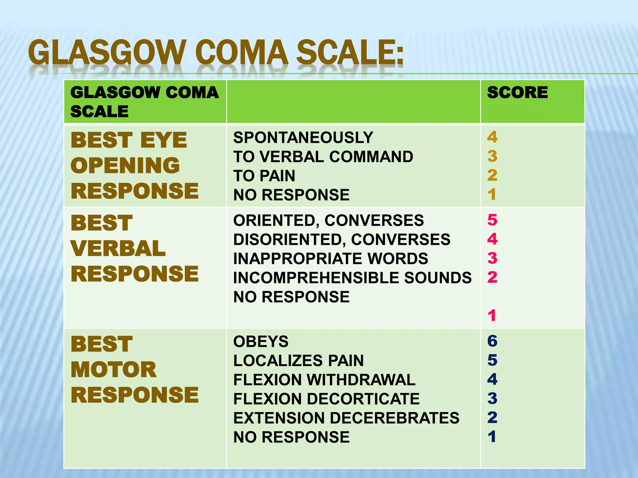
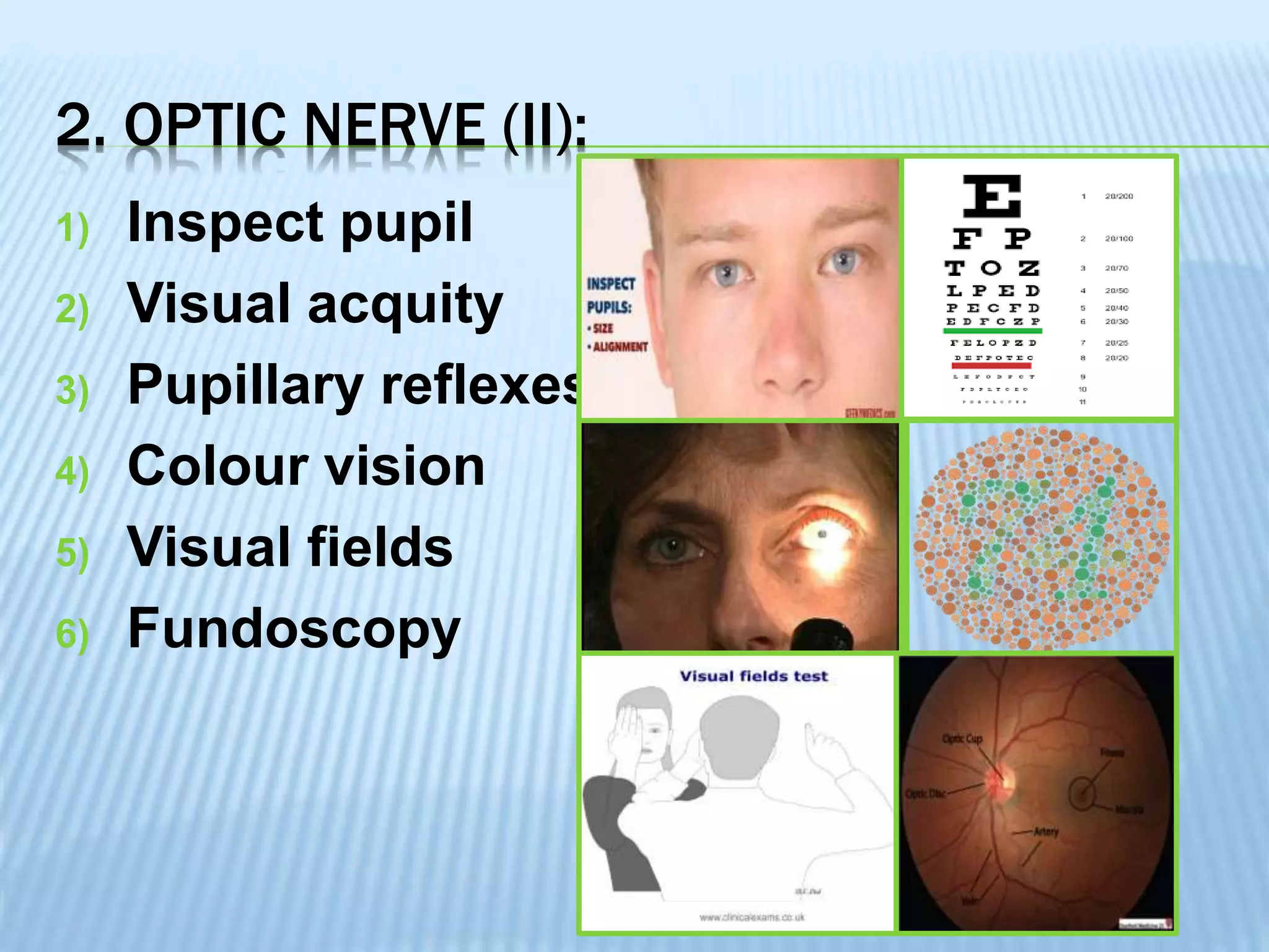
PREPARED BY,
VIPIN
Neurologicalassessmenthelps to:
1) Identify whichcomponentof the neurological
system are affected
2) If possible, determine the precise location of
the problem.
3) Screeningfor the presenceof discrete abnormalities in
patients at risk for the development of neurological
disorders
INTRODUCTION
COMPONENTS OF NEUROLOGICAL
EXAMINATION:
“PL MC MSC”
1) Patient history
2) Level of Consciousness
3) Mental Status Examination
4) Cranial Nerve Function test
5) Motor Power
6) Sensory Power
7) Cerebellar Functions
INDICATION:
Patient suffering under such as:
1) Meningitis
2) Head Injury
3) Brain Tumor
4) Unconscious Patient
5) High Grade Fever
6) Tetanus
7) Coma
EQUIPMENTS REQUIRED :
1) Ophthalmoscope
2) Otoscope
3) Tongue blade
4) Tuning fork
5) Pin
6) Tape measure
7) Flash light
8) Reflex hammer
9) Cotton wisps
10) Small container having:
 Salt, pepper, coffee powder, soap, lime etc.
 2 Test tubes for warmed water and crushed ice.
1.PATIENT HISTORY:
An accurate and detailed history is
vital here, Special attention should
be paid to several details such as
age of onset of symptoms.
2.LEVEL OF CONSCIOUSNESS:
The various levels are Alert, Lethargic,
Stuporous, Semi comatose and Comatose.
Alert: The patient is awake, responds
immediately and appropriately to verbal
stimuli.
Lethargic: Patient is drowsy inattentive but
easily arousable.
Stuporous: Patient sleep most of the time, he
can be aroused with great difficulty and
cooperates minimally when stimulated.
Semi comatose: There is no response to
verbal stimuli. Non-purposeful motor activity
is seen when the patient is stimulated with
pain.
2.LEVEL OF CONSCIOUSNESS:
Comatose: There is no response to
verbal or painful stimuli, no motor
activity is seen.
GLASGOW COMA SCALE:
First published by Teasdale and Jennet In 1974
MINIMUM MAXIMUM
EYE
RESPONSE
1 4
VERBAL
RESPONSE
1 5
MOTOR
RESPONSE
1 6
TOTAL 3 15
A Score of 15 indicates best level of
consciousness and a score of 3 the least level of
consciousness.
GLASGOW COMA SCALE:
GLASGOW COMA
SCALE
SCORE
BEST EYE
OPENING
RESPONSE
SPONTANEOUSLY
TO VERBAL COMMAND
TO PAIN
NO RESPONSE
4
3
2
1
BEST
VERBAL
RESPONSE
ORIENTED, CONVERSES
DISORIENTED, CONVERSES
INAPPROPRIATE WORDS
INCOMPREHENSIBLE SOUNDS
NO RESPONSE
5
4
3
2
1
BEST
MOTOR
RESPONSE
OBEYS
LOCALIZES PAIN
FLEXION WITHDRAWAL
FLEXION DECORTICATE
EXTENSION DECEREBRATES
NO RESPONSE
6
5
4
3
2
1
MNEUMONICS:
1.EYE RESPONSE:
2.VERBAL RESPONSE: Our Country WIN
3. MOTOR RESPONSE: Can’t Live Without
FAN’s
3. MENTAL STATUS EXAMINATION
Patient awareness and higher cognitive
functioning can be assessed by examining
orientation, attention and concentration, memory,
judgment, calculation and abstract reasoning.
a) Appearance and Behavior (Attire &
Grooming)
b) Attention span (5 or 6 digit backward &
forward)
c) Orientation (Name, Time, Month, Year, Place)
d) Memory(Immediate, Recent, Remote)
e) General knowledge( Question to current
events)
f) Mood and Behavior(Reaction to the topic
being discussed)
4. CRANIAL NERVE FUNCTION TEST:
12 pair of cranial nerve:
1) Olfactory Nerve
2) Optic Nerve
3) Occulomotor Nerve
4) Trochlear Nerve
5) Trigeminal Nerve
6) Abducens Nerve
7) Facial Nerve
8) Auditory or Acoustic Nerve
9) Glossopharyngeal Nerve
10) Vagus Nerve
11) Acessory Nerve
12) Hypoglossal Nerve
OOOTTA
FAG
VAH
4. CRANIAL NERVE FUNCTION:
INTRODUCTION:
1) Wash hands
2) Introduce yourself
3) Confirm patient details
4) Explain the examination
5) Gain consent
6) Position patient on a chair at eye level
7) Ask if the patient currently has any pain.
GENERAL INSPECTION:
1) General appearance
2) Obvious facial asymmetries
3) Position of eyes
4) Ptosis
5) Abnormality of speech or voice
1.OLFACTORY NERVE (I)
Olfaction can be tested more formally using
different odors, e.g. lemon or coffee
2. OPTIC NERVE (II):
1) Inspect pupil
2) Visual acquity
3) Pupillary reflexes
4) Colour vision
5) Visual fields
6) Fundoscopy
2. OPTIC NERVE (II):
Inspect pupils:
Size: normal size varies between individual
and depend on lighting conditions
(smaller in bright light, larger in dark).
Shape: pupils should be round- abnormal
shapes can be congenital or due to
pathology.
Symmetry: anisocoria (asymmetry between
the pupils)
2. OPTIC NERVE (II):
Assessment of Visual acuity:
 Stand the patient at 6 meters from the snellen chart
 Ask the patient to cover one eye and read the lowest
line they are able to
 Visual acuity is recorded as chart distance in meters
(numerator) over the number of the lowest line read
(denominator).
 Record the lowest line the patient was able to read.
 If the patient read 6/6 line, but gets 2 letter incorrect,
you would record as 6/6 (-2).
 If the patient gets more than 2 letters wrong, then the
previous line should be recorded as their acuity.
 When recording the vision it should state whether this
vision was (UA) OR (PH).
 Repeat above steps with the other eye.
2. OPTIC NERVE (II):
Pupillary reflexes:
Swinging light test
1) Move the pen torch rapidly between two
pupils, shining the light for three
seconds in each eye.
This test may detect a Relative Afferent
Pupillary
Defect (RAPD)- caused by damage to the
tract
between the optic nerve and optic chiasm.
3. OCULOMOTOR(III), TROCHLEAR (IV),
ABDUCEN (VI) CRANIAL NERVES:
Eye movements:
1) Hold your finger about 30cm directly in
front of the patient’s eyes and ask them to
look at it.
2) Ask the patient to keep their head still and
follow your finger with their eyes.
3) Ask the patient to report any double vision.
4) Move your finger through the various axes
of eye movements (“H” shape)
5) Observe for restriction of eye movements
and note any nystagmus.
3. OCULOMOTOR(III), TROCHLEAR (IV),
ABDUCEN (VI) CRANIAL NERVES:
4. TRIGEMINAL NERVE (V):
1) Sensory
2) Motor
3) Reflexes
5. FACIAL NERVE (VII):
1) Raised eyebrow.
2) Closed eyes
3) Blown out cheeks
4) Smiling
6. VESTIBULOCOCHLEAR NERVE (VIII):
1) Gross hearing testing
2) Rinne’s test
6. VESTIBULOCOCHLEAR NERVE (VIII):
Gross hearing testing:
1) Explain to the patient that you are going to say a
word or number and you’d like them to repeat it
back to you.
2) With your mouth approximately 15cm from the
ear, whisper a number or word.
3) Mask the ear not being tested by rubbing the
tragus.
4) Ask the patient to repeat the number or word back
to you.
5) If the patient repeats the correct word or number,
repeat the test at an arm’s length from the ear.
6) Assess the other ear in the same way.
6. VESTIBULOCOCHLEAR NERVE (VIII):
Rinne’s test:
1) Tap a 512 Hz tunning fork and place its base on
the mastoid process.
2) Ask the patient if they are able to hear it
3) If they are able to hear it, ask them to let you
know when they can no longer hear it.
4) Once the patient is unable to hear the sound via
the mastoid process move the tuning fork to
approximately 1 inch from the external auditory
meatus.
5) Ask the patient if they are able to hear the tuning
fork
6. VESTIBULOCOCHLEAR NERVE (VIII):
Rinne’s test:
6) If the patient is able to hear the tuning fork via air
conduction (after they were no longer able to hear
via bone conduction) it suggests their air
conduction is better than bone conduction
(Rinne’s positive).
Summary:
Normal= Air conduction>Bone conduction (+)
Neural deafness= Air conduction>Bone conduction
Conductive deafness= Bone conduction>Air
conduction(-)
6. VESTIBULOCOCHLEAR NERVE (VIII):
3) Weber’s test
4) Vestibular testing
6. VESTIBULOCOCHLEAR NERVE (VIII):
Weber’s test:
1) Tap a 512 Hz tuning fork and place in the
midline of the forehead.
2) Ask the patient where they can hear the
sound:
 Normal=Sound is heard equally in both
ears.
 Neural deafness=Sound is heard louder on
the side of the intact ear.
 Conductive deafness= Sound is heard
6. VESTIBULOCOCHLEAR NERVE (VIII):
Vestibular testing: “Unterberger” or “Turning
test”
Ask the patient to march on the spot with
arms outstretched and eyes closed:
 Normal- patient remains in the same
position.
 Vestibular lesion- patient will turn towards
the side of the lesion.
7. GLOSSOPHARYNGEAL & VAGUS NERVE (IX &
X):
7. GLOSSOPHARYNGEAL & VAGUS NERVE (IX &
X):
Assess soft palate and uvula:
 Symmetry- note any obvious deviation of
the uvula
 Ask patient to say “ahhhh”- observe
uvula moving upwards
 Ask patient to cough
 Swallow- ask patient to take a sip of
water- note any coughing/ delayed
swallow.
8. ACCESSORY NERVE (XI) :
1) Ask patient to shrug shoulders and
resist you pushing down.
2) Ask patient to turn head to one side
and resist you pushing it to the other.
9. HYPOGLOSSAL NERVE (XII) :
1) Inspect tongue for washing and
fasciculation's at rest
2) Ask patient to protrude tongue
3) Place your fingers on the patient’s cheek
and ask to push their tongue against it.
5. ASSESSMENT OF MOTOR FUNCTION:
There are few simple tests to find
Muscle strength both distal and
Proximal muscles of the arm and legs
Must be test in the following manner:
1) Apply maximum force to the extremity
while the patient pushes against the
observers fixed resistance. grade
accordingly using the following
abbreviations when referring each limb:
D - Distal
P - Proximal
RUE - Right upper extremity
RLE - Right lower extremity
LUE - Left upper extremity
LLE - Left lower extremity
MOTOR POWER GRADING:
5/5 Full range of motion against gravity with extreme
resistance
4/5 Full range of motion against gravity with some
resistance
3/5 Full range of motion against gravity no resistance
2/5 Full range of motion with gravity eliminated
1/5 Slight contraction visible
0/5 No movement
6. ASSESSMENT OF SENSORY FUNCTION:
Touch, Pain, Temperature and
Position sense.
Various senses are tested as follows:
1) Touch is assessed by passing a wisp of
cotton gently down various areas on the
body and asking the patient to identify the
area.
2) To assess the pain, gently touch various
parts of the body with a sterile needle and
request the patient to identify where the
sensation is felt.
CONT…….!
3) Temperature response is evaluated by
touching the patient with test tubes filled
with warm water and cracked ice
alternatively.
4) Sense of position is evaluated by
instructing the patient to close eyes, the
examiner grasps the patient index finger
at the last joint from the side and gently
raises or lowers it. The patient is asked
whether the index finger is positioned up
or down.
7. ASSESSMENT OF CEREBELLAR FUNCTION:
The Finger to Finger
Finger to nose
Romberg
Tandem walking
Patting test
Neurological examination 1

Neurological examination 1

  • 1.
  • 2.
    Neurologicalassessmenthelps to: 1) Identifywhichcomponentof the neurological system are affected 2) If possible, determine the precise location of the problem. 3) Screeningfor the presenceof discrete abnormalities in patients at risk for the development of neurological disorders INTRODUCTION
  • 3.
    COMPONENTS OF NEUROLOGICAL EXAMINATION: “PLMC MSC” 1) Patient history 2) Level of Consciousness 3) Mental Status Examination 4) Cranial Nerve Function test 5) Motor Power 6) Sensory Power 7) Cerebellar Functions
  • 4.
    INDICATION: Patient suffering undersuch as: 1) Meningitis 2) Head Injury 3) Brain Tumor 4) Unconscious Patient 5) High Grade Fever 6) Tetanus 7) Coma
  • 5.
    EQUIPMENTS REQUIRED : 1)Ophthalmoscope 2) Otoscope 3) Tongue blade 4) Tuning fork 5) Pin 6) Tape measure 7) Flash light 8) Reflex hammer 9) Cotton wisps 10) Small container having:  Salt, pepper, coffee powder, soap, lime etc.  2 Test tubes for warmed water and crushed ice.
  • 6.
    1.PATIENT HISTORY: An accurateand detailed history is vital here, Special attention should be paid to several details such as age of onset of symptoms.
  • 7.
    2.LEVEL OF CONSCIOUSNESS: Thevarious levels are Alert, Lethargic, Stuporous, Semi comatose and Comatose. Alert: The patient is awake, responds immediately and appropriately to verbal stimuli. Lethargic: Patient is drowsy inattentive but easily arousable. Stuporous: Patient sleep most of the time, he can be aroused with great difficulty and cooperates minimally when stimulated. Semi comatose: There is no response to verbal stimuli. Non-purposeful motor activity is seen when the patient is stimulated with pain.
  • 8.
    2.LEVEL OF CONSCIOUSNESS: Comatose:There is no response to verbal or painful stimuli, no motor activity is seen.
  • 9.
    GLASGOW COMA SCALE: Firstpublished by Teasdale and Jennet In 1974 MINIMUM MAXIMUM EYE RESPONSE 1 4 VERBAL RESPONSE 1 5 MOTOR RESPONSE 1 6 TOTAL 3 15 A Score of 15 indicates best level of consciousness and a score of 3 the least level of consciousness.
  • 10.
    GLASGOW COMA SCALE: GLASGOWCOMA SCALE SCORE BEST EYE OPENING RESPONSE SPONTANEOUSLY TO VERBAL COMMAND TO PAIN NO RESPONSE 4 3 2 1 BEST VERBAL RESPONSE ORIENTED, CONVERSES DISORIENTED, CONVERSES INAPPROPRIATE WORDS INCOMPREHENSIBLE SOUNDS NO RESPONSE 5 4 3 2 1 BEST MOTOR RESPONSE OBEYS LOCALIZES PAIN FLEXION WITHDRAWAL FLEXION DECORTICATE EXTENSION DECEREBRATES NO RESPONSE 6 5 4 3 2 1
  • 11.
    MNEUMONICS: 1.EYE RESPONSE: 2.VERBAL RESPONSE:Our Country WIN 3. MOTOR RESPONSE: Can’t Live Without FAN’s
  • 12.
    3. MENTAL STATUSEXAMINATION Patient awareness and higher cognitive functioning can be assessed by examining orientation, attention and concentration, memory, judgment, calculation and abstract reasoning. a) Appearance and Behavior (Attire & Grooming) b) Attention span (5 or 6 digit backward & forward) c) Orientation (Name, Time, Month, Year, Place) d) Memory(Immediate, Recent, Remote) e) General knowledge( Question to current events) f) Mood and Behavior(Reaction to the topic being discussed)
  • 13.
    4. CRANIAL NERVEFUNCTION TEST: 12 pair of cranial nerve: 1) Olfactory Nerve 2) Optic Nerve 3) Occulomotor Nerve 4) Trochlear Nerve 5) Trigeminal Nerve 6) Abducens Nerve 7) Facial Nerve 8) Auditory or Acoustic Nerve 9) Glossopharyngeal Nerve 10) Vagus Nerve 11) Acessory Nerve 12) Hypoglossal Nerve OOOTTA FAG VAH
  • 14.
  • 15.
    INTRODUCTION: 1) Wash hands 2)Introduce yourself 3) Confirm patient details 4) Explain the examination 5) Gain consent 6) Position patient on a chair at eye level 7) Ask if the patient currently has any pain.
  • 16.
    GENERAL INSPECTION: 1) Generalappearance 2) Obvious facial asymmetries 3) Position of eyes 4) Ptosis 5) Abnormality of speech or voice
  • 17.
    1.OLFACTORY NERVE (I) Olfactioncan be tested more formally using different odors, e.g. lemon or coffee
  • 18.
    2. OPTIC NERVE(II): 1) Inspect pupil 2) Visual acquity 3) Pupillary reflexes 4) Colour vision 5) Visual fields 6) Fundoscopy
  • 19.
    2. OPTIC NERVE(II): Inspect pupils: Size: normal size varies between individual and depend on lighting conditions (smaller in bright light, larger in dark). Shape: pupils should be round- abnormal shapes can be congenital or due to pathology. Symmetry: anisocoria (asymmetry between the pupils)
  • 20.
    2. OPTIC NERVE(II): Assessment of Visual acuity:  Stand the patient at 6 meters from the snellen chart  Ask the patient to cover one eye and read the lowest line they are able to  Visual acuity is recorded as chart distance in meters (numerator) over the number of the lowest line read (denominator).  Record the lowest line the patient was able to read.  If the patient read 6/6 line, but gets 2 letter incorrect, you would record as 6/6 (-2).  If the patient gets more than 2 letters wrong, then the previous line should be recorded as their acuity.  When recording the vision it should state whether this vision was (UA) OR (PH).  Repeat above steps with the other eye.
  • 21.
    2. OPTIC NERVE(II): Pupillary reflexes: Swinging light test 1) Move the pen torch rapidly between two pupils, shining the light for three seconds in each eye. This test may detect a Relative Afferent Pupillary Defect (RAPD)- caused by damage to the tract between the optic nerve and optic chiasm.
  • 22.
    3. OCULOMOTOR(III), TROCHLEAR(IV), ABDUCEN (VI) CRANIAL NERVES: Eye movements: 1) Hold your finger about 30cm directly in front of the patient’s eyes and ask them to look at it. 2) Ask the patient to keep their head still and follow your finger with their eyes. 3) Ask the patient to report any double vision. 4) Move your finger through the various axes of eye movements (“H” shape) 5) Observe for restriction of eye movements and note any nystagmus.
  • 23.
    3. OCULOMOTOR(III), TROCHLEAR(IV), ABDUCEN (VI) CRANIAL NERVES:
  • 24.
    4. TRIGEMINAL NERVE(V): 1) Sensory 2) Motor 3) Reflexes
  • 25.
    5. FACIAL NERVE(VII): 1) Raised eyebrow. 2) Closed eyes 3) Blown out cheeks 4) Smiling
  • 26.
    6. VESTIBULOCOCHLEAR NERVE(VIII): 1) Gross hearing testing 2) Rinne’s test
  • 27.
    6. VESTIBULOCOCHLEAR NERVE(VIII): Gross hearing testing: 1) Explain to the patient that you are going to say a word or number and you’d like them to repeat it back to you. 2) With your mouth approximately 15cm from the ear, whisper a number or word. 3) Mask the ear not being tested by rubbing the tragus. 4) Ask the patient to repeat the number or word back to you. 5) If the patient repeats the correct word or number, repeat the test at an arm’s length from the ear. 6) Assess the other ear in the same way.
  • 28.
    6. VESTIBULOCOCHLEAR NERVE(VIII): Rinne’s test: 1) Tap a 512 Hz tunning fork and place its base on the mastoid process. 2) Ask the patient if they are able to hear it 3) If they are able to hear it, ask them to let you know when they can no longer hear it. 4) Once the patient is unable to hear the sound via the mastoid process move the tuning fork to approximately 1 inch from the external auditory meatus. 5) Ask the patient if they are able to hear the tuning fork
  • 29.
    6. VESTIBULOCOCHLEAR NERVE(VIII): Rinne’s test: 6) If the patient is able to hear the tuning fork via air conduction (after they were no longer able to hear via bone conduction) it suggests their air conduction is better than bone conduction (Rinne’s positive). Summary: Normal= Air conduction>Bone conduction (+) Neural deafness= Air conduction>Bone conduction Conductive deafness= Bone conduction>Air conduction(-)
  • 30.
    6. VESTIBULOCOCHLEAR NERVE(VIII): 3) Weber’s test 4) Vestibular testing
  • 31.
    6. VESTIBULOCOCHLEAR NERVE(VIII): Weber’s test: 1) Tap a 512 Hz tuning fork and place in the midline of the forehead. 2) Ask the patient where they can hear the sound:  Normal=Sound is heard equally in both ears.  Neural deafness=Sound is heard louder on the side of the intact ear.  Conductive deafness= Sound is heard
  • 32.
    6. VESTIBULOCOCHLEAR NERVE(VIII): Vestibular testing: “Unterberger” or “Turning test” Ask the patient to march on the spot with arms outstretched and eyes closed:  Normal- patient remains in the same position.  Vestibular lesion- patient will turn towards the side of the lesion.
  • 33.
    7. GLOSSOPHARYNGEAL &VAGUS NERVE (IX & X):
  • 34.
    7. GLOSSOPHARYNGEAL &VAGUS NERVE (IX & X): Assess soft palate and uvula:  Symmetry- note any obvious deviation of the uvula  Ask patient to say “ahhhh”- observe uvula moving upwards  Ask patient to cough  Swallow- ask patient to take a sip of water- note any coughing/ delayed swallow.
  • 35.
  • 36.
    8. ACCESSORY NERVE(XI) : 1) Ask patient to shrug shoulders and resist you pushing down. 2) Ask patient to turn head to one side and resist you pushing it to the other.
  • 37.
  • 38.
    9. HYPOGLOSSAL NERVE(XII) : 1) Inspect tongue for washing and fasciculation's at rest 2) Ask patient to protrude tongue 3) Place your fingers on the patient’s cheek and ask to push their tongue against it.
  • 39.
    5. ASSESSMENT OFMOTOR FUNCTION: There are few simple tests to find Muscle strength both distal and Proximal muscles of the arm and legs Must be test in the following manner: 1) Apply maximum force to the extremity while the patient pushes against the observers fixed resistance. grade accordingly using the following abbreviations when referring each limb:
  • 40.
    D - Distal P- Proximal RUE - Right upper extremity RLE - Right lower extremity LUE - Left upper extremity LLE - Left lower extremity MOTOR POWER GRADING: 5/5 Full range of motion against gravity with extreme resistance 4/5 Full range of motion against gravity with some resistance 3/5 Full range of motion against gravity no resistance 2/5 Full range of motion with gravity eliminated 1/5 Slight contraction visible 0/5 No movement
  • 41.
    6. ASSESSMENT OFSENSORY FUNCTION: Touch, Pain, Temperature and Position sense. Various senses are tested as follows: 1) Touch is assessed by passing a wisp of cotton gently down various areas on the body and asking the patient to identify the area. 2) To assess the pain, gently touch various parts of the body with a sterile needle and request the patient to identify where the sensation is felt.
  • 42.
    CONT…….! 3) Temperature responseis evaluated by touching the patient with test tubes filled with warm water and cracked ice alternatively. 4) Sense of position is evaluated by instructing the patient to close eyes, the examiner grasps the patient index finger at the last joint from the side and gently raises or lowers it. The patient is asked whether the index finger is positioned up or down.
  • 43.
    7. ASSESSMENT OFCEREBELLAR FUNCTION: The Finger to Finger Finger to nose Romberg Tandem walking Patting test