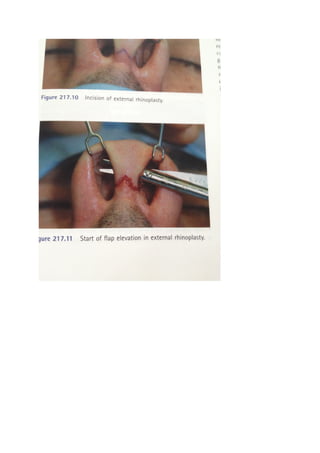
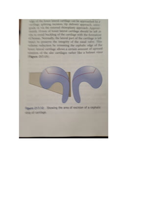
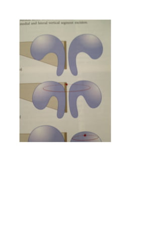
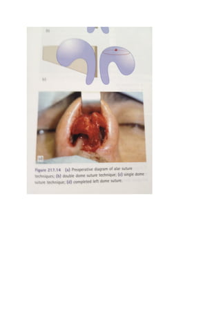
The nasal tip & nasolabial angle 
Anatomy : the nose can be considered to be divided into subunits consisting of the nasal dorsum, 
nasal sidewalls, the nasal tip, the alar lobules& the alar facets. 
Anatomically, nasal tip & nasolabial angle is formed from the alar cartilages& cartilagenuos nasal 
septum with muscular & fascial layers with skin overlying. Tha nasal vestibule is lined with skin in the 
lower part& with mucosa in the upper part. The skin-lined area contains hairs(vibrissae). 
Skin 
The skin overlying the nasal tip is usually thicker with more sebaceous tissue than the skin overlying 
the upper part of the nasal dorsum. 
Thin skin will not be camouflaged. However , thin skin will tend to heal more quickly & with less 
oedema. 
Conversely , subtle changes to the nasal skeleton will not be apparent in thick sebaceous skin, & this 
type of skin will tend to heal more slowly & with more postoperative oedema. 
Subcutaneous tissue of the nose contains the facial mimetic muscles & their investing fibrous 
aponeurosis. The plane between this & perichondrium/periosteum of the underlying nasal skeleton 
is the plane of dissection in rhinoplasty. 
Alar cartilage 
This U shaped cartilage has two processes: the medial & lateral crura. The intermediate domal area 
is the intermediate crus or transition area. 
Nasal spine 
The nasal spine is a midline anterior projection of the premaxillae at the lower border of the nasal 
aperture. The nasal septum is attached to the spine& maxillary crest by a strong fibrous attachment 
of periosteum & perichondrium. 
A prominant nasal spine can produce an open nasolabial angle, this can be corrected by resection of 
the nasal spine. Overresection can result in foreshortening of the nose & columellar retraction. 
Tip-supporting mechanisms 
Nasal tip as a tripod structure with the medial crura, lateral crura & septum forming the tripod. The 
major tip supporting mechanisms are: 
- The alar cartilages; 
- The attachment of the lower lateral cartilage to the upper lateral cartilage; 
- The attachment joining the medial crura to the septal cartilage. 
Minor tip –supporting mechanism are
1. Dorsal cartilaginous septum; 
2. Interdomal ligament; 
3. Membranous septum; 
4. The nasal spine; 
5. Investing skin & soft tissues; 
6. Alar side walls. 
Dividing the attachments between the upper & lower alar cartilages will allow the tip to drop whilst 
removing volume between the upper & lower lateral cartilages will allow elevation of the tip. 
Surgical approaches to the nasal tip 
Surgical approaches to the nasal tip include: 
1. Cartilage-splitting approach; 
2. Tip delivery approach; 
3. Retrograde approach; 
4. External rhinoplasty. 
Cartilage-spiltting incisions 
This is the least traumatic of the commonly used rhinoplasty incisions. A single incision is made 
through the vestibular skin over each lower lateral cartilage. The incision is made at the position that 
overlies the cartilaginous incision & where the cephalic strip of cartilage will be excised. 
Tip delivery 
The tip delivery approach delivers the alar cartilages with the underlying skin & mucosa as a bucket 
handle. The incision are made along the caudal margin of the alar cartilage & along the cephalic 
margin. The incision along the cephalic margin is known as the intercartilaginous incision, lying as it 
does between the upper & lower lateral cartilages. 
The incision along the caudal edge of the alar cartilage is called the rim incision. This is inaccurate 
because the inferior edge of the lower lateral is not parallel to the nasal rim, however, the incision 
inferior to the medial crus is usually referred to as a rim incision. 
After making these incisions, the overlying soft tissue & skin is dissected off the alar cartilage leaving 
the cartilage attached to its underlying vestibular skin & mucosa. By mobilizing this bucket handle 
the alar cartilage can be delivered still attached to its underlying vestibular skin. This gives excellent 
access to most of the alar cartilage so that a number of procedures can be used to modify the 
cartilages.
External rhinoplasty 
The external rhinoplasty approach gives the greatest exposure of any the rhinoplasty incisions. 
Advantages of the procedure are: 
1. Direct vision & complete exposure of the alar cartilage; 
2. It allows two-handed surgery; 
3. It enables accurate graft fixation; 
4. It is useful in teaching because of exposure; 
5. It is excellent for analysis of surgical problems particularly in revision & post-traumatic cases. 
6. It enables good exposure for surgery of the nasal septum( both in difficult septal deformities 
& in septal perforations.) 
The incision is a bilateral rim incision as used in the tip delivery. The medial crura rim incision are 
joined by a transverse incision which has a step or a V-shaped notch to allow accurate apposition of 
the skin edges. 
The skin flap is elevated & dissected off the underlying medial & lateral crura. The dissection can be 
continued cephalically to expose the nasal dorsum. The medial crura can be separated to give 
excellent exposure of the caudal end & dorsal area of the nasal septum. 
Fine monofilament nylon is ideal to close the incision on the columella. One can use rapidly 
absorbing sutures to close the intranasal part of the incision. 
Tip-defining procedures 
Usually , in cosmetic rhinoplasty, there is a need to narrow the nasal tip to achieve tip definition & 
refinement. This is normally associated with a need to elevate the nasal tip to (apparently shorten 
the nose & give it a more youthful appearance. Methods of altering tip definition include: 
1. Removal of cephalic strip of lower lateral cartilage; 
2. Vertical division +/- strip excision of lower lateral cartilage; 
3. Tip suturing; 
4. Tip grafts. 
A modest amount of tip narrowing & cephalic tip rotation can be achieved by excision of a 
cephalic strip from the lower lateral cartilage. The lateral part of the lower lateral cartilage is left 
intact to matintain the integrity of the nasal valve. The cephalic edge of the lower lateral 
cartilage can be approached by cartilage splitting incision/tip delivery approach/external 
rhinoplasty approach. 
Approximately 10mm of lower lateral cartilage should be left in situ to avoid buckling of the 
cartilage with the formation of bossae. Normally, lateral part of the cartilage is left intact to 
preserve the integrity of the nasal valve.
This volume reduction by trimming the cephalic edge of the lower lateral cartilage allows a 
certain amount of upward rotation of the alar cartilage rather like a helmet visor. 
Tip-suturing techniques 
Suturing methods of contouring the nasal tip have become more popular in the attempt to find 
predictable methods of modifying the nasal tip without the complications that may be the result of 
excessive cartilage resection. 
1. A single suture passing through both lateral & intermediate crura to produce a double dome 
unit(double dome suture technique). 
2. Single dome unit can be used in combination with this method to achieve greater & more 
precise narrowing of the nasal tip(single dome suture technique). 
Suture contouring of the nasal tip is often used with support grafts, such as columellar struts to 
strengthen the medial crura & to enable some tip projection by advancement of the medial crura on 
the strut. 
These techniques are most useful in achieving norrowing & rotation of the nasal tip. The main 
advantage of this technique is that it is reversible & does not require surgical resection. However , 
suture techniques are more applicable to mild & moderate tip deformities. They are not very useful 
in dealing with either the underprojected or overprojected nasal tip. 
Management of the overprojected tip 
Tip overprojection may be caused by 
-alar cartilage overproduction. 
- nasal spine overdevelopment; 
-caudal septal overdevelopment; 
- overdevelopment of the quadrangular cartilage dorsum; 
- elongated columella; 
- iatrogenic overproduction. 
Reduction of tip projection can be achieved by: 
- Transfixion incision; 
- Vertical dome division(Goldman); 
- Medial & lateral crura vertical segment excision. 
Transfixing incision 
Complete transfixion of the membranous septum & the attachment of the medial crural footplate 
allows the alar cartilages to be repositioned in relation to the nasal septum. This gives some tip set
back but will not be effective in the grossly overprojected nasal tip. When the tip is setback using 
this method it must be held in place with absorbable sutures. 
Vertical dome division(Goldman) 
The procedure involves a tip delivery approach followed by vertical division of the alar domes 
approximately 1mm lateral to the highest point of the dome. The cartilage & its underlying mucosa 
are incised using scissors or a scalpel blade. Intermediate crura are rotated anteriorly & they & 
medial crura can then be sutured together using an absorbable suture . when the medial crura are 
stabilised in this way, their height can be trimmed to an appropriate level. If the medial crura are 
weak , they can be supported by a columella strut graft of septal cartilage. 
Kridel & Konior have described a variation on vertical dome division. In this procedure, the nasal 
dome is exposed with an external approach. The medial crura are sutured together to support each 
other & overprojecting dome is resected. The lower lateral cartilage are not sutured to the medial 
crura. 
Adamson describe a vertical incision medial to the high point of the dome & overlapping of the 
lower lateral cartilage. 
The interrupted strip with cartilage excision 
A greater degree of tip setback achieved by excising a vertical portion of either medial or lateral crus 
or combination of two. 
The underprojected nasal tip 
Tip projection can be increased by the following techniques: 
1. Goldman tip & medial crural strut; 
2. Onlay graft; 
3. Lateral crural steal; 
4. Shield graft. 
1)Goldman tip & medial crural strut 
Vertical dome division with division of the dome 2mm or so lateral to the apex of the dome will 
allow some increased tip projection by lengthening the medial crural segment of the nose. If this 
procedure is used, a medial crural strut is helpful to provide some strengthening of the medial crura. 
This is straight piece of cartilage, preferably septal, alternatively conchal & approximately 3-4mm 
wide & as long as is necessary. The cartilage strut is placed between the medial crura & sutured 
between the medial crura using an absorbable suture. 
2) onlay grafts 
It can be used as umbrella graft to augment the tip.
3) lateral crural steal 
The alar cartilages are dissected off the underlying vestibular skin in the intermediate crural area & 
the alar cartilages may be delivered. The lateral crura are then advanced to the medial crura & 
sutured with parmanant sutures. 
4) shield grafts 
The main indication for the shield graft is to increase tip projection in the underprojected tip with a 
short columella & weak lower lateral cartilages. The graft material used is usually septal cartilage but 
conchal cartilage may also be used although this is not such a rigid material. 
The graft can be inserted either through a marginal incision or via an external rhinoplasty 
approach,& sutured to the medial crura. 
The broad nasal tip 
The appearance of a box-shaped or broad nasal tip may be because the nasal skin is thick & 
sebaceous, because of the shape or thickness of the alar cartilages or because of an abnormality of 
the nasal septum causing widening of the nasal tip. 
Goldman tip technique or nasal tip suturing to produce a narrowing effect. 
The nasal septum& nasolabial angle 
Usually some degree f tip rotation is needed in reduction rhinoplasty to achieve a smaller & more 
youthful nose. Much of this can be achieved with volume reduction from the cephalic strip removal 
from the lower lateral cartilage/ removal a small triangle of septal cartilage from the caudal edge to 
allow futher tip rotation. This will open the nasolabial angle. 
If this overdone there will be over-rotation of the nasal tip, this part of rhinoplasty should be carried 
out judiciously. 
Complications of operations 
1) Deformity; 
2) Retracted 
3) Alar asymmetry 
4) Retracted columella; 
5) Bossae
The nasal tip & nasolabial angle

The nasal tip & nasolabial angle

  • 1.
    The nasal tip& nasolabial angle Anatomy : the nose can be considered to be divided into subunits consisting of the nasal dorsum, nasal sidewalls, the nasal tip, the alar lobules& the alar facets. Anatomically, nasal tip & nasolabial angle is formed from the alar cartilages& cartilagenuos nasal septum with muscular & fascial layers with skin overlying. Tha nasal vestibule is lined with skin in the lower part& with mucosa in the upper part. The skin-lined area contains hairs(vibrissae). Skin The skin overlying the nasal tip is usually thicker with more sebaceous tissue than the skin overlying the upper part of the nasal dorsum. Thin skin will not be camouflaged. However , thin skin will tend to heal more quickly & with less oedema. Conversely , subtle changes to the nasal skeleton will not be apparent in thick sebaceous skin, & this type of skin will tend to heal more slowly & with more postoperative oedema. Subcutaneous tissue of the nose contains the facial mimetic muscles & their investing fibrous aponeurosis. The plane between this & perichondrium/periosteum of the underlying nasal skeleton is the plane of dissection in rhinoplasty. Alar cartilage This U shaped cartilage has two processes: the medial & lateral crura. The intermediate domal area is the intermediate crus or transition area. Nasal spine The nasal spine is a midline anterior projection of the premaxillae at the lower border of the nasal aperture. The nasal septum is attached to the spine& maxillary crest by a strong fibrous attachment of periosteum & perichondrium. A prominant nasal spine can produce an open nasolabial angle, this can be corrected by resection of the nasal spine. Overresection can result in foreshortening of the nose & columellar retraction. Tip-supporting mechanisms Nasal tip as a tripod structure with the medial crura, lateral crura & septum forming the tripod. The major tip supporting mechanisms are: - The alar cartilages; - The attachment of the lower lateral cartilage to the upper lateral cartilage; - The attachment joining the medial crura to the septal cartilage. Minor tip –supporting mechanism are
  • 2.
    1. Dorsal cartilaginousseptum; 2. Interdomal ligament; 3. Membranous septum; 4. The nasal spine; 5. Investing skin & soft tissues; 6. Alar side walls. Dividing the attachments between the upper & lower alar cartilages will allow the tip to drop whilst removing volume between the upper & lower lateral cartilages will allow elevation of the tip. Surgical approaches to the nasal tip Surgical approaches to the nasal tip include: 1. Cartilage-splitting approach; 2. Tip delivery approach; 3. Retrograde approach; 4. External rhinoplasty. Cartilage-spiltting incisions This is the least traumatic of the commonly used rhinoplasty incisions. A single incision is made through the vestibular skin over each lower lateral cartilage. The incision is made at the position that overlies the cartilaginous incision & where the cephalic strip of cartilage will be excised. Tip delivery The tip delivery approach delivers the alar cartilages with the underlying skin & mucosa as a bucket handle. The incision are made along the caudal margin of the alar cartilage & along the cephalic margin. The incision along the cephalic margin is known as the intercartilaginous incision, lying as it does between the upper & lower lateral cartilages. The incision along the caudal edge of the alar cartilage is called the rim incision. This is inaccurate because the inferior edge of the lower lateral is not parallel to the nasal rim, however, the incision inferior to the medial crus is usually referred to as a rim incision. After making these incisions, the overlying soft tissue & skin is dissected off the alar cartilage leaving the cartilage attached to its underlying vestibular skin & mucosa. By mobilizing this bucket handle the alar cartilage can be delivered still attached to its underlying vestibular skin. This gives excellent access to most of the alar cartilage so that a number of procedures can be used to modify the cartilages.
  • 3.
    External rhinoplasty Theexternal rhinoplasty approach gives the greatest exposure of any the rhinoplasty incisions. Advantages of the procedure are: 1. Direct vision & complete exposure of the alar cartilage; 2. It allows two-handed surgery; 3. It enables accurate graft fixation; 4. It is useful in teaching because of exposure; 5. It is excellent for analysis of surgical problems particularly in revision & post-traumatic cases. 6. It enables good exposure for surgery of the nasal septum( both in difficult septal deformities & in septal perforations.) The incision is a bilateral rim incision as used in the tip delivery. The medial crura rim incision are joined by a transverse incision which has a step or a V-shaped notch to allow accurate apposition of the skin edges. The skin flap is elevated & dissected off the underlying medial & lateral crura. The dissection can be continued cephalically to expose the nasal dorsum. The medial crura can be separated to give excellent exposure of the caudal end & dorsal area of the nasal septum. Fine monofilament nylon is ideal to close the incision on the columella. One can use rapidly absorbing sutures to close the intranasal part of the incision. Tip-defining procedures Usually , in cosmetic rhinoplasty, there is a need to narrow the nasal tip to achieve tip definition & refinement. This is normally associated with a need to elevate the nasal tip to (apparently shorten the nose & give it a more youthful appearance. Methods of altering tip definition include: 1. Removal of cephalic strip of lower lateral cartilage; 2. Vertical division +/- strip excision of lower lateral cartilage; 3. Tip suturing; 4. Tip grafts. A modest amount of tip narrowing & cephalic tip rotation can be achieved by excision of a cephalic strip from the lower lateral cartilage. The lateral part of the lower lateral cartilage is left intact to matintain the integrity of the nasal valve. The cephalic edge of the lower lateral cartilage can be approached by cartilage splitting incision/tip delivery approach/external rhinoplasty approach. Approximately 10mm of lower lateral cartilage should be left in situ to avoid buckling of the cartilage with the formation of bossae. Normally, lateral part of the cartilage is left intact to preserve the integrity of the nasal valve.
  • 4.
    This volume reductionby trimming the cephalic edge of the lower lateral cartilage allows a certain amount of upward rotation of the alar cartilage rather like a helmet visor. Tip-suturing techniques Suturing methods of contouring the nasal tip have become more popular in the attempt to find predictable methods of modifying the nasal tip without the complications that may be the result of excessive cartilage resection. 1. A single suture passing through both lateral & intermediate crura to produce a double dome unit(double dome suture technique). 2. Single dome unit can be used in combination with this method to achieve greater & more precise narrowing of the nasal tip(single dome suture technique). Suture contouring of the nasal tip is often used with support grafts, such as columellar struts to strengthen the medial crura & to enable some tip projection by advancement of the medial crura on the strut. These techniques are most useful in achieving norrowing & rotation of the nasal tip. The main advantage of this technique is that it is reversible & does not require surgical resection. However , suture techniques are more applicable to mild & moderate tip deformities. They are not very useful in dealing with either the underprojected or overprojected nasal tip. Management of the overprojected tip Tip overprojection may be caused by -alar cartilage overproduction. - nasal spine overdevelopment; -caudal septal overdevelopment; - overdevelopment of the quadrangular cartilage dorsum; - elongated columella; - iatrogenic overproduction. Reduction of tip projection can be achieved by: - Transfixion incision; - Vertical dome division(Goldman); - Medial & lateral crura vertical segment excision. Transfixing incision Complete transfixion of the membranous septum & the attachment of the medial crural footplate allows the alar cartilages to be repositioned in relation to the nasal septum. This gives some tip set
  • 5.
    back but willnot be effective in the grossly overprojected nasal tip. When the tip is setback using this method it must be held in place with absorbable sutures. Vertical dome division(Goldman) The procedure involves a tip delivery approach followed by vertical division of the alar domes approximately 1mm lateral to the highest point of the dome. The cartilage & its underlying mucosa are incised using scissors or a scalpel blade. Intermediate crura are rotated anteriorly & they & medial crura can then be sutured together using an absorbable suture . when the medial crura are stabilised in this way, their height can be trimmed to an appropriate level. If the medial crura are weak , they can be supported by a columella strut graft of septal cartilage. Kridel & Konior have described a variation on vertical dome division. In this procedure, the nasal dome is exposed with an external approach. The medial crura are sutured together to support each other & overprojecting dome is resected. The lower lateral cartilage are not sutured to the medial crura. Adamson describe a vertical incision medial to the high point of the dome & overlapping of the lower lateral cartilage. The interrupted strip with cartilage excision A greater degree of tip setback achieved by excising a vertical portion of either medial or lateral crus or combination of two. The underprojected nasal tip Tip projection can be increased by the following techniques: 1. Goldman tip & medial crural strut; 2. Onlay graft; 3. Lateral crural steal; 4. Shield graft. 1)Goldman tip & medial crural strut Vertical dome division with division of the dome 2mm or so lateral to the apex of the dome will allow some increased tip projection by lengthening the medial crural segment of the nose. If this procedure is used, a medial crural strut is helpful to provide some strengthening of the medial crura. This is straight piece of cartilage, preferably septal, alternatively conchal & approximately 3-4mm wide & as long as is necessary. The cartilage strut is placed between the medial crura & sutured between the medial crura using an absorbable suture. 2) onlay grafts It can be used as umbrella graft to augment the tip.
  • 6.
    3) lateral cruralsteal The alar cartilages are dissected off the underlying vestibular skin in the intermediate crural area & the alar cartilages may be delivered. The lateral crura are then advanced to the medial crura & sutured with parmanant sutures. 4) shield grafts The main indication for the shield graft is to increase tip projection in the underprojected tip with a short columella & weak lower lateral cartilages. The graft material used is usually septal cartilage but conchal cartilage may also be used although this is not such a rigid material. The graft can be inserted either through a marginal incision or via an external rhinoplasty approach,& sutured to the medial crura. The broad nasal tip The appearance of a box-shaped or broad nasal tip may be because the nasal skin is thick & sebaceous, because of the shape or thickness of the alar cartilages or because of an abnormality of the nasal septum causing widening of the nasal tip. Goldman tip technique or nasal tip suturing to produce a narrowing effect. The nasal septum& nasolabial angle Usually some degree f tip rotation is needed in reduction rhinoplasty to achieve a smaller & more youthful nose. Much of this can be achieved with volume reduction from the cephalic strip removal from the lower lateral cartilage/ removal a small triangle of septal cartilage from the caudal edge to allow futher tip rotation. This will open the nasolabial angle. If this overdone there will be over-rotation of the nasal tip, this part of rhinoplasty should be carried out judiciously. Complications of operations 1) Deformity; 2) Retracted 3) Alar asymmetry 4) Retracted columella; 5) Bossae