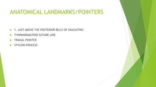
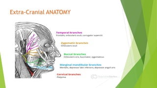
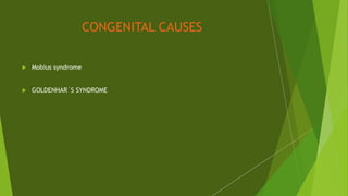
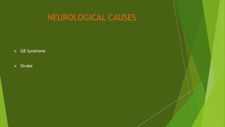
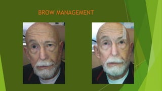
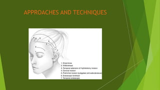
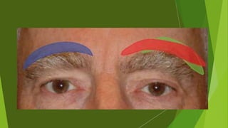
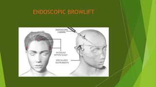
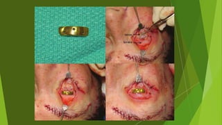
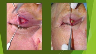
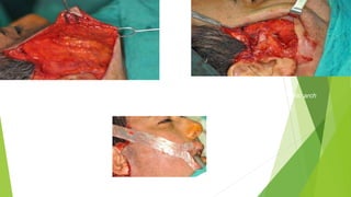
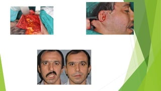
The document discusses facial nerve palsy and facial reanimation. It begins with an introduction to the facial nerve and its functions. It then covers anatomy of the facial nerve, classifications and etiologies of facial nerve palsy, presentation and evaluation, non-surgical and surgical management options. For surgical management it discusses various nerve repair techniques including cable grafting and nerve transfers. It also covers approaches to managing specific areas like the eyebrow, eyelids, and lips which may be paralyzed. Both static and dynamic reconstruction techniques are outlined.
This document discusses techniques for reanimating facial paralysis. It begins by outlining general principles, including reinnervating muscles early, separately reanimating the upper and lower face, and tailoring the procedure to the patient's needs and assessment. Surgical techniques are then described, including neural methods like nerve grafting and transfers, musculofacial transpositions, and static procedures. The timing of different techniques depends on whether the paralysis is acute (<3 weeks), intermediate (3 weeks to 2 years), or chronic (>2 years). Assessment involves evaluating the cause and extent of paralysis along with patient factors. The goal is to restore facial symmetry, competence, protection, and dynamic smile.
This document discusses intraoperative neurophysiological monitoring during surgery. It describes monitoring brain and nerve pathways to reduce risks from surgery and protect the brain, nerves, and spinal cord. Specific techniques are covered, including electrical stimulation of pathways to identify changes from surgery and ensure integrity. Placement of recording electrodes and how they are used to monitor facial and other cranial nerves is also summarized.
Local flaps in head & neack reconstructionMd Roohia
A flap is a unit of tissue transferred from one site to another while maintaining its own blood supply. Flaps are classified based on their location as local or distant. Local flaps can be random, which rely on subdermal vessels, or axial, relying on named vessels. Flaps are also classified based on their composition, such as skin, muscle, or bone flaps. When planning local flaps, goals include color and thickness match, sensory preservation, and minimal secondary defects. Common local flap techniques include advancement, rotation, and transposition flaps to close facial defects. Complications can be prevented through proper planning to avoid tension and including a flap margin.
The document discusses the embryology, anatomy, components, causes of injury, grading systems, evaluation, and treatment of the facial nerve. It covers the development of the facial nerve from the embryonic stage through maturity and describes the various parts of the nerve and their functions. The document also outlines different classification systems for nerve injuries, approaches for evaluating facial nerve paralysis, and surgical and non-surgical techniques for treating injuries or reanimating paralysis of the facial nerve.
1. Flaps are used in reconstructive surgery to repair structural defects following procedures like cancer surgery. They involve transferring tissue from one part of the body to another while maintaining or reconnecting its blood supply.
2. There are many types of flaps classified by their blood supply, tissue type, and location. Common flaps used in head and neck reconstruction include local flaps like nasolabial and advancement flaps as well as regional and distant flaps like pectoralis major and radial forearm flaps.
3. Proper flap selection and design is important to replace tissue "like with like" and adhere to anatomical borders and units for optimal cosmetic and functional outcomes.
The facial nerve originates from the pons and has both motor and sensory components. It passes through the internal acoustic meatus, facial canal within the temporal bone, and exits at the stylomastoid foramen. Within the facial canal it has several segments separated by genu. Important surgical landmarks along the facial canal include the geniculate ganglion, cochleariform process, and digastric ridge. The main trunk divides into 5 terminal branches in the parotid gland which innervate muscles of facial expression. Careful knowledge of the anatomy is important for safe middle ear, mastoid, and parotid surgery.
Micro vascular free flaps used in head and neck reconstruction /certified fi...Indian dental academy
Welcome to Indian Dental Academy
The Indian Dental Academy is the Leader in continuing dental education , training dentists in all aspects of dentistry and offering a wide range of dental certified courses in different formats.
Indian dental academy has a unique training program & curriculum that provides students with exceptional clinical skills and enabling them to return to their office with high level confidence and start treating patients
State of the art comprehensive training-Faculty of world wide repute &Very affordable.
STOMAL RECURRENCE AFTER LARYNGECTOMY-1.pptxSendhil Kumar
Stomal recurrence after laryngectomy occurs in 1.7-15% of patients and is usually fatal. Risk factors include subglottic tumor location, advanced T and N stage, pre-operative tracheostomy, and positive margins. Surgery is the main treatment for localized recurrence, involving wide excision and mediastinal lymph node dissection. Post-operative radiation may prevent recurrence in high risk patients by sterilizing the area. Close follow-up is important to detect recurrence early.
The document discusses the use of the Hadad-Bassagasteguy (HB) flap in reconstructing anterior skull base defects after endonasal skull base surgery. The HB flap uses the vascularized nasal septal mucoperiosteum to repair defects. In a study of 53 patients who underwent HB flap reconstruction, only 2 patients (3.8%) experienced post-operative cerebrospinal fluid leaks. The study found the HB flap to be effective at preventing post-operative CSF leaks across a variety of patient profiles and skull base surgery types. The HB flap is becoming a standard technique for reconstructing anterior skull base defects due to its high success rate and versatility.
This document discusses various flap techniques used in ENT reconstruction. It begins with a brief history of flaps and then covers principles of mucosal, skin, bony, lip, nasal, and pinna reconstruction. Different types of flaps are described such as local advancement flaps, rotational flaps, transposition flaps, interpolated flaps, myocutaneous flaps, and examples such as forehead, nasolabial, pectoralis major, deltopectoral, and temporoparietal flaps. Design, vascular supply, and advantages of local flaps are also summarized.
This document provides an overview of various osteotomy approaches for accessing lesions in the skull base and neck. It discusses the history, classification, advantages, and disadvantages of different osteotomies. Key approaches mentioned include fronto-nasal-orbital osteotomy, Lefort I osteotomy, zygomatic osteotomy, and mandibulotomy. The document emphasizes that the choice of osteotomy depends on factors like the location and extent of the lesion as well as involvement of surrounding structures. Modifications to standard approaches are also described to optimize exposure and resection of different pathologies.
The document discusses different incision techniques for lip splitting surgeries. It describes various incisions used for lower and upper lip splitting, including the Roux-Trotter, McGregor, Robson, Bhatt, Hayter, Rassekh, Weber-Ferguson, and Altemir incisions. It emphasizes that incisions should follow relaxed skin tension lines to minimize scarring, protect important structures like nerves and vessels, and allow for adequate exposure while achieving good functional and cosmetic outcomes. The Hayter incision, which incorporates a chevron shape into the vermilion border, is highlighted as one of the best techniques.
This document discusses maxillectomy, which is the surgical removal of part or all of the maxilla bone. It provides a history of maxillectomy and describes the anatomy of the maxilla bone. It also discusses different classifications of maxillectomy procedures based on the extent of bone removed. The common indications for maxillectomy are malignant tumors like squamous cell carcinoma. The approaches used include lateral rhinotomy, Weber-Ferguson, and transoral-transpalatal. Reconstruction options involve dental prosthetics, maxillofacial prosthetics, and titanium implants.
Maxillectomy and craniofacial resection Mamoon Ameen
all maxillectomy types in detail and maxillofacial resection ,indications ,contraindications ,preoperative asssessment and detail techniques and rehabilitations
Intraoperative neuromonitoring (IONM) allows surgeons to monitor vulnerable nerves like the facial, recurrent laryngeal, and vagus nerves during head and neck surgery. IONM is done using electromyography to provide real-time information about the functional integrity of nerves. Electrodes are placed on muscles innervated by the nerves of interest and the nerves can be stimulated during surgery to ensure their function is being preserved. IONM helps reduce patient morbidity from nerve injuries during surgery.
This document discusses the anatomy, etiology, evaluation, and management of iatrogenic facial nerve injury. It describes the segments of the facial nerve and provides surgical landmarks. Common causes of facial nerve palsy include Bell's palsy, trauma, infection, tumors, and iatrogenic injury during procedures like mastoidectomy or parotid surgery. Evaluation involves tests like ENoG and EMG to determine the severity of injury. Management depends on the timing and location of injury, and may involve supportive care, steroids, nerve decompression, repair, grafting, or muscle transposition procedures. The goal is to restore facial muscle function through reinnervation of the nerve.
This document discusses various techniques for reconstructing skin and soft tissue defects in the head and neck region. It describes different types of grafts, local flaps, regional flaps, and free flaps that can be used including split thickness grafts, full thickness grafts, axial pattern flaps, transposition flaps, pedicled flaps like pectoralis major and latissimus dorsi flaps. Careful preoperative planning is important when using local flaps for head and neck reconstruction.
facial nerve- pathophysiology, electrodiagnostic and imagingDr Ranjeet Kumar Lal
This document discusses the pathophysiology, electrodiagnostic tests, and imaging of the facial nerve. It begins by describing the anatomy and components of the facial nerve. It then discusses the classification systems used to grade facial nerve injuries based on the degree and type of injury. Various electrodiagnostic tests are described that can help evaluate facial nerve dysfunction and prognosis for recovery, including nerve excitability testing, maximal stimulation testing, nerve conduction velocity testing, and electromyography. Imaging may also be used to identify causes of facial nerve injury or pathology.
This document discusses the forehead flap procedure. It provides background on the history and anatomy of the forehead flap, which is based on the superficial temporal artery and its branches. The forehead flap can be used to reconstruct large defects in the nose, eyelids, cheeks, mouth, chin, and tongue. The technique involves outlining the flap based on the eyebrows and behind the ear. A tunnel is constructed to pass the flap to the defect site, either directly through the cheek or deep to the zygomatic arch. The donor site is closed primarily while the flap is monitored, with a second surgery needed to divide and close the bridge of the flap. Complications are rare given the rich blood supply but include infection, nerve injury, and
This document discusses neck dissection, including:
1. The history and evolution of neck dissection from the late 19th century to modern functional neck dissections.
2. The surgical anatomy of the neck relevant to neck dissection, including lymphatic drainage patterns and the classification of lymph node levels.
3. The technique of neck dissection, including exposure, resection of lymph node levels, and hemostasis.
4. Potential complications of neck dissection like hematoma, wound infections, and neural injuries.
This document describes various approaches to the infratemporal fossa (ITF) for tumors. It discusses anterior approaches like transoral, transantral, and transmaxillary which provide access to the anteromedial ITF. Lateral approaches like transzygomatic access the lateral ITF. Inferior approaches like transmandibular reach the ITF from below. The document outlines the pioneers who developed different ITF approaches and describes in detail the postauricular infratemporal fossa approach developed by Fisch, involving types A, B, and C. It discusses the advantages, disadvantages, and variations of different ITF approaches.
- Patients with delayed-onset facial paralysis from temporal bone fractures are generally treated conservatively with corticosteroids unless contraindicated.
- Patients with immediate-onset complete paralysis undergo nerve stimulator testing between 3-7 days to determine if surgical exploration is needed.
- Two surgical approaches are used for otic capsule-sparing fractures - transmastoid/supralabyrinthine or transmastoid/middle cranial fossa, depending on mastoid aeration and ability to fully decompress the nerve.
- Translabyrinthine approach is used for otic capsule-disrupting fractures to fully expose the facial nerve from geniculate ganglion to stylomastoid for
Acoustic rhinometry uses sound pulses transmitted into the nose to measure nasal cavity cross-sectional areas along its length. Reflected sound pulses are detected and analyzed to generate area-distance graphs. It provides more detailed anatomic information than rhinomanometry. Measurements correlate well with CT scans and nasal resistance. However, accuracy decreases in the posterior nose, especially when congested. Normal minimum cross-sectional areas are around 0.7 cm2 increasing to 0.9 cm2 after decongestion. Results graphs show areas before and after decongestion with recorded CSA values compared to norms.
This document discusses various incisions and techniques used in rhinoplasty surgery. It describes the main incisions used including caudal septal, intercartilaginous, vestibular, infracartilaginous, and transcolumellar incisions. It then discusses techniques for accessing and mobilizing the bony nasal pyramid including different types of osteotomies. Other topics covered include hump removal, saddle nose correction, tip surgery, and correcting various tip abnormalities.
Local and regional flaps in head and neck reconstructionSaleh Bakry
Local and regional flaps are used for head and neck reconstruction. There are several types of flaps classified by blood supply, location, configuration, tissue content, and transfer method. Local flaps use adjacent tissue while regional flaps are near but not adjacent. Free flaps completely detach tissue and reconnect blood vessels microsurgically. Examples of local and regional flaps discussed are advancement, rotation, and transposition flaps as well as pedicled and free myocutaneous flaps.
This document discusses ossiculoplasty procedures for reconstructing the ossicular chain in the middle ear. It describes the history of different prosthetic materials used, including wires, homografts, and plastics. An ideal prosthesis is said to be durable, biocompatible, and easy to manipulate. Current prostheses discussed include plastipore covered in cartilage to prevent extrusion, ceramic implants which have higher extrusion rates, and hydroxylapatite which can directly contact the tympanic membrane. Surgical techniques are explained for different ossicular discontinuities classified by Austin and Wullstein. Key factors for success are described as infection control, tissue health, and eustachian tube function.
This document discusses the management of Bell's palsy, which is an acute, unilateral paralysis of the facial nerve. It describes the causes, symptoms, diagnosis, and various treatment options for Bell's palsy. For initial treatment, oral steroids and antiviral drugs are often prescribed within 72 hours. Surgical options are also discussed, including nerve grafts and transfers to restore facial function. Prognosis depends on factors like age and severity, with about 71% recovering fully within 6 months. Residual effects may include muscle contractures or synkinesis that can sometimes require additional surgery.
Bell's palsy is the most common cause of acute facial paralysis. It results in weakness or paralysis of muscles on one side of the face. While the exact cause is unclear, it is thought to be due to viral infection of the facial nerve. Diagnosis is clinical and treatment involves corticosteroids to reduce inflammation. Most patients recover fully within 6 months. Surgical options are considered for incomplete or delayed recovery and include nerve grafts and transfers to restore muscle function. Static procedures correct asymmetry at rest while dynamic reanimation aims to restore symmetry during smiling.
The document provides information on the facial nerve (CN VII) including its anatomy, branches, segments, development and disorders. Some key points:
- The facial nerve has motor and sensory components and contains fibers for facial expression, taste, and lacrimal/salivary glands.
- It exits the skull via the stylomastoid foramen and has segments within the skull, internal auditory canal, middle ear and mastoid.
- Injuries are classified using Seddon or Sunderland systems based on the level of nerve disruption.
- Evaluation includes tests of lacrimal, stapedius, taste and salivary functions to localize the lesion. Electrodiagnostic tests assess prognosis
This document discusses facial palsy and its management. It begins with an overview of grading scales used to assess recovery from facial nerve paralysis. It then describes various clinical tests that can localize facial nerve injuries, including taste tests, salivation tests, tearing tests, and blink reflex tests. Surgical management options for long-standing facial palsy are discussed, including nerve grafts, hypoglossal-facial nerve anastomoses, cross-facial nerve grafts, and the babysitter technique. Static and dynamic reanimation procedures are also summarized, such as sling plasties, temporalis muscle transfers, and free muscle transfers. The document concludes with a case example of Bell's pals
The facial nerve can be divided into six segments as it travels from the brainstem to the face. The intrameatal segment, as it travels through the internal auditory canal, is the narrowest point where the nerve is most susceptible to entrapment from inflammation. Bell's palsy, the most common form of facial paralysis, presents with retroauricular pain followed by unilateral peripheral facial paralysis affecting the frontal branch. It is diagnosed based on symptoms in the absence of an identifiable cause. Treatment involves corticosteroids while protecting the cornea from issues like lagophthalmos.
This document discusses nerve injuries and their classification. It begins by describing the basic structure and physiology of neurons and nerves. It then classifies nerve injuries based on Seddon's and Sunderland's classifications, ranging from neurapraxia involving a conduction block to neurotmesis with complete nerve disruption. Specific conditions like trigeminal neuralgia, Bell's palsy, and traumatic neuromas are explained. Surgical and medical management strategies are outlined for different nerve injuries and disorders.
The document discusses various topics related to eyelid and facial reconstruction:
- It describes the different layers of the eyelid and techniques for repairing full-thickness eyelid lacerations.
- Entropion, ectropion, canalicular injuries, and dacryocystorhinostomy procedures are summarized.
- Facial nerve anatomy and various modalities for facial reanimation including nerve grafts, muscle transfers, and static procedures are outlined.
- Techniques for managing eyelid dysfunction in facial paralysis cases such as tarsorrhaphy and gold weights are also mentioned.
BLOCKS AND MODERN IOL different types and iolsManjunathN95
Local anesthesia is commonly used for ocular surgery due to its safety advantages compared to general anesthesia. Various techniques can be employed to achieve globe and conjunctival anesthesia, orbicularis muscle paralysis, and low intraocular pressure. Modern intraocular lenses (IOLs) feature improved designs, materials, and optics to provide both distance and near vision. Aspheric IOLs reduce spherical aberration, while multifocal IOLs contain multiple focal points through designs like concentric rings, annular sections, or diffraction patterns to enable reading without glasses.
Prognostic test in facial nerve palsy in( ENT )haneen ayad
The document summarizes key information about the facial nerve (cranial nerve VII), including its anatomy, branches, paralysis, causes, signs, and prognostic tests. It notes that the facial nerve has motor, sensory and parasympathetic fibers. It can be divided into intracranial and extracranial parts. Causes of facial nerve paralysis include Bell's palsy, infection, trauma, tumors and stroke. Signs include facial asymmetry and inability to close the eye. Prognostic tests discussed include Schirmer test, stapedial reflex testing, electrogustometry, and electromyography.
This document describes the anatomy of the eye and techniques for local anesthesia. The eye orbits contain the globe, extraocular muscles, orbital fat, nerves and blood vessels. The extraocular muscles allow eye movement and are innervated by three cranial nerves. Local anesthesia aims to anesthetize the eye, paralyze the muscles, block the facial nerve to open the eyelids, and block vision. This is typically achieved via a retrobulbar or peribulbar block, using lidocaine with epinephrine injected near the optic nerve and muscles.
This document discusses the assessment and management of head, neck, and spinal injuries. It covers examining various structures of the head and neck to check for injuries. CT scans are recommended for patients with a Glasgow Coma Scale under 14 or those on blood thinners. Injuries discussed include hematomas, hemorrhages, and fractures. Management involves monitoring intracranial pressure and administering medications. Surgical intervention may be needed to remove blood or repair fractures. The document also discusses assessing spinal cord injuries, various injury syndromes, and treatments which may involve spinal fusion or decompression surgery.
1) Ocular anesthesia techniques include topical, local infiltration, retrobulbar, peribulbar, and sub-Tenon's blocks. General anesthesia is also used for children, uncooperative patients, or lengthy procedures.
2) Local anesthesia is commonly used for cataract surgery, glaucoma surgery, and other minor anterior segment procedures as it avoids risks of general anesthesia but requires a cooperative patient.
3) Retrobulbar block provides the most effective akinesia but carries risks of optic nerve or retinal damage if not performed correctly. Modern techniques aim to reduce these risks while still achieving adequate akinesia.
Evaluation and Management of Facial Nerve Palsy by Dr.Aditya Tiwari.Aditya Tiwari
The document discusses the evaluation and management of facial nerve palsy. It begins with an introduction and overview of causes, evaluation of nerve function, and goals of management. It then discusses factors governing the timing and treatment of facial nerve palsy, assessment and planning, and specific management techniques. Surgical options including nerve decompression, repair, grafting and transfers are outlined. Non-surgical treatments like physical therapy are also summarized.
This document discusses nasal fractures, including:
- Nasal fractures make up about 40% of all facial fractures.
- They are commonly caused by lateral impacts in young men.
- Nasal fractures can be classified based on the nature, extent of deformity, and pattern of the fracture.
- Treatment depends on the classification but may include closed or open reduction techniques. Complications can include poor cosmetic results if not treated properly.
The document summarizes the anatomy and clinical applications of the facial nerve. It begins with the nuclear origin and functional components of the facial nerve. It then describes the intra cranial and extra cranial course of the nerve, its branches including the greater petrosal, chorda tympani, and terminal branches. Applications including facial nerve palsy, Bell's palsy, and preventing injury during dental procedures are discussed. Clinical testing and special tests of facial nerve function are also outlined.
Traumatic optic neuropathy occurs when the optic nerve is injured from blunt force trauma anywhere along its path. While high-dose steroids and optic canal decompression surgery have been used as treatments, the evidence for their efficacy is limited. For non-transected injuries, observation is typically recommended, as primary damage to the optic nerve fibers is often permanent. Effective treatment options are extremely limited, and patients should be informed of the uncertainties regarding any proposed interventions.
This document provides information on ophthalmic anatomy, physiology, and anesthesia. It discusses the layers of the eye, intraocular pressure regulation, types of local anesthesia including peribulbar and retrobulbar blocks, and considerations for general anesthesia. Key factors discussed include maintaining stable intraocular pressure during surgery and preventing complications.
This document discusses various techniques for anesthesia during cataract surgery. It begins by explaining the importance of good anesthesia and then provides a brief history of anesthesia methods. Local anesthetics commonly used include lidocaine and bupivacaine. Retrobulbar and peribulbar blocks are described in detail, including complications. Topical anesthesia is also discussed. The document concludes by summarizing different techniques for temporarily paralyzing the orbicularis oculi muscle through facial nerve blocks.
This document provides information about the anterolateral thigh (ALT) flap, including its history, anatomy, harvesting technique, advantages, and disadvantages. It was introduced in 1984 for reconstruction and can include skin, fat, fascia, and muscle. The primary blood supply comes from the descending branch of the lateral circumflex femoral artery. The flap can be harvested with a skin paddle up to 35cm long and 25cm wide based on a single dominant perforator. It provides a reliable option for reconstruction in the head and neck, breast, extremities, and other areas due to its long pedicle and versatility. Potential disadvantages include color mismatch, hair inclusion, and bulk.
Anatomy Of Eyelid And Blepharoplasty.pptxGautam Kalra
This document discusses the anatomy and procedures involved in blepharoplasty of the eyelids. It begins with the anatomy of the eyelid, including the layers, muscles and tendons. It then discusses various blepharoplasty procedures like simple skin blepharoplasty, anchor blepharoplasty, orbital fat excision, and procedures for blepharoptosis and lower eyelid blepharoplasty. Techniques like orbital fat transposition and septal or capsular plication are also summarized. The document provides an overview of the anatomical structures and surgical approaches involved in eyelid rejuvenation.
The radial forearm flap is based on the radial artery and its venae comitantes. It has a long history of use since the 1970s for reconstructing various head and neck, facial, and extremity defects. The radial forearm flap has reliable anatomy, can be harvested as a pedicle or free flap, and provides a thin, pliable skin that is well-suited for reconstruction. The flap is elevated along a fascial plane, with the radial artery and venae comitantes preserved. While it is very reliable, complications can include tendon tethering, infection, and donor site morbidity like weakness or pain.
The forehead flap is used to reconstruct large facial defects. It has a reliable blood supply from the supratrochlear, supraorbital, and superficial temporal arteries. The flap can be raised to reach most oromaxillary defects. The technique involves outlining the flap based on the defect, elevating the flap in the subgaleal plane, and tunneling it to the recipient site. It is then sutured in place. The donor site is closed primarily or with a skin graft. The flap pedicle is divided in a second surgery after 3 weeks once the flap is fully vascularized. Complications can include infection, poor cosmetic outcome, and facial nerve injury.
This document discusses various non-surgical facial rejuvenation techniques, including chemical peels, dermabrasion, lasers, and injectables. It begins by describing age-related facial changes and skin types. It then covers patient and physician directed treatments such as chemical peels, dermabrasion, lasers, fillers, and platelet rich plasma. Specific techniques like glycolic acid peels, TCA peels, microdermabrasion, and laser resurfacing are explained in detail. Complications and appropriate candidates for different techniques are also outlined.
This document discusses nutritional considerations in burn patients. It notes that burns greater than 20% total body surface area result in a massive metabolic injury due to increased caloric demands and systemic catabolism. Proper nutrition is critical for wound healing and recovery. The document outlines macronutrient and micronutrient requirements, emphasizing adequate protein intake of 1.5-2 g/kg/day for adults and 2.5-4 g/kg/day for children. Early enteral nutrition is preferred but parenteral nutrition may be needed for patients with gastrointestinal issues. Close monitoring is required to prevent complications like refeeding syndrome or electrolyte abnormalities that can arise from nutritional support.
Flexor tendon injury final edit with picturesGautam Kalra
This document discusses flexor tendon injuries and their management. It covers the anatomy of flexor tendons and pulley system, zones of injury, tendon healing process, and approaches to repairing different types of injuries. For zone I injuries of the finger, which involve a single tendon in the osteofacial tunnel, the document recommends end-to-end repair if sufficient length is available, or transosseous techniques if the stump is too short. Avulsion injuries are classified and recommendations are given for repair timing based on the classification and presence of the vincular system.
Adhd Medication Shortage Uk - trinexpharmacy.comreignlana06
The UK is currently facing a Adhd Medication Shortage Uk, which has left many patients and their families grappling with uncertainty and frustration. ADHD, or Attention Deficit Hyperactivity Disorder, is a chronic condition that requires consistent medication to manage effectively. This shortage has highlighted the critical role these medications play in the daily lives of those affected by ADHD. Contact : +1 (747) 209 – 3649 E-mail : sales@trinexpharmacy.com
Does Over-Masturbation Contribute to Chronic Prostatitis.pptxwalterHu5
In some case, your chronic prostatitis may be related to over-masturbation. Generally, natural medicine Diuretic and Anti-inflammatory Pill can help mee get a cure.
These lecture slides, by Dr Sidra Arshad, offer a quick overview of the physiological basis of a normal electrocardiogram.
Learning objectives:
1. Define an electrocardiogram (ECG) and electrocardiography
2. Describe how dipoles generated by the heart produce the waveforms of the ECG
3. Describe the components of a normal electrocardiogram of a typical bipolar lead (limb II)
4. Differentiate between intervals and segments
5. Enlist some common indications for obtaining an ECG
6. Describe the flow of current around the heart during the cardiac cycle
7. Discuss the placement and polarity of the leads of electrocardiograph
8. Describe the normal electrocardiograms recorded from the limb leads and explain the physiological basis of the different records that are obtained
9. Define mean electrical vector (axis) of the heart and give the normal range
10. Define the mean QRS vector
11. Describe the axes of leads (hexagonal reference system)
12. Comprehend the vectorial analysis of the normal ECG
13. Determine the mean electrical axis of the ventricular QRS and appreciate the mean axis deviation
14. Explain the concepts of current of injury, J point, and their significance
Study Resources:
1. Chapter 11, Guyton and Hall Textbook of Medical Physiology, 14th edition
2. Chapter 9, Human Physiology - From Cells to Systems, Lauralee Sherwood, 9th edition
3. Chapter 29, Ganong’s Review of Medical Physiology, 26th edition
4. Electrocardiogram, StatPearls - https://www.ncbi.nlm.nih.gov/books/NBK549803/
5. ECG in Medical Practice by ABM Abdullah, 4th edition
6. Chapter 3, Cardiology Explained, https://www.ncbi.nlm.nih.gov/books/NBK2214/
7. ECG Basics, http://www.nataliescasebook.com/tag/e-c-g-basics
Muktapishti is a traditional Ayurvedic preparation made from Shoditha Mukta (Purified Pearl), is believed to help regulate thyroid function and reduce symptoms of hyperthyroidism due to its cooling and balancing properties. Clinical evidence on its efficacy remains limited, necessitating further research to validate its therapeutic benefits.
Here is the updated list of Top Best Ayurvedic medicine for Gas and Indigestion and those are Gas-O-Go Syp for Dyspepsia | Lavizyme Syrup for Acidity | Yumzyme Hepatoprotective Capsules etc
ABDOMINAL TRAUMA in pediatrics part one.drhasanrajab
Abdominal trauma in pediatrics refers to injuries or damage to the abdominal organs in children. It can occur due to various causes such as falls, motor vehicle accidents, sports-related injuries, and physical abuse. Children are more vulnerable to abdominal trauma due to their unique anatomical and physiological characteristics. Signs and symptoms include abdominal pain, tenderness, distension, vomiting, and signs of shock. Diagnosis involves physical examination, imaging studies, and laboratory tests. Management depends on the severity and may involve conservative treatment or surgical intervention. Prevention is crucial in reducing the incidence of abdominal trauma in children.
share - Lions, tigers, AI and health misinformation, oh my!.pptxTina Purnat
• Pitfalls and pivots needed to use AI effectively in public health
• Evidence-based strategies to address health misinformation effectively
• Building trust with communities online and offline
• Equipping health professionals to address questions, concerns and health misinformation
• Assessing risk and mitigating harm from adverse health narratives in communities, health workforce and health system
4. ANATOMY OF THE FACIAL NERVE
Unique as it has:
Sensory fibres
Motor fibres
Autonomic fibres
Special Sensory fibres
5. ANATOMY OF THE FACIAL NERVE
Facial Nucleus - lie in PONTINE REGION OF BRAIN STEM
Intratemporal anatomy
Extratemporal anatomy
6. NOTE THE CLOSE PROXIMITY OF CN VII AND CN VIII
THE CP ANGLE
The cell bodies giving rise to the frontal
branch of the facial nerve receive
bilateral cortical input explaining why
an upper motor neurone lesion results in
contralateral facial paralysis with sparing
of the frontalis muscle.
36. LOWER FACE
Drooping of lower lip.
Sagging of cheek.
Disability of articulation and deglutition.
Constant drooling
37.
38. SYNKINESIS
Aberrant regeneration of nerve after injury.
6 weeks after injury
Most common:
Eyelid closure with smiling.
Brow wrinkling with mouth movements.
Mouth grimacing when eyes are closed.
39. GLOBAL ISSUES
Underlying disease process.
Nutrition.
Social isolation.
Physical pain.
Depression.
41. MANAGEMENT OF FACIAL PALSY
SHOULD be tailored to the individual.
It is important to assess what the aim is-
To restore function
Regain resting symmetry
Achieve dynamic spontaneous movement
43. EYE PROTECTION- FIRST PRIORITY
Lubrication with artificial tears
Protective glasses
Goggles and sunglasses
Moisture chamber
TAPING OF EYE LIDS
44. BOTULINUM TOXIN
Use on normal side to improve symmetry.
Paralyses selected muscles by disrupting acetyl choline release.
Eg. If marginal mandibular branch is damaged then the DEPRESSOR ANGULI
ORIS denervation can result in asymmetrical lower lip. (esp on smiling)
BOTULINUM WEAKENS THE NORMAL MUSCLE
51. SURGICAL MANAGEMENT
AIM OF OPERATIVE TREATMENT IS TO –
1. Protect the eye
2. Restore facial symmetry
3. Facilitate a spontaneous dynamic
smile
4. Improve speech
52. NERVE REPAIR
Depending on NATURE AND LOCATION of injury
Nerve repair be attempted at the time or as soon as possible after the event
Exploration within 72 hours - identification of distal nerve stump by using a
nerve stimulator.
53. NERVE REPAIR INDICATIONS AND
TECHNIQUES
Most effective way of reanimation.
End to end is distal end has been identified.
Epineural repair
Key principle is to have no tension at the coaptation site (breakage of 8-0 suture is
the rule)
Minimally debride the ends to expose epineurium
54. CABLE GRAFTING
Potential donor- Gr. Auricular N. , Sural N. , Branches of cervical plexus
Done when there is tension at the repair site.
>2cm distance or breakage of 8-0 suture
Grafting may be done 3 weeks to 1 year after the injury
55. Nerve transfers
Require nerve regeneration only over a single neurorrhaphy.
Potential indications for nerve transfers include:
• The distal stump is present
• Proximal, ipsilateral facial nerve stump is unavailable for grafting
• Facial muscles are capable of useful function after reinnervation
The distal facial nerve stump may be coapted to:
• Hypoglossal nerve (most common)
• Nerve to masseter
• Glossopharyngeal nerve
• Accessory nerve
• Phrenic nerve
56. However, nerve transfer may produce
mass facial movement and synkinesis
palliated with botulinum toxin
function of the donor nerve is also sacrificed.
57. XII –VII SUBSTITUTION
best suited to providing input to the facial
nerve
When successful, intentionally manipulating
the tongue causes facial movement
(permits intentional facial movement)
Unlike CFNG, changes in facial expression
are not spontaneous
Recovery generally occurs over 6-24 months
and may be observed for up to 5 years
provide excellent tone, a normal
appearance at rest in 90% of patients and
protection of the eye
58. time interval between initial denervation and nerve transfer is the primary marker of success.
reinnervation must occur <2 years after injury (otherwise atrophy and neuro-muscular fibrosis)
Paralysis and atrophy of the ipsilateral tongue occurs due to its denervation. (In 25% of patients leading
to speech and swallowing impairment).
59. CONTRAINDICATION
Hypoglossal nerve transfer is contraindicated in patients at risk of developing
other cranial neuropathies e.g. NF type II
patients with concomitant, ipsilateral low cranial nerve dysfunction (CN IX, X,
XI) palsies.
61. MASSETERIC FACIAL NERVE TRANSFER
The motor nerve to the masseter
muscle is a branch of the
mandibular division of the
trigeminal nerve
minimal donor morbidity
Its anatomical location is
consistent
62. CROSS FACIAL NERVE GRAFT
Harnesses neuronal activity from the uninjured facial nerve activity to the
contralateral side to power a free muscle transfer.
gold standard to accomplish symmetrical, spontaneous facial movement
63. Indications for CFNG include:
• A distal stump is present
• Complete transection when the ipsilateral proximal facial nerve stump is
unavailable for grafting
• The facial muscles are capable of useful function following reinnervation (probably
<1year post-injury)
However, a CFNG alone is usually not powerful enough to produce an adequate
smile.
64. useful in association with other reanimation techniques to address a single territory e.g. in isolated marginal
mandibular nerve paralysis.
useful with a partial facial palsy to enhance residual function.
Less success has been reported with reinnervation of the marginal mandibular or temporal branches, although it
may provide adequate tone (Fattah et al. 2012).
Limited success -facial musculature usually reinnervates poorly with time.
65. CFNG may be done as a one- or two stage procedure
One-stage CFNG: Both ends are repaired at the same operation.
TWO STAGE CFNG-
66. 2nd stage
This is often performed 9-12 months following the 1st stage
A positive Tinel’s sign can be elicited at the end of the nerve graft (indicates the presence axonal
regeneration)
Resect the terminal neuroma on the sural nerve and Suture the graft to distal (paralysed) stump of facial
nerve
68. MANAGEMENT OF PARALYZED BROW
BROW LIFT
ELEVATES PTOTIC EYEBROW due to
paralysis of frontalis
Improves aesthetic appearance
Removes upper visual field obstruction
93. Upper lip andcheek
Paralysis of the oral musculature, including drooling of saliva and
speech difficulties.
lead to difficulties with chewing food, cheek biting, and pocketing
of food in the buccal sulcus due to paralysis of the buccinators.
The main emphasis of surgery is usually centered on reconstruction
of a smile.
94. SmileAnalysis
Itisrecognized that the unopposed smile on the normal side in
unilateral facial paralysis will be an exaggerated expression of the
same movement after reconstruction of the paralyzed side.
95. SmileAnalysis
The preoperative plan : The two arrows on
the left cheek illustrate the direction of
movement of the left commissure and
upper lip when smiling.
The location of the cross-facial nerve
graft is outlined in the upper lip.
On the right side of the face is the intended
location of the transferred muscle.
96. Upper lip andcheek
Ifthe concern is primarily for asymmetry at rest, then a static
procedure with slings can be quite beneficial.
For the patient who is willing to apply conscious effort and desires
static correction as well as the ability to achieve a smile, dynamic
correction isrequired .
97. Static reconstruction
Autologous :made of fascia such as tensor fascia lata or a tendon,
preferably the plantaris, Ifthis tendon isnot available, the extensor
tendon of the second or third toe can be used.
immune compatible .
incorporated into the surrounding tissues and closely maintains its
pre-surgical length.
98. Static reconstruction
Alloplastic materials :polytetrafluoroethylene , polypropylene mesh,
and silicone rods that tend to form granulomata,
Granulomata formation.
have a high rate of extrusion .
are easily stretchable.
99. Photo 1: Subdermal dissection to expose SMAS Photo 2: Skeletonization of zygomatic arch
Photo 3: Orientation of Fascia lata
100. Photo 4: Insertion of Fascia lata Photo 5: Suspension with Fascia lata
Photo 6: Pre and post operative photos
103. REGIONAL MUSCLE TRANSFER
Local muscle flaps e.g. masseter and temporalis flaps
Used when there is
absence of suitable mimetic muscles after long-standing atrophy
No further potential for useful function after reinnervation.
104. MASSETER MUSCLE TRANSFER
All or part of the masseter can be used as a local muscle flap for facial
reanimation
INTRA-ORAL APPROACH - the muscle’s insertion can be detached from the
lower mandibular border, transposed anteriorly, divided into three slips and
inserted into the dermis above the lip, at the oral commissure and below the
lip
105. Masseter muscle transplantation
Baker and Conley : Transplanting the entire muscle
Rubin : separating the most anterior half of the muscle only
and transposing it to the upper and lower lip.
Rubin : transplanting the temporalis and masseter muscles
together
The temporalis provides motion to the upper lip and nasolabial
fold; the masseter provides support to the corner of the mouth
and
lower lip.
106. Temporalis muscletransfer
• Retrograde temporalis muscle transplantation:Gillis
• Itinvolves detaching the origin of the muscle from the temporal
fossa and turning it over the zygomatic arch to extend to the oral
commissure.
107. Temporalis muscletransfer
Advantage :
excellent static positioning as well as voluntary activity.
Itis capable of producing an oblique lift to the mouth
Diadvantage :significant hollowing in the temporal region .
The bulge of muscle present where it passes over the arch of the
zygoma.
No control of the direction of movement.
108. Temporalis muscletransfer
Hollowing in the temporal region that can be filled with an implant.
Baker and Conley :recommend leaving the anterior portion of the
temporalis behind to partially camouflage the temporal hollowing.
109. Temporalis muscletransfer
McLaughlin : Antegrade temporalistransplantation
The temporalis muscle is detached from the coronoid
process of the mandible and brought forward.
Fascial grafts are used to reach the angle of the
mouth
112. Two Staged Free muscletransplantation
Cross-facial nerve graft followed by the muscle transplantation.
Suitable approach is to pare down a muscle to the desired size
before transplantation.
Muscle can be used are : Gracilis , P.Minor ,rectus abdominis,LD,
ECRB,SA,RF , Abductor haullicis.
Gracilis muscle is suitable for facialparalysis reconstruction
because:
113. Free muscle transplantation :TwoStaged
Gracilis muscle issuitable for facialparalysis reconstruction because:
• The neurovascular pedicle is reliable and relatively easy to prepare.
A segment of muscle can be cut to any desired size based on the
neurovascular pedicle. Thisallows the surgeon to customize the
muscle to the patient's facial requirements.
There is no functional loss in the leg.
Because the scar is in the medial aspect of the thigh, it is reasonably
well hidden.
The thigh is far enough removed from the face that a simultaneous
preparation of the muscle and the face is easily accomplished
114. Free muscle transplantation :TwoStaged
The muscle issplit longitudinally & the anterior portion of the muscle
is used.
The amount of muscle that is taken varies from 30%to 70%of the
cross section of the muscle, depending on the muscle size and
needs of theface.
After facial measurements are taken, a piece of muscle with a little
extra length isremoved.
Itis usually inserted into the fibers of the paralyzed orbicularis oris
above and below the commissure and along the upper lip .
Preoperative smile analysis determines the points of insertion.
115. Free muscle transplantation :TwoStaged
The gracilis is positioned so that its hilum is close to the mouth and
the motor nerve can be tunneled into the upper lip.
The upper buccal sulcus incision is reopened, and the free end of
the nerve graft isidentified and coapted to the gracilis muscle
motor nerve.
vascular Anastomosis : facial vessels, superficial temporal vessels,
transverse facial vein .
116. Free muscle transplantation :TwoStaged
Movement :6 months or more haveelapsed
Maximal movement :gained by18 months.
At this stage, an assessment is made of the resting tension in the
muscle andits excursion with smiling.
Third procedure to adjust the muscle :either tightening or loosening.
This can be combined with other touch-up procedures such as
debulking or an adjustment of the insertion of origin.
117. Free muscle transplantation :TwoStaged
With this procedure, patients usually gain around 50%as much
movement on the paralyzed side as on the non paralyzed side.
118. 1st Stage
A “short” cross-facial nerve graft is seen lying on the
cheek in the position that it will be in when inserted End of sural nerve
119. 2nd Stage
Gracilis muscle with nerveand
vascular pedicle
Inset into orbicularis oris Gracilis muscle sutured to deep temporal fascia
120. Single-stage muscletransfers
Innervation :contralateral facial nerve.
Technique requires :muscle with a long nerve segment, such as the
latissimus dorsi or rectus abdominis,gracilis.
The nerve is tunneled across the lip and coapted to the facial nerve
branches on the opposite side of the face.
Advantages :
1. only one operation
2. only one site of coaptation for regenerating axons to cross.
3. There does not appear to be any significant denervation atrophy
of the muscle while it awaits reinnervation.
121. Single-stage muscletransfers
Disadvantage :
The muscle may function with facial movement, it may not contract
when the patientsmiles.
This is because the facial nerve branches that are used are close to
the mouth and are usually found through a nasolabial incision on
the unaffected side. This approach does not allow thorough facial
nerve mapping to be performed; thus, the most appropriate nerve
branches may not be recruited.
123. Lower Lip
The lower lip deformity caused by marginal mandibular nerve palsy .
The marginal mandibular nerve consists of one to three branches :
supplies the depressor labii inferioris, depressor anguli oris, mentalis,
and portions of the lower lip orbicularis oris.
The muscle function that is missed most by the patient is that of the
depressor labii inferioris.
Paralysis of this muscle results in the inability to depress, lateralize,
and evert the lower lip.
124. Lower Lip
In the normal resting position :the deformity is not usually noticeable
as the lips are closed and the depressors are relaxed.
However, when the patient is talking, the paralyzed side stays in an
elevated position, whereas the nonparalyzed side isable to move
inferiorly and away from the teeth.
The deformity is most accentuated when the patient attempts a full
smile, showing his or herteeth
125. Muscle Transplantation
Edgerton : transplantation of the anterior belly of the digastric muscle.
The insertion of the digastric muscle to the mandible on the
paralyzed side is divided and attached to a fascia lata graft that is
then secured to the mucocutaneous border of the involved lip.
Conley : modified this technique by leaving the mandibular insertion
intact but divided the tendon between anterior and posterior bellies,
rotated the muscle, and reattached the tendon to the lateral aspect
of the lowerlip.
digastric transplantation tends to act more as a passive restraint on
the lower lip rather than as an active depressor
126. Muscle Transplantation
T
erzis: has further modified the digastric transplantation by
combining it with a cross-facial nerve graft coapted to a marginal
mandibular nerve branch on the unaffected side, thereby allowing
the possibility of spontaneous activation with smiling.
127. Selective Myectomy
Achieves symmetry both at rest and with expression.
Selective myectomy of the depressor labii inferioris of the
nonparalyzed side.
Depressor resection can be performed as an outpatient procedure
under local anesthetic.
Simple myotomy will not produce long-standing results, whereas
results from myectomy have been permanent.
128. Selective Myectomy
Injection of either long-acting local anesthetic or botulinum toxin
into the depressor labi inferioris.
Thisinjection allows the patient a chance to decide whether to
proceed with the muscle resection based on the loss of function of
the depressor.
As a result of this operation, the shape of the smile is altered on the
normal side, and the lower lip isnow symmetric with the opposite
side.
129. Selective Myectomy
Depressor labii inferioris :marked preoperatively by asking the
patient to show the teeth and palpating over the lower lip.
The muscle can be felt as a band passing from the lateral aspect of
the lower lip inferiorly and laterally to the chin.
Incision : intraoral buccal sulcus incision.
130. The muscle is identified; it is partly hidden by the
orbicularis oris, whose fibers must be elevated to
reveal the more vertically and obliquely oriented
fibers of the depressor labii inferioris, which measures
approximately 1 cm inwidth
Care must be taken to preserve the branches of the
mental nerve during thedissection
Once the muscle has been identified, the central
portion of the muscle belly is resected
MASTOID SEGMENT-1. TYMPANIC NERVE-small sensory branch to the external auditory canal
Injury may cause hypoaesthesia of the part of EAC- HITSELBERGER`s SIGN
2. NERVE TO STAPEDIUS- STAPEDIUS MUSCLE DAMPENS LOUD NOICE hence hyperacusis occurs in facial nerve injury
Interestingly, cell bodies of the motor nerve are not located in the facial nucleus hence stapedius muscle is unaffected in mobius syndrome
3. CHORDA TYMPANI – joins lingual nerve to provide special sense of taste
Parasympathetic supply to submandibular and sublingual salivary glands
Posterior auricular nerve is the first nerve branch after the exit from stylomastoid foramen
The facial nerve then enters the parotid gland and arborises between the deep and superficial lobes of the parotid gland. The nerve first divides into temporozygomatic and cervicofacial divisions These divisions divide, rejoin and divide again to form the pes anserinus (Goose’s foot) to ultimately give the terminal bran-ches, namely, temporal (frontal), zygo-matic (malar and infraorbital), buccal, man-dibular and cervical nerves
Temporal (frontal) branch: This is the terminal branch of the superior division and travels along Pitanguy’s line which extends from 0.5cm below the tragus to 1.5cm above and lateral to the eyebrow. The nerve becomes increasingly super-ficial as it travels cephalad and lies just deep to the temporoparietal (superficial temporal) facia at the temple. At the level of zygomatic arch, it arborises into two to four branches to innervate the frontalis muscle from its inferior aspect. Temporal branch injury causes ipsila-teral frontalis muscle paralysis.
Zygomatic branch. This is arguably the most important branch of the facial nerve as it supplies orbicularis oculi, which enables eye protection. Conse-quently, injury to the zygomatic branch may cause lagophthalmos (inability to completely close the eye) with risks of exposure keratitis, corneal ulceration and scarring.
Buccal branch: This divides into multiple branches travelling at the level of the parotid duct. The surgical landmark to locate these branches is 1cm or one fingerbreadth below the zygomatic arch. The buccal branch innervates the buccinator and upper lip musculature. It is also important for lower eyelid function, as the medial canthal fibres of buccal branch innervate the inferior and medial orbicularis oculi. Injury to the buccal branch causes difficulty emptying food from the cheek and an impaired ability to smile. However, due to the high degree of arborisation (buccal branch always receives input from both the superior and inferior divisions of the facial nerve) damage to this branch is less likely to result in a functional deficit. The zygomatic/ buccal motor branch, that innervates the zygomaticus major can consistently be found at the midpoint (Zuker’s point) of a line drawn between the helical root and the lateral oral commissure
MARGINAL MANDIBULAR
This is a terminal branch of inferior division and runs just below the border of mandible, deep to platysma and super-ficial to the facial vein and artery. It supplies the muscles of the lower lip (depressor anguli oris). Injury results in ipsilateral lack of depression of the lower lip and asymmetry of open mouth smiling or crying
Cervical branch: This is a terminal branch of the inferior division of the facial nerve. It runs down into the neck to supply platysma (from its deep surface)
Interconnections exist between buccal and zygomatic facial nerve branches in 70-90% of patients; hence injury to these branches may be clinically compensated for by these interconnections. This is not true for the frontal and marginal mandibular branches which are terminal branches without signi-ficant crossover. Hence injuries to the fron-tal and marginal mandibular branches are less likely to clinically recover.
MOBIUS SYNDROME- Unknown origin
- Present with facial and ocular symptoms- incomplete eye closure, mask like facies, drooling of saliva & difficulty in sucking
FACIAL NERVE always involved
ABDUCENS AND HYPOGLOSSAL NERVE MAY ALSO BE INVOLVED.
OTHER CN MAY ALSO BE INVOLVED (III, V, IX, XI)
CAN BE U/L OR B/L
ASSOCIATED ABNORMALITIES-CVS, LIMB ABNORMALITIES (CLUB FEET, SYNDACTYLY), POLANDS SYNDROME
GOLDENHAR`s SYNDROME- HEMIFACIAL MICROSOMIA, EPIBULBAR DERMOID CYST AND VERTEBRAL ANOMALIES
MELKERSON ROSENTHAL SYNDROME-
TRIAD OF NON INFLAMMATORY FACIAL EDEMA
CONGENITAL TONGUE FISSURES (LINGUA PLICATA)
FACIAL PALSY.
Ramsay Hunt Syndrome - paralysis of the facial nerve (facial palsy) and rash affecting the ear or mouth.
Ear abnormalities such as ringing in the ears (tinnitus) and hearing loss may also be present.
BELLS PALSY- AETIOLOGY UNKNOWN, LATENT HERPES VIRUS MAY BE THE CAUSE.
IMMUNE REACTION CAUSING SWELLING OF THE NERVE WITHIN THE FACIAL CANAL resulting in microcirculation disruption and demyelination.
INCIDENCE 1 in 5000.
MORE COMMON IN DIABETICS
SUDDEN ONSET FACIAL PALSY OR PARESIS
PRODROMAL TASTE DISTURBANCES DUE TO CONDUCTION ANOMALY IN THE CHORDA TYMPANI BRANCH, HYPERACUSIS AND EXCESSIVE LACRIMATION.
INITIAL MANAGEMENT CORTICOSTEROIDS AND ANTIVIRALS
RECOVERY BEGIN BY 6th Month
COMMONEST AMONG THE FACIAL PALSY IN NEW BORNS-RECOVER WITHIN MONTH
Palpebral fissure 12 mm
Orbital width 29 mm
Marginal reflex distance-2 (M~) is the distance measured
between the light reflex and central portion of the
lower lid when a patient's eye is in the neutral position.
Greater auricular nerve: a. well-matched to the facial nerve diameter,
b. is in the same surgical field
c. leaves patients only with sensory loss of the inferior 2/3 of the auricle and over the angle of the mandible.
d. It is found just deep to platysma, and runs superiorly over the sternomastoid muscle from Erb’s point (one-third of the distance from either the mastoid process or the external auditory canal to the clavicular origin of the sternomastoid muscle) parallel and 1-2cm posterior to the external jugular vein
Sural nerve: a. Being distant to the face
b. it facilitates a two-team approach
c. well-matched to the facial nerve diameter
d. leaves minimal donor site morbidity (scars are often inconspicuous and the patients are usually left with sensory loss on the lateral border of the foot
d. is of greater length that the greater auricular nerve making it better suited to bridging longer defects and for grafting to more peripheral branches
A combined CN X-XII deficit may cause profound swallowing dysfunction.
This is identical to hypoglossal nerve transfer, except that it involves partial sectioning of the hypoglossal nerve, and performing an end-to-side neurorrhaphy between the hypoglossal nerve and a donor nerve graft which is then connected to the distal facial nerve, thereby preserving ipsilateral hypo-glossal function. It can be used when there is ipsilateral lower cranial nerve dysfunction or if the patient is unwilling to accept tongue dysfunction.
A cranial nerve is transferred to achieve quicker reinnervation and to preserve musculature and potentially the denervated stump while axons migrate across the CFNG