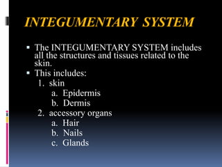
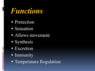
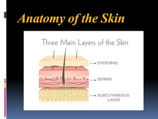
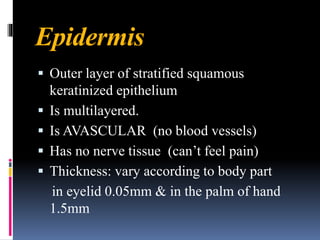
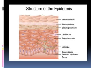
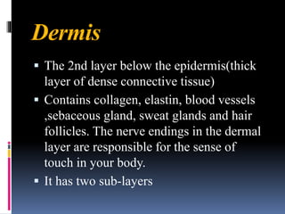
SHAZIA REHMAN
Integumentary system
INTEGUMENTARY SYSTEM
▪ The INTEGUMENTARY SYSTEM includes
all the structures and tissues related to the
skin.
▪ This includes:
1. skin
a. Epidermis
b. Dermis
2. accessory organs
a. Hair
b. Nails
c. Glands
Skin is the largest organ of the body. It
has an area of 2 square metres (22
square feet) in adults, and weighs
about 3 to 5 kg. The thickness of skin
varies from 0.5mm thick on the
eyelids to 4.0mm thick on the heels of
your feet.
Functions
▪ Protection
▪ Sensation
▪ Allows movement
▪ Synthesis
▪ Excretion
▪ Immunity
▪ Temperature Regulation
Anatomy of the Skin
Epidermis
▪ Outer layer of stratified squamous
keratinized epithelium
▪ Is multilayered.
▪ Is AVASCULAR (no blood vessels)
▪ Has no nerve tissue (can’t feel pain)
▪ Thickness: vary according to body part
in eyelid 0.05mm & in the palm of hand
1.5mm
Stratum Corneum
▪ This layer consists of 15-30 layers of dead
keratinized cells.
▪ This dry, dead layer helps prevent the
penetration of microbes and provides a
mechanical protection against abrasion for the
more delicate, underlying layers.
▪ Cells in this layer are shed periodically and
are replaced by cells pushed up from the
stratum granulosum
▪ They are shed every four weeks.
Stratum Lucidum
▪ This layer has closely packed
keratinocyte cells with seleiden, a
clear protein rich in lipids.
▪ This is the thick skin that you find in
your palms and feet and its
transparency is due to seleiden.
Stratum Granulosum
▪ This layer is composed of 3-5 layers of
Keratinocytes. It appears grainy due to the
changes in keratinocytes that are being pushed
to this layer.
▪ The cells become flatter, their cell membranes
thicken, and they generate large amounts of
the proteins keratin.
Stratum Spinosum
▪ This layer is composed of daughter
keratinocytes and dendritic cells,
which fight infections in the body.
▪ Stratum spinosum is shiny in
appearance due to protruding
structures called desmosomes.
Stratum Basale
▪ This is the lowest layer of epidermis and is
composed of keratinocytes, melanocytes and
tactile cells.
▪ Keratinocytes produced here constantly
undergo cell division and are pushed to the
upper layers of the epidermis.
▪ Melanocytes produce melanin, a brown
pigment that is responsible for skin coloration
and protecting against the harmful effecte of
UV light.
Dermis
▪ The 2nd layer below the epidermis(thick
layer of dense connective tissue)
▪ Contains collagen, elastin, blood vessels
,sebaceous gland, sweat glands and hair
follicles. The nerve endings in the dermal
layer are responsible for the sense of
touch in your body.
▪ It has two sub-layers
The Papillary Layer
▪ The upper, papillary layer, contains a
thin arrangement of collagen fibers.
The papillary layer supplies nutrients
to select layers of the epidermis and
regulates temperature.
▪ This layer forms blunt conical
projection, called Dermal papillae.
The Reticular Layer
▪ The lower, reticular layer, is thicker
and made of thick collagen fibers that
are arranged in parallel to the surface
of the skin.
▪ It supports other components of the
skin, such as hair follicles, sweat
glands, and sebaceous glands.
Hypodermis
▪ The consists of well-vascularized, loose
connective tissue and adipose tissue. The
deeper tissues including muscle, tendon,
ligament, joint capsule and bone that lie
beneath the hypodermis.
▪ This layer maintains the temperature and acts
like a cushion or shock absorber. The layer
also helps attach dermis to the bones and
muscles.
Hair
▪ Hair is an accessory organ of the skin made
of columns of tightly packed dead
keratinocytes /cornified found in most
regions of the body.
▪ The few hairless parts of the body include
the palmar surface of the hands, plantar
surface of the feet, lips, labia minora,
and penis.
Hair Shaft: part of your hair that can be seen
above your scalp.
Hair follicle :A tunnel-shaped structure in the
epidermis of the skin. Hair starts growing at
the bottom of a hair follicle.
Bulb: Forms the base of the hair follicle
Hair Papilla: A knoblike vascular indentation
of the bottom of a hair follicle, on which
the hair bulb fits.
Arrector pili muscle
These are small smooth muscles attached to hair
follicles. Contraction of these Muscles causes
the hairs to stand on end.
Life span of hair
Lifespan varies in different region
Scalp hair :3-5 years
Eye brow and eyelash :3-5 months
Nails
Nails are accessory organs of
the skin made of sheets of
hardened keratinocytes and
found on the distal ends of the
fingers and toes.
Parts of nail
Nail plate: the visible hard part of the nail
Nail folds: the skin that frames the nail plate
on three sides
Nail bed: (epithelial layer)the skin beneath the
nail plate
Cuticle: the tissue that overlaps your nail plate
at the base of your nail - it protects the new
keratin cells that slowly emerge from the nail
bed
Lunula: the whitish, half-moon shape at the
base of your nail underneath the plate
Nail matrix
Root of the nail. Forms nail plate from base of nail
bed
Eponychium
Skin just proximal to cuticle, Binds nail to
underlying skin at proximal margin
Hyponychium
Area beneath free edge of nail, Junction where nail
bed meets tip of digit skin
Perionychium
the tissue bordering the root and sides of a fingernail
or toenail.
Sebaceous glands
▪ Sebaceous glands are exocrine glands found in
the dermis of the skin that produce an oily
secretion known as sebum.
▪ Sebum is produced in the sebaceous glands
and carried through ducts to the surface of the
skin or to hair follicles.
Sebaceous glands are found in every part of
The skin except for the thick skin of the
palms of the hands and soles of the feet.
Function:
▪ Lubricates and protects the skin
Sweat glands (sudoriferous gland )
The body of the gland is made up of a coiled tube,
surrounded by a good blood supply, and a duct,
which opens onto the skin surface through a
pore(sweat pore).
Sweat glands are found throughout the skin but are
more numerous in areas such as the soles of the feet,
palms of the hand, armpits and groin.
Sweat that helps flush out toxins from the body,
keeping it cool.

Integumentary system(Skin & its assessory organs)

  • 1.
  • 2.
    INTEGUMENTARY SYSTEM ▪ TheINTEGUMENTARY SYSTEM includes all the structures and tissues related to the skin. ▪ This includes: 1. skin a. Epidermis b. Dermis 2. accessory organs a. Hair b. Nails c. Glands
  • 3.
    Skin is thelargest organ of the body. It has an area of 2 square metres (22 square feet) in adults, and weighs about 3 to 5 kg. The thickness of skin varies from 0.5mm thick on the eyelids to 4.0mm thick on the heels of your feet.
  • 4.
    Functions ▪ Protection ▪ Sensation ▪Allows movement ▪ Synthesis ▪ Excretion ▪ Immunity ▪ Temperature Regulation
  • 5.
  • 6.
    Epidermis ▪ Outer layerof stratified squamous keratinized epithelium ▪ Is multilayered. ▪ Is AVASCULAR (no blood vessels) ▪ Has no nerve tissue (can’t feel pain) ▪ Thickness: vary according to body part in eyelid 0.05mm & in the palm of hand 1.5mm
  • 8.
    Stratum Corneum ▪ Thislayer consists of 15-30 layers of dead keratinized cells. ▪ This dry, dead layer helps prevent the penetration of microbes and provides a mechanical protection against abrasion for the more delicate, underlying layers. ▪ Cells in this layer are shed periodically and are replaced by cells pushed up from the stratum granulosum ▪ They are shed every four weeks.
  • 9.
    Stratum Lucidum ▪ Thislayer has closely packed keratinocyte cells with seleiden, a clear protein rich in lipids. ▪ This is the thick skin that you find in your palms and feet and its transparency is due to seleiden.
  • 10.
    Stratum Granulosum ▪ Thislayer is composed of 3-5 layers of Keratinocytes. It appears grainy due to the changes in keratinocytes that are being pushed to this layer. ▪ The cells become flatter, their cell membranes thicken, and they generate large amounts of the proteins keratin.
  • 11.
    Stratum Spinosum ▪ Thislayer is composed of daughter keratinocytes and dendritic cells, which fight infections in the body. ▪ Stratum spinosum is shiny in appearance due to protruding structures called desmosomes.
  • 12.
    Stratum Basale ▪ Thisis the lowest layer of epidermis and is composed of keratinocytes, melanocytes and tactile cells. ▪ Keratinocytes produced here constantly undergo cell division and are pushed to the upper layers of the epidermis. ▪ Melanocytes produce melanin, a brown pigment that is responsible for skin coloration and protecting against the harmful effecte of UV light.
  • 13.
    Dermis ▪ The 2ndlayer below the epidermis(thick layer of dense connective tissue) ▪ Contains collagen, elastin, blood vessels ,sebaceous gland, sweat glands and hair follicles. The nerve endings in the dermal layer are responsible for the sense of touch in your body. ▪ It has two sub-layers
  • 14.
    The Papillary Layer ▪The upper, papillary layer, contains a thin arrangement of collagen fibers. The papillary layer supplies nutrients to select layers of the epidermis and regulates temperature. ▪ This layer forms blunt conical projection, called Dermal papillae.
  • 15.
    The Reticular Layer ▪The lower, reticular layer, is thicker and made of thick collagen fibers that are arranged in parallel to the surface of the skin. ▪ It supports other components of the skin, such as hair follicles, sweat glands, and sebaceous glands.
  • 16.
    Hypodermis ▪ The consistsof well-vascularized, loose connective tissue and adipose tissue. The deeper tissues including muscle, tendon, ligament, joint capsule and bone that lie beneath the hypodermis. ▪ This layer maintains the temperature and acts like a cushion or shock absorber. The layer also helps attach dermis to the bones and muscles.
  • 17.
    Hair ▪ Hair isan accessory organ of the skin made of columns of tightly packed dead keratinocytes /cornified found in most regions of the body. ▪ The few hairless parts of the body include the palmar surface of the hands, plantar surface of the feet, lips, labia minora, and penis.
  • 19.
    Hair Shaft: partof your hair that can be seen above your scalp. Hair follicle :A tunnel-shaped structure in the epidermis of the skin. Hair starts growing at the bottom of a hair follicle. Bulb: Forms the base of the hair follicle Hair Papilla: A knoblike vascular indentation of the bottom of a hair follicle, on which the hair bulb fits.
  • 20.
    Arrector pili muscle Theseare small smooth muscles attached to hair follicles. Contraction of these Muscles causes the hairs to stand on end. Life span of hair Lifespan varies in different region Scalp hair :3-5 years Eye brow and eyelash :3-5 months
  • 21.
    Nails Nails are accessoryorgans of the skin made of sheets of hardened keratinocytes and found on the distal ends of the fingers and toes.
  • 22.
  • 23.
    Nail plate: thevisible hard part of the nail Nail folds: the skin that frames the nail plate on three sides Nail bed: (epithelial layer)the skin beneath the nail plate Cuticle: the tissue that overlaps your nail plate at the base of your nail - it protects the new keratin cells that slowly emerge from the nail bed Lunula: the whitish, half-moon shape at the base of your nail underneath the plate
  • 24.
    Nail matrix Root ofthe nail. Forms nail plate from base of nail bed Eponychium Skin just proximal to cuticle, Binds nail to underlying skin at proximal margin Hyponychium Area beneath free edge of nail, Junction where nail bed meets tip of digit skin Perionychium the tissue bordering the root and sides of a fingernail or toenail.
  • 25.
    Sebaceous glands ▪ Sebaceousglands are exocrine glands found in the dermis of the skin that produce an oily secretion known as sebum. ▪ Sebum is produced in the sebaceous glands and carried through ducts to the surface of the skin or to hair follicles.
  • 26.
    Sebaceous glands arefound in every part of The skin except for the thick skin of the palms of the hands and soles of the feet. Function: ▪ Lubricates and protects the skin
  • 27.
    Sweat glands (sudoriferousgland ) The body of the gland is made up of a coiled tube, surrounded by a good blood supply, and a duct, which opens onto the skin surface through a pore(sweat pore). Sweat glands are found throughout the skin but are more numerous in areas such as the soles of the feet, palms of the hand, armpits and groin. Sweat that helps flush out toxins from the body, keeping it cool.