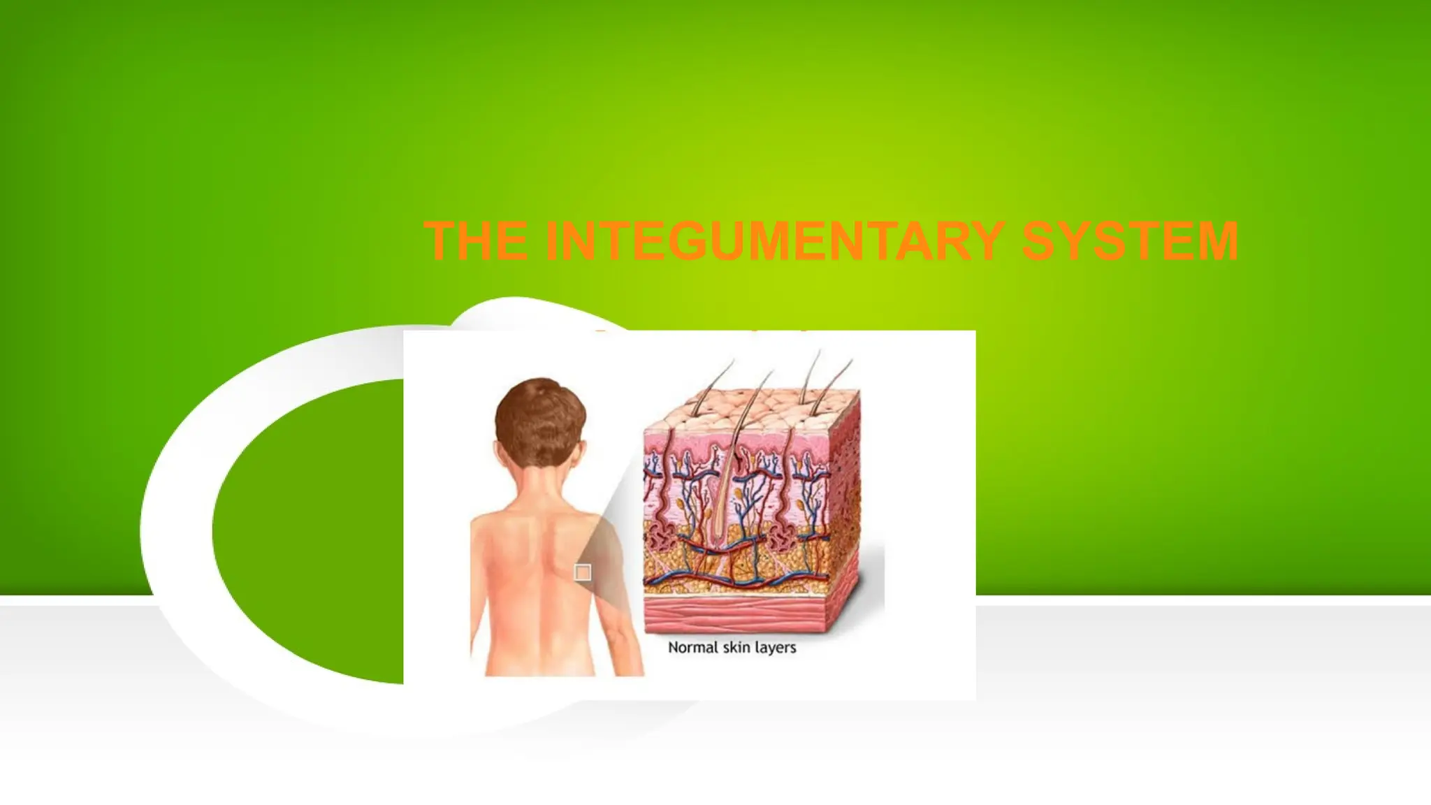
THE INTEGUMENTARY SYSTEM
the largest body
system
skin and the skin
derivatives - hair,
nails and skin
(exocrine) glands.
The integument
(skin) as an organ
3.
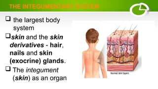
THE INTEGUMENTARY SYSTEM
Theintegument (skin)
the largest organ of the body
16% of body weight
1.5 to 2 m2
in area
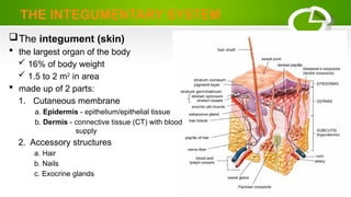
made up of 2 parts:
1. Cutaneous membrane
a. Epidermis - epithelium/epithelial tissue
b. Dermis - connective tissue (CT) with blood
supply
2. Accessory structures
a. Hair
b. Nails
c. Exocrine glands
4.
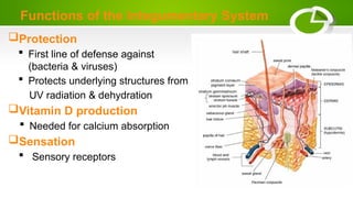
Functions of theIntegumentary System
Protection
First line of defense against
(bacteria & viruses)
Protects underlying structures from
UV radiation & dehydration
Vitamin D production
Needed for calcium absorption
Sensation
Sensory receptors
5.
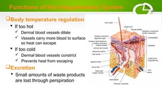
Body temperature regulation
If too hot
Dermal blood vessels dilate
Vessels carry more blood to surface
so heat can escape
If too cold
Dermal blood vessels constrict
Prevents heat from escaping
Excretion
Small amounts of waste products
are lost through perspiration
Functions of the Integumentary System
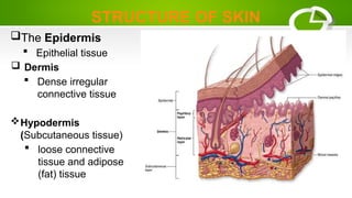
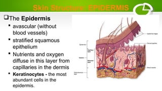
Skin Structure: EPIDERMIS
TheEpidermis
avascular (without
blood vessels)
stratified squamous
epithelium
Nutrients and oxygen
diffuse in this layer from
capillaries in the dermis
Keratinocytes - the most
abundant cells in the
epidermis.
8.
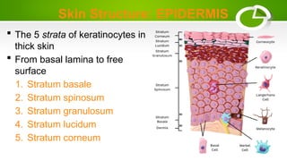
Skin Structure: EPIDERMIS
The 5 strata of keratinocytes in
thick skin
From basal lamina to free
surface
1. Stratum basale
2. Stratum spinosum
3. Stratum granulosum
4. Stratum lucidum
5. Stratum corneum
9.
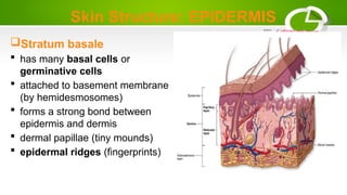
Skin Structure: EPIDERMIS
Stratumbasale
has many basal cells or
germinative cells
attached to basement membrane
(by hemidesmosomes)
forms a strong bond between
epidermis and dermis
dermal papillae (tiny mounds)
epidermal ridges (fingerprints)
10.
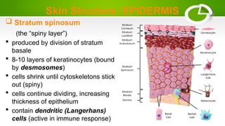
Skin Structure: EPIDERMIS
Stratum spinosum
(the “spiny layer”)
produced by division of stratum
basale
8-10 layers of keratinocytes (bound
by desmosomes)
cells shrink until cytoskeletons stick
out (spiny)
cells continue dividing, increasing
thickness of epithelium
contain dendritic (Langerhans)
cells (active in immune response)
11.
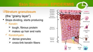
Skin Structure: EPIDERMIS
Stratumgranulosum
(the “grainy layer”)
Stops dividing, starts producing
Keratin
tough, fibrous protein
makes up hair and nails
Keratohyalin
dense granules
cross-link keratin fibers
12.
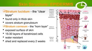
Skin Structure: EPIDERMIS
Stratumlucidum - the “clear
layer”
found only in thick skin
covers stratum granulosum
Stratum corneum - the “horn layer”
exposed surface of skin
15-30 layers of keratinized cells
water resistant
shed and replaced every 2 weeks
13.
Skin Structure: EPIDERMIS
ThinSkin
Covers most of the body
Has 4 layers of keratinocytes
Thick Skin
Covers the palms of the hands
and soles of the feet
Has 5 layers of keratinocytes
14.
Skin Structure: DERMIS
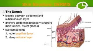
TheDermis
located between epidermis and
subcutanoues layer
anchors epidermal accessory structure
(hair follicles, sweat glands)
two components
1. outer papillary layer
2. deep reticular layer
15.
Skin Structure: DERMIS
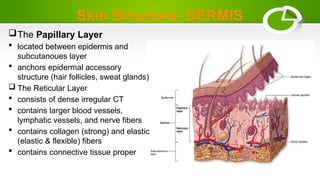
ThePapillary Layer
located between epidermis and
subcutanoues layer
anchors epidermal accessory
structure (hair follicles, sweat glands)
The Reticular Layer
consists of dense irregular CT
contains larger blood vessels,
lymphatic vessels, and nerve fibers
contains collagen (strong) and elastic
(elastic & flexible) fibers
contains connective tissue proper
16.
DERMATITIS
An inflammation ofthe papillary
layer.
Caused by:
infection, radiation, mechanical irritation
or chemicals (e.g. poison ivy)
Characterized by itch or pain
17.
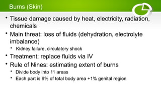
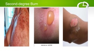
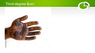
Burns (Skin)
• Tissuedamage caused by heat, electricity, radiation,
chemicals
• Main threat: loss of fluids (dehydration, electrolyte
imbalance)
• Kidney failure, circulatory shock
• Treatment: replace fluids via IV
• Rule of Nines: estimating extent of burns
• Divide body into 11 areas
• Each part is 9% of total body area +1% genital region
Skin Structure: HYPODERMIS
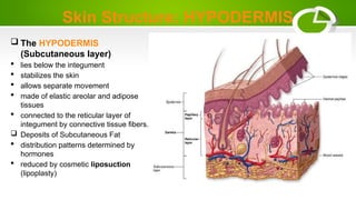
The HYPODERMIS
(Subcutaneous layer)
lies below the integument
stabilizes the skin
allows separate movement
made of elastic areolar and adipose
tissues
connected to the reticular layer of
integument by connective tissue fibers.
Deposits of Subcutaneous Fat
distribution patterns determined by
hormones
reduced by cosmetic liposuction
(lipoplasty)
27.
Skin Structure
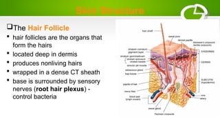
The HairFollicle
hair follicles are the organs that
form the hairs
located deep in dermis
produces nonliving hairs
wrapped in a dense CT sheath
base is surrounded by sensory
nerves (root hair plexus) -
control bacteria
28.
Structure of Hair
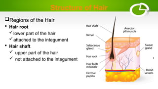
Regionsof the Hair
Hair root
lower part of the hair
attached to the integument
Hair shaft
upper part of the hair
not attached to the integument
29.
Hair Function
Head
UVprotection
cushion from trauma
insulation
Nostrils, Ear canals and Eyelashes
prevent entry of foreign material
Root hair plexus
sensory nerves at base of hair follicle that detect slight
movement of hair
Arrector pili muscle
attached to every hair follicle
contract to stand hair perpendicular to skin surface
30.
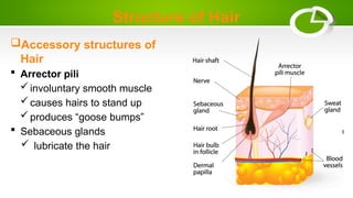
Structure of Hair
Accessorystructures of
Hair
Arrector pili
involuntary smooth muscle
causes hairs to stand up
produces “goose bumps”
Sebaceous glands
lubricate the hair
31.
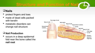
Structure and Functionof Nail
Nails
protect fingers and toes
made of dead cells packed
with keratin
metabolic disorders can
change nail structure
Nail Production
occurs in a deep epidermal
fold near the bone called the
nail root


![NURS1108_Lecture_4_-_Integumentary[1].pptx](https://cdn.slidesharecdn.com/ss_thumbnails/nurs1108lecture4-integumentary1-250824175356-397255ad-thumbnail.jpg?width=640&height=640&fit=bounds)


