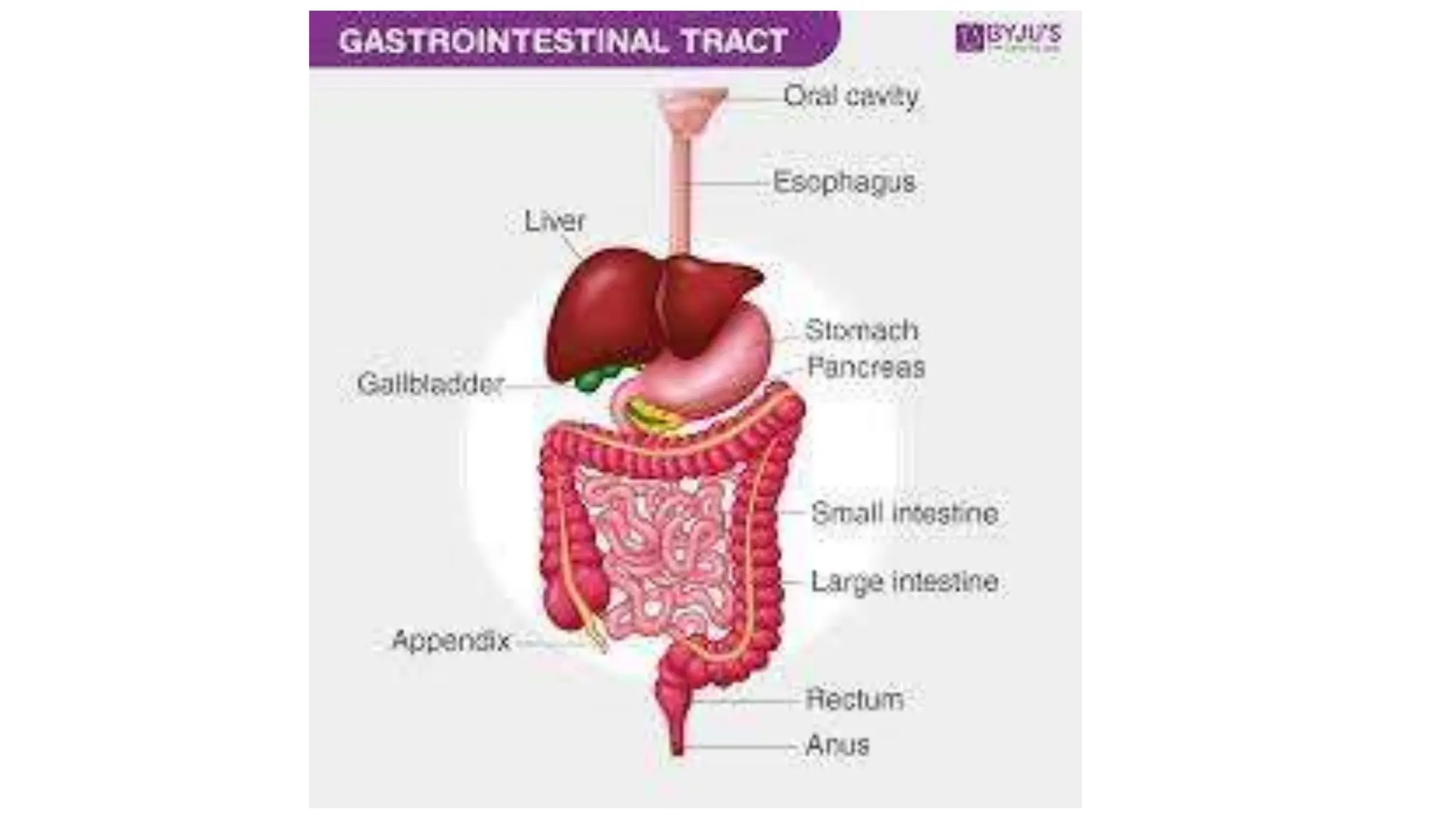
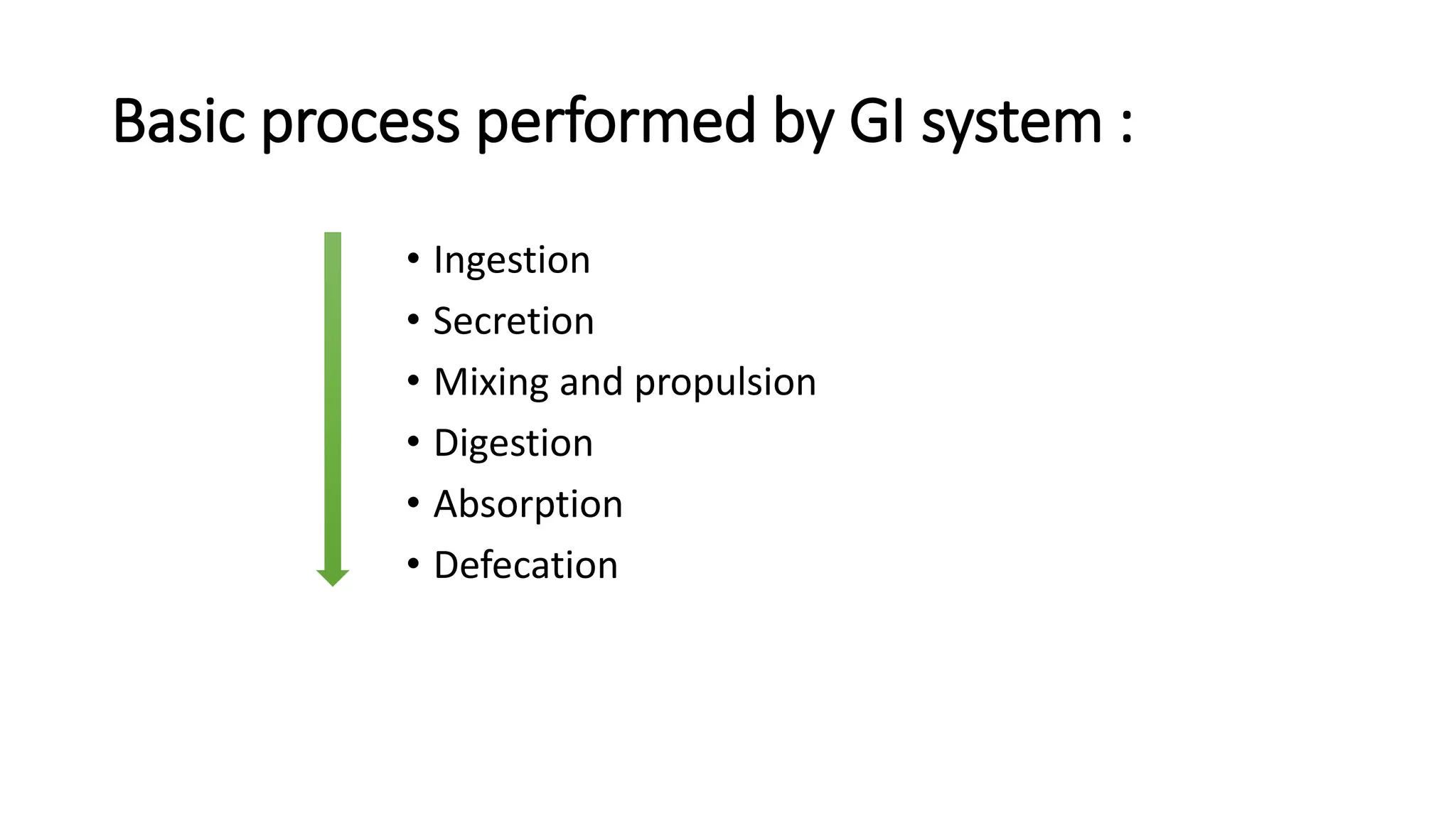
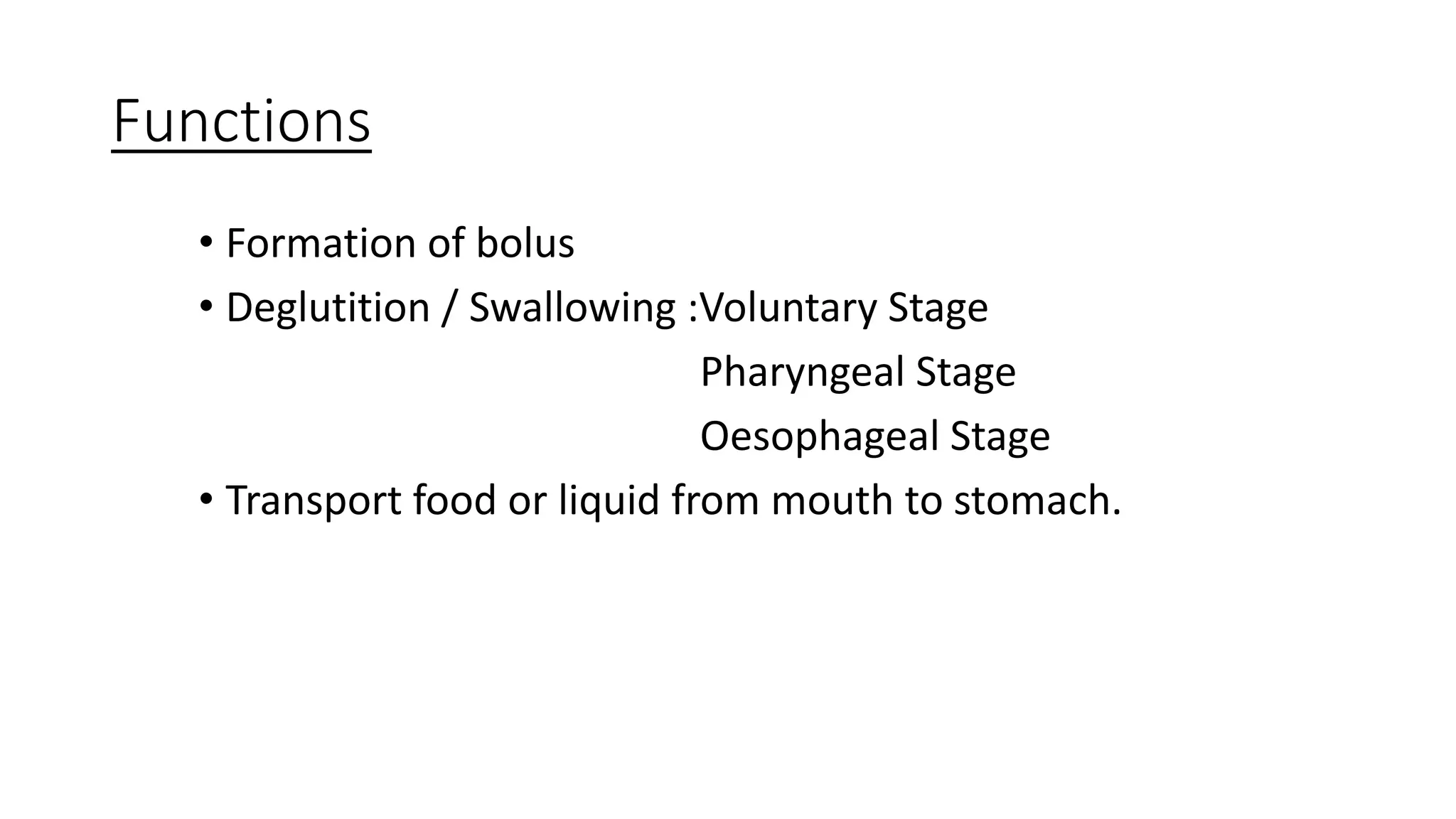
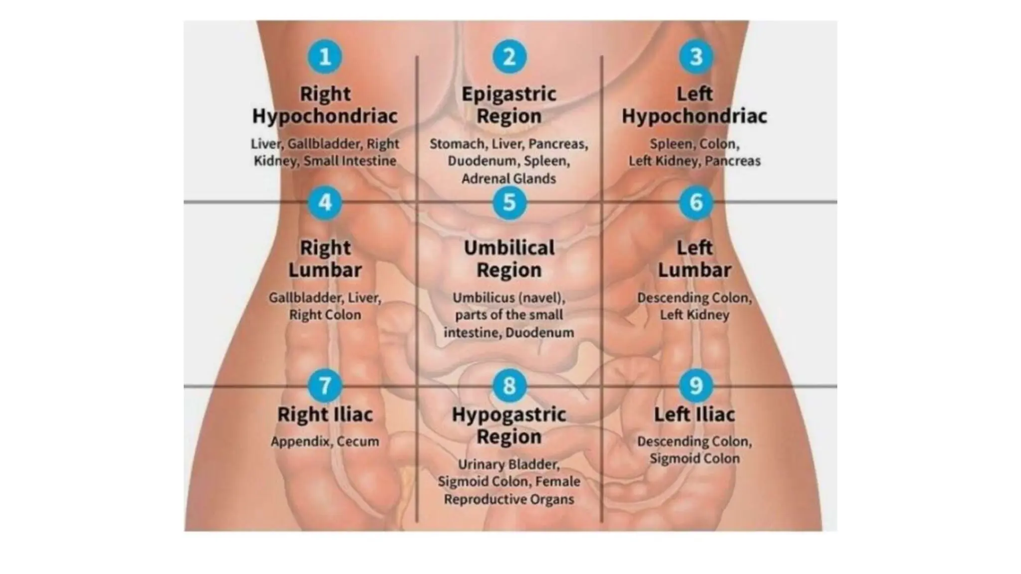
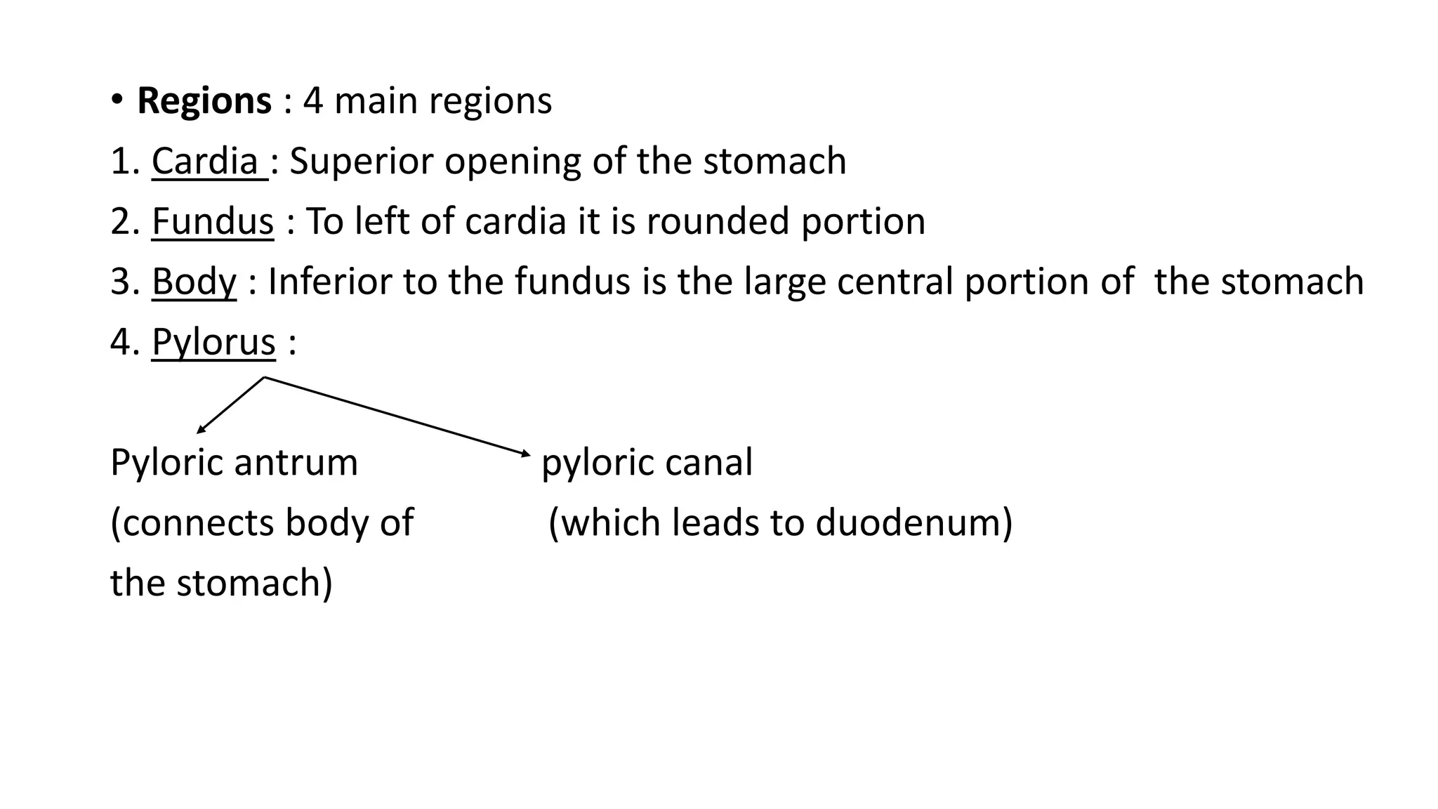
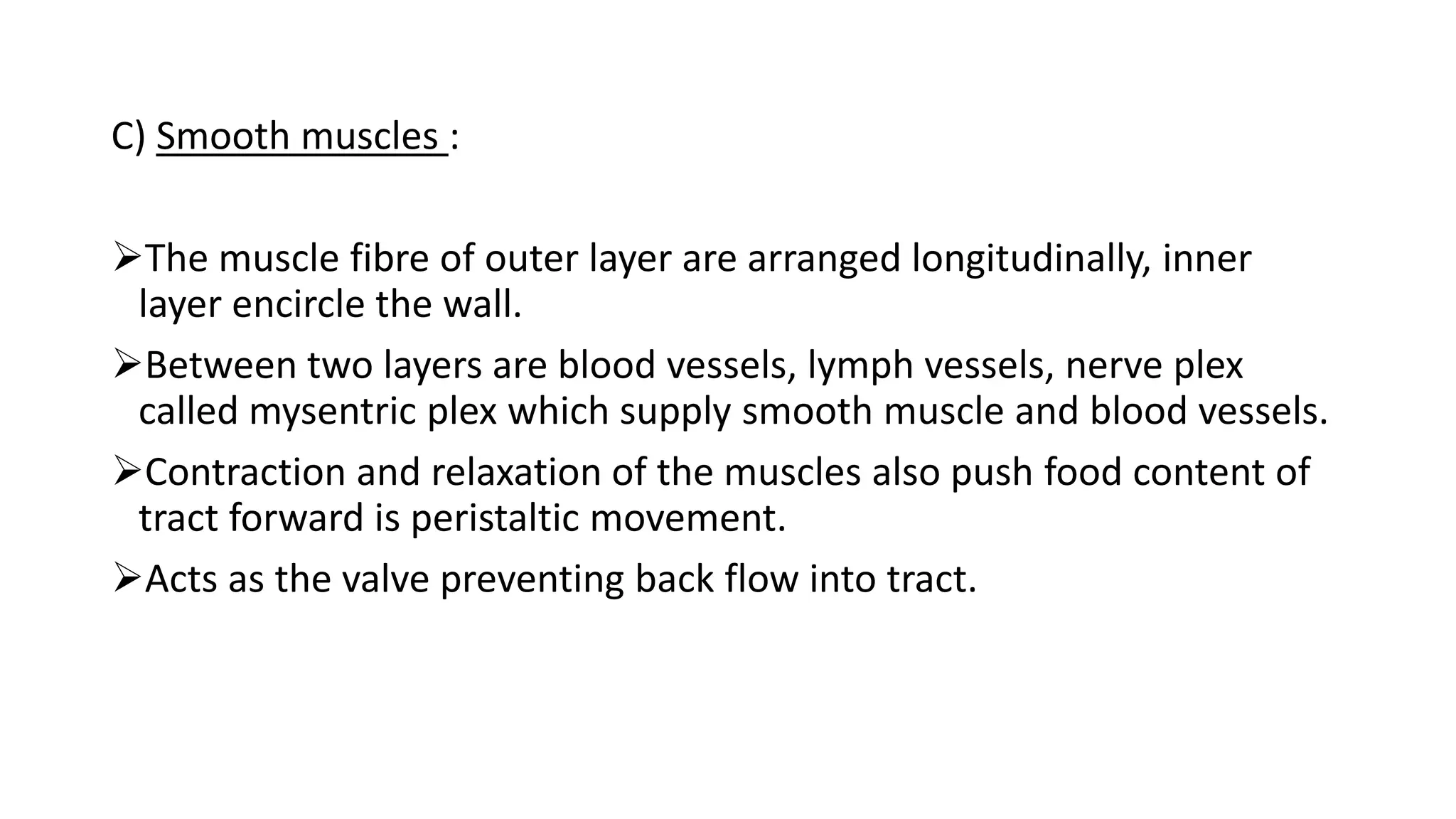
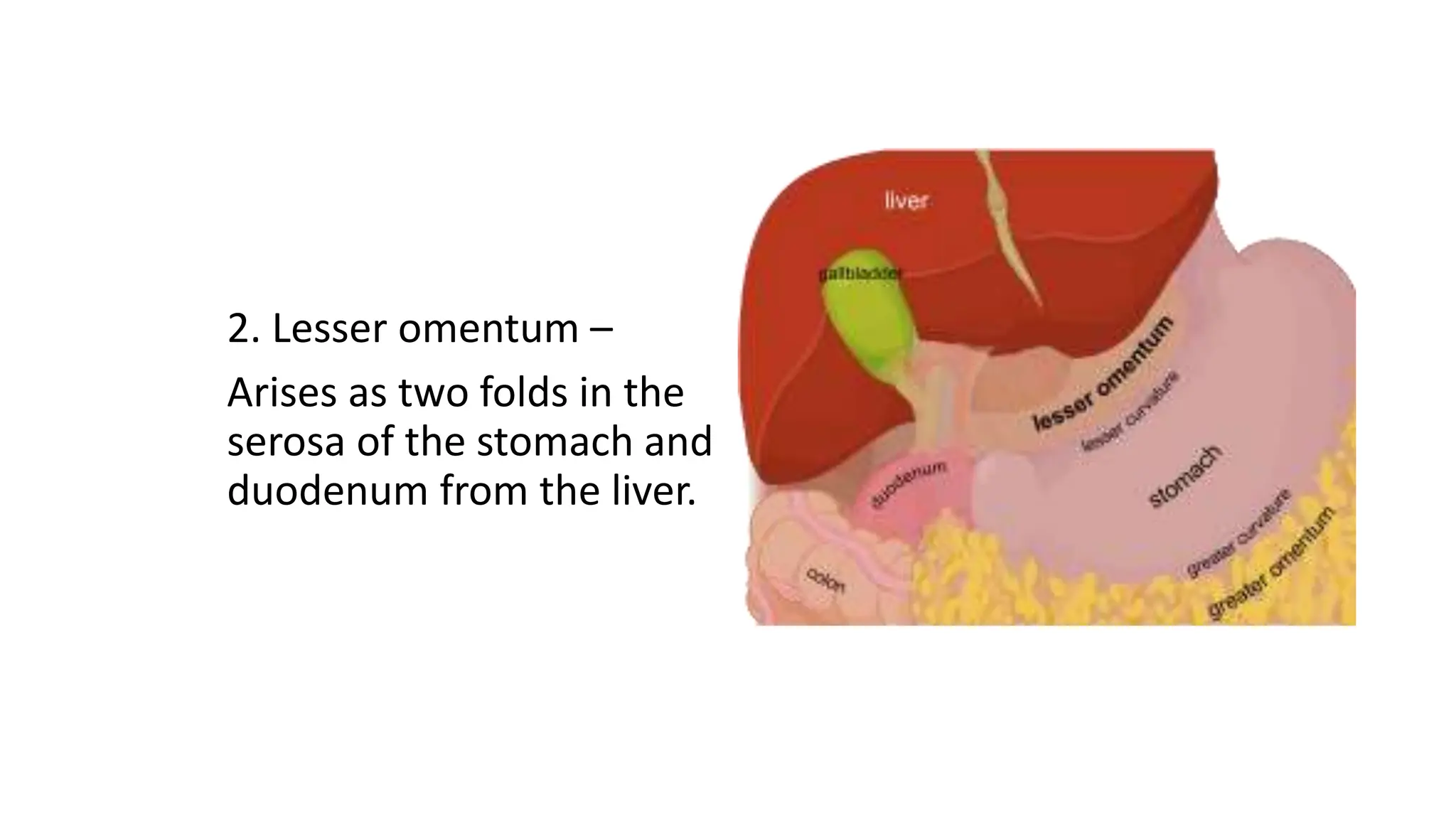
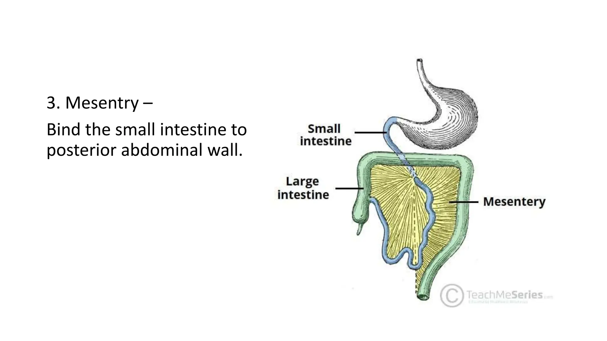
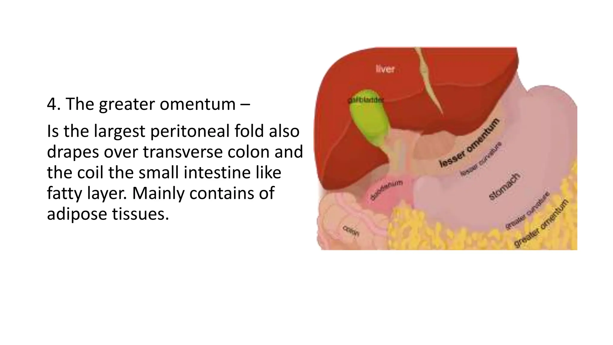
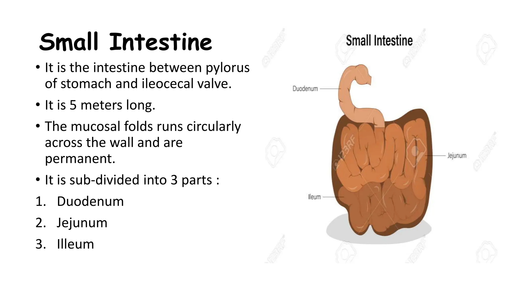
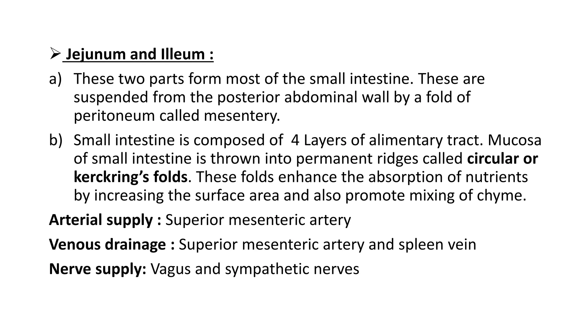
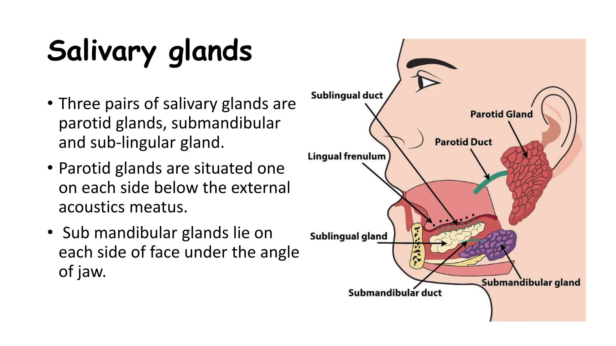
THE DIGESTIVE
SYSTEM
MISS CHARUTA KUNJEER
TUTOR
SANCHETI INSTITUTE OF NURSING EDUCATION
ORGAN OF DIGESTIVE SYSTEM
GASTRO INTESTINAL TRACT OR
ALIMENTARY CANAL
• Mouth
• Pharynx
• Oesophagus
• Stomach
• Small intestine
• Large intestine
ACCESSORY DIGESTIVE ORGAN
• Teeth
• Tongue
• Salivary gland
• Liver
• Gall bladder
• Pancreas
Basic process performed by GI system :
• Ingestion
• Secretion
• Mixing and propulsion
• Digestion
• Absorption
• Defecation
Oesophagus
• It is collapsible muscular tube and 25
cm long.
• It lies in median plain of thorax, in
front of vertebral column behind
trachea and heart.
• The oesophagus ends into superior
part of stomach known as
oesophageal hiatus.
• This oesophagus passes through
diaphragm.
Structure
It is made up of 4 layers Superficial layer adventitia (outer layer)
Mucosa (lining of body canals)
Sub mucosa (layer of connective tissues)
Muscularis (muscular layer that surrounds
the hollow organ)
Blood supply :
Oesophageal branch of aorta
Abdominal region is supplied by inferior phrenic and left gastric branch of
celiac artery
Venous drainage :
Azygous (unilateral vessel that ascends in the right posterior mediastinum
along with the thoracic vertebrae) and hemiazygous (left posterior
mediastinum with the thoracic vertebrae) vein.
Nerve supply :
Sympathetic and para sympathetic nerve.
Functions
• Formation of bolus
• Deglutition / Swallowing :Voluntary Stage
Pharyngeal Stage
Oesophageal Stage
• Transport food or liquid from mouth to stomach.
Stomach
• Stomach : Most dilated part of digestive tube, J shaped
enlargement, of GI tract directly inferior to diaphragm.
• Situation : Epigastric, Left Hypochondria And Umbilical Region.
• Parts : 2 orifices (cardiac at oesophagus and pyloric at duodenum)
• Curvatures : 2 curvatures (lesser-superior border and greater-
inferior border)
• Parts : 1. Fundus
2. Body
3. Pylorus
• Regions : 4 main regions
1. Cardia : Superior opening of the stomach
2. Fundus : To left of cardia it is rounded portion
3. Body : Inferior to the fundus is the large central portion of the stomach
4. Pylorus :
Pyloric antrum pyloric canal
(connects body of (which leads to duodenum)
the stomach)
• Blood Supply :
Left Gastric Artery (Branch Of Celiac
Trunk)
Right Gastric Artery (Branch Of Hepatic
Trunk)
Right Gastroepiploic (Branch Of Gastro
Duodenal )
Left Gastroepiploic (Branch Of Splenic
Artery)
• Nerve supply :
Sympathetic supply by celiac trunk
Para-sympathetic from vagus nerve
Sympathetic decrease the mobility
of stomach and secretions of gastric
juice
Vagal stimulation has opposite effect
• Functions :
1. Reservoir of food
2. Peristaltic movement it softens and mix food with gastric
juices.
3. Gastric gland produce gastric juice contains enzyme that
play important role in digestion of food.
4. Gastric gland produce HCL which destroy many organism
present in food and drink.
5. Absorption of water, glucose, alcohol and salt.
Alimentary Canal
Alimentary Canal
• The wall of GI tract from the lower oesophagus to anal canal has basic
4 layered arrangement of tissues : Mucosa, submucosa, muscularis,
serosa.
A ) Mucosa is mainly made of three layers of tissues :
1. Mucosa membrane
2. Lamina propria
3. Muscularis mucosa
Made up of stratified squamous epithelium
with mucus secreting gland.
Mucus lubricates & protects from digestive
enzymes.
Secretions are saliva, gastric juice, pancreatic
juice, bile juice are called as digestive juice
and contains enzymes which chemically
breaks the food.
B) Submucosa :
It is made up of elastic fibres.
With in this layers there are plexus of blood vessels and
nerve and lymph vessels and lymphoid tissues.
Blood vessels are arteriole, venule, capillaries.
Nerve supply is mesenteric plex
C) Smooth muscles :
The muscle fibre of outer layer are arranged longitudinally, inner
layer encircle the wall.
Between two layers are blood vessels, lymph vessels, nerve plex
called mysentric plex which supply smooth muscle and blood vessels.
Contraction and relaxation of the muscles also push food content of
tract forward is peristaltic movement.
Acts as the valve preventing back flow into tract.
D) Serous or peritoneum :
• The peritoneum is the largest serous membrane of the body.
• Some organs lies in the peritoneal cavity they are kidney, pancreas.
• It contains a large fold that weave between viscera, these contain blood
vessels, lymphatic vessels, nerves
5 Major Peritoneal Folds
1. Faliciform ligament-
which attaches liver to anterior
abdominal wall
2. Lesser omentum –
Arises as two folds in the
serosa of the stomach and
duodenum from the liver.
3. Mesentry –
Bind the small intestine to
posterior abdominal wall.
4. The greater omentum –
Is the largest peritoneal fold also
drapes over transverse colon and
the coil the small intestine like
fatty layer. Mainly contains of
adipose tissues.
5. Mesocolon –
Binds large intestine and
posterior wall of abdominal
wall
Small Intestine
• It is the intestine between pylorus
of stomach and ileocecal valve.
• It is 5 meters long.
• The mucosal folds runs circularly
across the wall and are
permanent.
• It is sub-divided into 3 parts :
1. Duodenum
2. Jejunum
3. Illeum
Duodenum :
It extends from pylorus to ileocecal junction. It is about 6 m long(the
length is greater in male then female)
Features : Large surface area for digestion of food which is achieved by
its greater length, presence of circular folds of mucus membrane and
villi and not obliterated by distention. Villi are finger like projections.
Supply : Sympathetic and Para-sympathetic nerve.
Function : Digestion and Absorption of food.
 Jejunum and Illeum :
a) These two parts form most of the small intestine. These are
suspended from the posterior abdominal wall by a fold of
peritoneum called mesentery.
b) Small intestine is composed of 4 Layers of alimentary tract. Mucosa
of small intestine is thrown into permanent ridges called circular or
kerckring’s folds. These folds enhance the absorption of nutrients
by increasing the surface area and also promote mixing of chyme.
Arterial supply : Superior mesenteric artery
Venous drainage : Superior mesenteric artery and spleen vein
Nerve supply: Vagus and sympathetic nerves
Large Intestine
• It is the last part of the digestive
tube and follows the small
intestine.
• It starts at ileocaecal junction.
• Parts are :
1) Caecum
2) Vermiform appendix
3) Colon
4) Rectum
5) Anal canal
1) Caecum :
Lies in the right iliac fossa. The ileocaecal valve guards the opening of
ileum into caecum, 2 cms below this opening of the vermiform
appendix. It continues with the ascending colon.
2) Vermiform Appendix:
It is the narrowest part of the digestive tube. It is usually 8 cms long
and 0.5 cm wide. It has a base and a tip. The tip may occupy different
positions.
Arterial supply : Appendicular artery and one of the caecal branches of
mesenteric artery.
3) Colon :
It has three parts
a) Ascending colon
b) Transverse colon
c) Descending colon
In colon three thick bands called taeniae coli,
situated at regular interval around it.
Small pouches of visceral peritoneum filled
with fat are attached to taeniae coli and are
called Epiploic appendages.
Artery supply : superior mesenteric artery
Venous drainage : portal vein, para-aortic lymph-nodes
Nerve supply :
1. parasympathetic nerve : Ascending and most of the transverse
colon
2. Pelvic nerve : Small part of transverse colon and pelvic colon
3. Sympathetic nerve : Relax smooth muscle and constrict ileocaecal
and internal anal sphintcher.
4) Rectum and Anus :
Rectum starts at the middle of sacrum and is 15 cm long. It follows
forward curvature of sacrum and coccyx and ends 2.5 cms below and
in front of coccyx where it continues as anal canal.
Anal canal is the last 4 cms of digestive tube. From the anorectal
junction it passes downward and backward to the anus. There are two
layers/ walls they are internal and external anal sphincter of skeletal
muscle.
Thus anal canal is site of porto-systemic anastomosis.
Arterial Supply : Superior rectal artery, middle and inferior artery
Venous drainage : Superior rectal artery end in portal vein.
Liver
• The liver is the largest gland in the
body.
• Weight : 1500 g also receives 1500
ml of the blood per minute.
• Lies : under diaphragm, right
hypochondriac/ epigastric and
partly into left hypochondriac
regions. It extends into the
thoracic cavity upto 5 th rib and
receives protection from costal
cartilages.
• Relations :
1. Anterior : Abdominal wall and diaphragm
2. Posterior : Oesophagus, gallbladder aorta, inferior vena cava
3. Below : Stomach, duodenum, right kidney, suprarenal gland
4. Laterally : Lower ribs
• Liver biopsy is done through right 8th intercostal space in full
expiration to avoid injury to lung.
• Liver has inferior or visceral surface which is related to various
abdominal viscera.
• Fissure : It is 5 cm known as portal hepatitis (gateway of liver).
• Attached to the margins of porta hepatitis is a fold of peritoneum the
lesser omentum which extends from the liver to the lesser curvature
of stomach.
• Lobes : 4 lobes
1. Right lobe
2. Left lobe
3. Caudate lobe
4. Quadrate lobe
• Arterial supply :
Common hepatic artery of coeliac trunk of aorta (20%), digestive tract
from portal vein(80%).
• Venous drainage :
Hepatic vein into inferior vena cava.
• Nerve supply :
Sympathetic nerve enters the liver with hepatic artery to control the
lumen of the vessels.
Functions of liver –
•Metabolism -The liver is a hub for many physiological processes, including
macronutrient metabolism, blood volume regulation, and lipid and cholesterol
homeostasis.
•Immunity- The liver supports the immune system and breaks down few
compounds, including many current drugs.
•Digestion -The liver produces bile, which helps carry away waste and break
down fats in the small intestine during digestion.
•Detoxification -The liver stores vitamins and minerals (iron and copper), and
releases them into the blood when needed.
•Vitamin storage- The liver makes up about 2% of an adult's body weight.
Gall Bladder
• Gall bladder is pyriform organ 7 cm long with a capacity of 50 ml.
• It is present in a depression on the inferior surface of the liver.
• Function: It acts as a storage and concentrating organ of bile.
• Parts : Fundus, Body
• Arterial Supply : Right Hepatic Artery
• Venous Drainage : Extrahepatic Biliary Apparatus Biliary Apparatus
Drains Into Lymph Nodes In The Porta Hepatitis.
• Nerve Supply : Sympathetic Nerve Supply
• Epithelial Cells : Simple Columnar Epithelium
Pancreas
• Pancreas is a gland present across the upper part of posterior
abdominal wall from right to the left side.
• It contains both exocrine secretion from digestion of carbohydrates,
fats and protein as well as endocrine secretion, from Islets of
Langerhans, which act on the carbohydrates metabolism.
• Pancreas is 15 cms long and also consists of head, neck, body and tail.
• An process from the lowest part of head passes to left behind
superior mesenteric vessels.
• Neck : A small segment behind
which superior mesenteric vein and
splenic vein join to form the portal
vein.
• Body : Triangular in shape and is
related to the splenic vein join to
form the portal vein.
• Tail : The tail is the part of lying
anterior to left kidney reaching up to
the hilum of spleen.
Spleen
• Discussed in unit 5
Mouth
• Mouth is the beginning of
digestive tract and is guarded by
two lips with cheeks at the sides.
• It contains tongue and teeth.
• It opens posteriorly in pharynx.
• Roof of the mouth is formed by
hard palate and soft palate.
• Floor of the mouth is formed by mylohyoid muscles.
• Vestibule of the mouth : It is a part of mouth cavity inside the
lips and cheeks and outside the gums and teeths.
• Mouth cavity proper : Thus is the cavity lying within the teeth
and gums all around. It contains the mobile tongue, in the
midline the floor of front of oral cavity is a fold mucous
membrane, the frenulum of tongue.
Salivary glands
• Three pairs of salivary glands are
parotid glands, submandibular
and sub-lingular gland.
• Parotid glands are situated one
on each side below the external
acoustics meatus.
• Sub mandibular glands lie on
each side of face under the angle
of jaw.
• The duct of submandibular gland opens on the side of the frenulum.
• Sublingual glands lies under mucous membrane of the floor of
mouth.
Nerve supply :
Autonomic Nervous System
Parasympathetic : Increases secretions with enzymes
Sympathetic : Decreases secretions
Tongue
• The tongue is muscular organ
with in the mouth cavity.
• Functions :
a) It helps in swallowing of food
b) It helps in speech
• Parts :
a) Root – attached to the mandible and hyoid bone and which helps
from swallowing.
b) Dorsal surface – is rough and divided into posterior 1/3 rd and
anterior 2/3 rd by ‘V’ shaped.
c) The ventral surface is smooth and a tip pointing anteriorly
• The dorsum of tongue has 3
types of papillae:
1. Filiform : Thin, pointed with no
taste buds also present on
anterior two thirds of the
tongue.
2. Fungiform : Mushrooms like
structure with a few taste buds
also present on one side and
the tip of the tongue.
3. Vallate papillae : 10-12 in
number lying in front of ‘V’
shaped sulcus.
• Muscles :
Intrinsic- change the shape of tongue.
Extrinsic – moves of tongue.
Genioglossus – prevents from blocking the airway
• Nerve supply :
Hypoglossal nerve which is 12 th cranial nerve.
• Arterial supply :
Lingual artery (a branch of external carotid artery).
• Venous drainage :
Lingual vein (internal jugular vein)
Teeth
• No visible teeths in newborn.
• Between 6 months and 3 years 20 temporary dedciduous teeth erupt.
• Between 6 to 24 years the number of teeths present are 8
Incisors Canines Molars
2 1 2
Incisors Canines Pre- Molars Molars
2 1 2 3
• Functions :
1. Incisors – cutting
2. Canines – tearing
3. Pre-molars and molars – grinding
• Parts :
Crown : hardest calcified tissue, the enamel
Root : buried in the jaw and held by the
cementum which anchors tooth to the
surrounding bone .
Neck : Gum margin
Pulp cavity : Centre of the dentine with
vessels and sensory nerves.
Gum or gingiva : Part of lining of mouth
• Nerve supply :
Upper teeth - Maxillary division of
trigeminal nerve (V cranial nerve)
Lower teeth – Mandibulary division of
trigeminal nerve (V cranial nerve)
Digestion and Metabolism
• Mouth:
About 1.5 litres of saliva is secreted by three sets of salivary glands
• Functions :
1. Salivary amylase digests the cooked starch maltose.
2. It acts as cleansing agent for the mouth.
3. It keeps the mouth moist and lubricated.
4. It helps in getting the taste of food.
5. It helps in swallowing.
• Stomach
1. It serves as a temporary store for food ingested.
2. It initiates the digestion of proteins.
3. Gastric secretions are highly acidic and kills harmful micro-
organisms carried out with food.
4. It secretes gastric juice which contains intrinsic factor for absorption
of vitamin B12
5. Mechanical breakdown of food contents by peristaltic movement of
stomach.
6. The addition of gastric juice liquifey the contents to chyme.
7. Gastric juice has three phases : cephalic, gastric and intestinal
phase.
• Pancreas
1. Daily 1.2-1.5 litres of pancreatic juice is secreted by the pancreas. The
stimulus for the secretion comes from the entry of chyme into
duodenum.
2. Pancreatic juices are alkaline in nature :
a) Amylase: converts starch into sugar
b) Lipase : breaks lipids into fatty acid, glycerol, monoglycerides
c) Proteases : digests proteins to polypeptides and amino acids.
d) Trypsin : acts on inactive precursors called chymotrypsinogen.
e) Secretin and cholecystokinin : presence of acidic chyme from stomach in
duodenum stimulates CCK hormones from endocrine cells in the wall of
duodenum.
• Liver
1. It converts glucose into glycogen for storage in presence of insulin.
2. Catabolises nutrients and produces heat.
3. Amino acids and nucleic acid into urea and uric acid.
4. Liver secretes bile which contains bile pigment that is bilirubin, bile salts
and cholesterol.
5. Gall bladder acts as store house for bile.
6. Stores iron and vitamins B12, A,D,E,K.
7. Major site for synthesis of plasma proteins like albumin and blood
clotting factors.
8. Metabolism of alcohol takes place in liver.
9. Thyroid hormones and steroids are metabolized in liver.
•Secretion of bile :
1. 800-1000 ml of bile is secreted on daily basis. The secretion of the
bile is continuous process. Increased blood flow increases bile
secretion and vice versa.
2. Bile is yellowish green liquid and has alkaline pH and has no
digestive enzymes.
3. Bile acids which are present like taurocholic acid, glycocholic acid,
sodium taurocholic acid, sodium glycocholic acid, cholesterol,
lecithin.
4. Bile enters in duodenum while digestion.
• Functions of bile :
1. Emulsification of fats, reduces the size of globules
increasing their areas and aborption.
2. Carrier for the elimination of bile pigments, bilirubin which
is formed from the breakdown of hemoglobin.
3. Bile acts as a medium foe excretion of toxins and
cholesterol.
4. Acts as fats absorbing agents vit A,D,E,K.
• Small intestine :
1. Daily secretion of 1-2 litres of intestinal juices.
2. Proteins are digested by proteases to amino acids.
3. Carbohydrates are digested by amylase to sugar.
4. Fats are hydrolysed by lipase to fatty acid and glycerol.
5. Lactose break down by lactase to glucose and galactose.
6. Sucrose digested by sucrase to glucose and fructose.
7. Maltose is broken by maltase to glucose.
• Absorption in small intestine :
Occurs by two process :
1. Diffusion : with the help of intestinal epithelial cells into intestinal
lumen.
2. Active transport: all the components are transported to villi. About
95 % is absorbed in Small Intestine.
• Large intestine :
1. The secretions of the large intestines does not contains any
enzymes but has mucin, water, salts and alkaline pH.
2. Water, minerals, drugs are absorbed by large intestine.
3. Contents of ileum are fluid but due to absorption of water,
semisolid consistency of faeces is achieved.
4. It contains bacteria like E. coli, Enterobacter aerogenes,
streptococcus faecalis.
5. Due to decompose of bilirubin gives brown color to faeces.
6. The friendly bacteria synthesizes useful substances such as B group
vitamins, vitamin K which gets absorbed in large intestine.
Digestion Process :
As the food passes it
fills caecum and
accumulates in
ascending colon
Peristalsis occurs in large
intestine shows haustral
churning after this the haustra
remains relaxed
haustra remains relaxed
and after the walls
contract and pushes food
in haustrum
Mass peristalsis occurs
with strong peristatlsis in
transverse colon passes
the content in rectum
Food initiates this
gastrocolic reflex in
colon and defecation
takes place
• Defecation
This irritates the
nerve endings
of rectum and
gives signals for
defecation
Stimulus starts
the peristaltic
actions and
feces enters into
rectum
Anal is guarded
by internal and
external
sphintcher
muscles
Thank you!!!

THE DIGESTIVE SYSTEM -digestive system anatomy and physiology

  • 1.
    THE DIGESTIVE SYSTEM MISS CHARUTAKUNJEER TUTOR SANCHETI INSTITUTE OF NURSING EDUCATION
  • 3.
    ORGAN OF DIGESTIVESYSTEM GASTRO INTESTINAL TRACT OR ALIMENTARY CANAL • Mouth • Pharynx • Oesophagus • Stomach • Small intestine • Large intestine ACCESSORY DIGESTIVE ORGAN • Teeth • Tongue • Salivary gland • Liver • Gall bladder • Pancreas
  • 4.
    Basic process performedby GI system : • Ingestion • Secretion • Mixing and propulsion • Digestion • Absorption • Defecation
  • 5.
    Oesophagus • It iscollapsible muscular tube and 25 cm long. • It lies in median plain of thorax, in front of vertebral column behind trachea and heart. • The oesophagus ends into superior part of stomach known as oesophageal hiatus. • This oesophagus passes through diaphragm.
  • 6.
    Structure It is madeup of 4 layers Superficial layer adventitia (outer layer) Mucosa (lining of body canals) Sub mucosa (layer of connective tissues) Muscularis (muscular layer that surrounds the hollow organ)
  • 8.
    Blood supply : Oesophagealbranch of aorta Abdominal region is supplied by inferior phrenic and left gastric branch of celiac artery Venous drainage : Azygous (unilateral vessel that ascends in the right posterior mediastinum along with the thoracic vertebrae) and hemiazygous (left posterior mediastinum with the thoracic vertebrae) vein. Nerve supply : Sympathetic and para sympathetic nerve.
  • 9.
    Functions • Formation ofbolus • Deglutition / Swallowing :Voluntary Stage Pharyngeal Stage Oesophageal Stage • Transport food or liquid from mouth to stomach.
  • 10.
    Stomach • Stomach :Most dilated part of digestive tube, J shaped enlargement, of GI tract directly inferior to diaphragm. • Situation : Epigastric, Left Hypochondria And Umbilical Region. • Parts : 2 orifices (cardiac at oesophagus and pyloric at duodenum) • Curvatures : 2 curvatures (lesser-superior border and greater- inferior border) • Parts : 1. Fundus 2. Body 3. Pylorus
  • 13.
    • Regions :4 main regions 1. Cardia : Superior opening of the stomach 2. Fundus : To left of cardia it is rounded portion 3. Body : Inferior to the fundus is the large central portion of the stomach 4. Pylorus : Pyloric antrum pyloric canal (connects body of (which leads to duodenum) the stomach)
  • 14.
    • Blood Supply: Left Gastric Artery (Branch Of Celiac Trunk) Right Gastric Artery (Branch Of Hepatic Trunk) Right Gastroepiploic (Branch Of Gastro Duodenal ) Left Gastroepiploic (Branch Of Splenic Artery) • Nerve supply : Sympathetic supply by celiac trunk Para-sympathetic from vagus nerve Sympathetic decrease the mobility of stomach and secretions of gastric juice Vagal stimulation has opposite effect
  • 16.
    • Functions : 1.Reservoir of food 2. Peristaltic movement it softens and mix food with gastric juices. 3. Gastric gland produce gastric juice contains enzyme that play important role in digestion of food. 4. Gastric gland produce HCL which destroy many organism present in food and drink. 5. Absorption of water, glucose, alcohol and salt.
  • 17.
  • 18.
    Alimentary Canal • Thewall of GI tract from the lower oesophagus to anal canal has basic 4 layered arrangement of tissues : Mucosa, submucosa, muscularis, serosa. A ) Mucosa is mainly made of three layers of tissues : 1. Mucosa membrane 2. Lamina propria 3. Muscularis mucosa Made up of stratified squamous epithelium with mucus secreting gland. Mucus lubricates & protects from digestive enzymes. Secretions are saliva, gastric juice, pancreatic juice, bile juice are called as digestive juice and contains enzymes which chemically breaks the food.
  • 19.
    B) Submucosa : Itis made up of elastic fibres. With in this layers there are plexus of blood vessels and nerve and lymph vessels and lymphoid tissues. Blood vessels are arteriole, venule, capillaries. Nerve supply is mesenteric plex
  • 20.
    C) Smooth muscles: The muscle fibre of outer layer are arranged longitudinally, inner layer encircle the wall. Between two layers are blood vessels, lymph vessels, nerve plex called mysentric plex which supply smooth muscle and blood vessels. Contraction and relaxation of the muscles also push food content of tract forward is peristaltic movement. Acts as the valve preventing back flow into tract.
  • 21.
    D) Serous orperitoneum : • The peritoneum is the largest serous membrane of the body. • Some organs lies in the peritoneal cavity they are kidney, pancreas. • It contains a large fold that weave between viscera, these contain blood vessels, lymphatic vessels, nerves
  • 22.
    5 Major PeritonealFolds 1. Faliciform ligament- which attaches liver to anterior abdominal wall
  • 23.
    2. Lesser omentum– Arises as two folds in the serosa of the stomach and duodenum from the liver.
  • 24.
    3. Mesentry – Bindthe small intestine to posterior abdominal wall.
  • 25.
    4. The greateromentum – Is the largest peritoneal fold also drapes over transverse colon and the coil the small intestine like fatty layer. Mainly contains of adipose tissues.
  • 26.
    5. Mesocolon – Bindslarge intestine and posterior wall of abdominal wall
  • 27.
    Small Intestine • Itis the intestine between pylorus of stomach and ileocecal valve. • It is 5 meters long. • The mucosal folds runs circularly across the wall and are permanent. • It is sub-divided into 3 parts : 1. Duodenum 2. Jejunum 3. Illeum
  • 28.
    Duodenum : It extendsfrom pylorus to ileocecal junction. It is about 6 m long(the length is greater in male then female) Features : Large surface area for digestion of food which is achieved by its greater length, presence of circular folds of mucus membrane and villi and not obliterated by distention. Villi are finger like projections. Supply : Sympathetic and Para-sympathetic nerve. Function : Digestion and Absorption of food.
  • 30.
     Jejunum andIlleum : a) These two parts form most of the small intestine. These are suspended from the posterior abdominal wall by a fold of peritoneum called mesentery. b) Small intestine is composed of 4 Layers of alimentary tract. Mucosa of small intestine is thrown into permanent ridges called circular or kerckring’s folds. These folds enhance the absorption of nutrients by increasing the surface area and also promote mixing of chyme. Arterial supply : Superior mesenteric artery Venous drainage : Superior mesenteric artery and spleen vein Nerve supply: Vagus and sympathetic nerves
  • 32.
    Large Intestine • Itis the last part of the digestive tube and follows the small intestine. • It starts at ileocaecal junction. • Parts are : 1) Caecum 2) Vermiform appendix 3) Colon 4) Rectum 5) Anal canal
  • 33.
    1) Caecum : Liesin the right iliac fossa. The ileocaecal valve guards the opening of ileum into caecum, 2 cms below this opening of the vermiform appendix. It continues with the ascending colon. 2) Vermiform Appendix: It is the narrowest part of the digestive tube. It is usually 8 cms long and 0.5 cm wide. It has a base and a tip. The tip may occupy different positions. Arterial supply : Appendicular artery and one of the caecal branches of mesenteric artery.
  • 34.
    3) Colon : Ithas three parts a) Ascending colon b) Transverse colon c) Descending colon In colon three thick bands called taeniae coli, situated at regular interval around it. Small pouches of visceral peritoneum filled with fat are attached to taeniae coli and are called Epiploic appendages.
  • 35.
    Artery supply :superior mesenteric artery Venous drainage : portal vein, para-aortic lymph-nodes Nerve supply : 1. parasympathetic nerve : Ascending and most of the transverse colon 2. Pelvic nerve : Small part of transverse colon and pelvic colon 3. Sympathetic nerve : Relax smooth muscle and constrict ileocaecal and internal anal sphintcher.
  • 36.
    4) Rectum andAnus : Rectum starts at the middle of sacrum and is 15 cm long. It follows forward curvature of sacrum and coccyx and ends 2.5 cms below and in front of coccyx where it continues as anal canal. Anal canal is the last 4 cms of digestive tube. From the anorectal junction it passes downward and backward to the anus. There are two layers/ walls they are internal and external anal sphincter of skeletal muscle. Thus anal canal is site of porto-systemic anastomosis. Arterial Supply : Superior rectal artery, middle and inferior artery Venous drainage : Superior rectal artery end in portal vein.
  • 37.
    Liver • The liveris the largest gland in the body. • Weight : 1500 g also receives 1500 ml of the blood per minute. • Lies : under diaphragm, right hypochondriac/ epigastric and partly into left hypochondriac regions. It extends into the thoracic cavity upto 5 th rib and receives protection from costal cartilages.
  • 38.
    • Relations : 1.Anterior : Abdominal wall and diaphragm 2. Posterior : Oesophagus, gallbladder aorta, inferior vena cava 3. Below : Stomach, duodenum, right kidney, suprarenal gland 4. Laterally : Lower ribs • Liver biopsy is done through right 8th intercostal space in full expiration to avoid injury to lung.
  • 39.
    • Liver hasinferior or visceral surface which is related to various abdominal viscera. • Fissure : It is 5 cm known as portal hepatitis (gateway of liver). • Attached to the margins of porta hepatitis is a fold of peritoneum the lesser omentum which extends from the liver to the lesser curvature of stomach. • Lobes : 4 lobes 1. Right lobe 2. Left lobe 3. Caudate lobe 4. Quadrate lobe
  • 40.
    • Arterial supply: Common hepatic artery of coeliac trunk of aorta (20%), digestive tract from portal vein(80%). • Venous drainage : Hepatic vein into inferior vena cava. • Nerve supply : Sympathetic nerve enters the liver with hepatic artery to control the lumen of the vessels.
  • 41.
    Functions of liver– •Metabolism -The liver is a hub for many physiological processes, including macronutrient metabolism, blood volume regulation, and lipid and cholesterol homeostasis. •Immunity- The liver supports the immune system and breaks down few compounds, including many current drugs. •Digestion -The liver produces bile, which helps carry away waste and break down fats in the small intestine during digestion. •Detoxification -The liver stores vitamins and minerals (iron and copper), and releases them into the blood when needed. •Vitamin storage- The liver makes up about 2% of an adult's body weight.
  • 42.
    Gall Bladder • Gallbladder is pyriform organ 7 cm long with a capacity of 50 ml. • It is present in a depression on the inferior surface of the liver. • Function: It acts as a storage and concentrating organ of bile. • Parts : Fundus, Body • Arterial Supply : Right Hepatic Artery • Venous Drainage : Extrahepatic Biliary Apparatus Biliary Apparatus Drains Into Lymph Nodes In The Porta Hepatitis. • Nerve Supply : Sympathetic Nerve Supply • Epithelial Cells : Simple Columnar Epithelium
  • 44.
    Pancreas • Pancreas isa gland present across the upper part of posterior abdominal wall from right to the left side. • It contains both exocrine secretion from digestion of carbohydrates, fats and protein as well as endocrine secretion, from Islets of Langerhans, which act on the carbohydrates metabolism. • Pancreas is 15 cms long and also consists of head, neck, body and tail. • An process from the lowest part of head passes to left behind superior mesenteric vessels.
  • 45.
    • Neck :A small segment behind which superior mesenteric vein and splenic vein join to form the portal vein. • Body : Triangular in shape and is related to the splenic vein join to form the portal vein. • Tail : The tail is the part of lying anterior to left kidney reaching up to the hilum of spleen.
  • 46.
  • 47.
    Mouth • Mouth isthe beginning of digestive tract and is guarded by two lips with cheeks at the sides. • It contains tongue and teeth. • It opens posteriorly in pharynx. • Roof of the mouth is formed by hard palate and soft palate.
  • 48.
    • Floor ofthe mouth is formed by mylohyoid muscles. • Vestibule of the mouth : It is a part of mouth cavity inside the lips and cheeks and outside the gums and teeths. • Mouth cavity proper : Thus is the cavity lying within the teeth and gums all around. It contains the mobile tongue, in the midline the floor of front of oral cavity is a fold mucous membrane, the frenulum of tongue.
  • 49.
    Salivary glands • Threepairs of salivary glands are parotid glands, submandibular and sub-lingular gland. • Parotid glands are situated one on each side below the external acoustics meatus. • Sub mandibular glands lie on each side of face under the angle of jaw.
  • 50.
    • The ductof submandibular gland opens on the side of the frenulum. • Sublingual glands lies under mucous membrane of the floor of mouth. Nerve supply : Autonomic Nervous System Parasympathetic : Increases secretions with enzymes Sympathetic : Decreases secretions
  • 51.
    Tongue • The tongueis muscular organ with in the mouth cavity. • Functions : a) It helps in swallowing of food b) It helps in speech
  • 52.
    • Parts : a)Root – attached to the mandible and hyoid bone and which helps from swallowing. b) Dorsal surface – is rough and divided into posterior 1/3 rd and anterior 2/3 rd by ‘V’ shaped. c) The ventral surface is smooth and a tip pointing anteriorly
  • 53.
    • The dorsumof tongue has 3 types of papillae: 1. Filiform : Thin, pointed with no taste buds also present on anterior two thirds of the tongue. 2. Fungiform : Mushrooms like structure with a few taste buds also present on one side and the tip of the tongue. 3. Vallate papillae : 10-12 in number lying in front of ‘V’ shaped sulcus.
  • 54.
    • Muscles : Intrinsic-change the shape of tongue. Extrinsic – moves of tongue. Genioglossus – prevents from blocking the airway • Nerve supply : Hypoglossal nerve which is 12 th cranial nerve. • Arterial supply : Lingual artery (a branch of external carotid artery). • Venous drainage : Lingual vein (internal jugular vein)
  • 55.
    Teeth • No visibleteeths in newborn. • Between 6 months and 3 years 20 temporary dedciduous teeth erupt. • Between 6 to 24 years the number of teeths present are 8 Incisors Canines Molars 2 1 2 Incisors Canines Pre- Molars Molars 2 1 2 3
  • 56.
    • Functions : 1.Incisors – cutting 2. Canines – tearing 3. Pre-molars and molars – grinding • Parts : Crown : hardest calcified tissue, the enamel Root : buried in the jaw and held by the cementum which anchors tooth to the surrounding bone .
  • 57.
    Neck : Gummargin Pulp cavity : Centre of the dentine with vessels and sensory nerves. Gum or gingiva : Part of lining of mouth • Nerve supply : Upper teeth - Maxillary division of trigeminal nerve (V cranial nerve) Lower teeth – Mandibulary division of trigeminal nerve (V cranial nerve)
  • 58.
    Digestion and Metabolism •Mouth: About 1.5 litres of saliva is secreted by three sets of salivary glands • Functions : 1. Salivary amylase digests the cooked starch maltose. 2. It acts as cleansing agent for the mouth. 3. It keeps the mouth moist and lubricated. 4. It helps in getting the taste of food. 5. It helps in swallowing.
  • 59.
    • Stomach 1. Itserves as a temporary store for food ingested. 2. It initiates the digestion of proteins. 3. Gastric secretions are highly acidic and kills harmful micro- organisms carried out with food. 4. It secretes gastric juice which contains intrinsic factor for absorption of vitamin B12 5. Mechanical breakdown of food contents by peristaltic movement of stomach. 6. The addition of gastric juice liquifey the contents to chyme. 7. Gastric juice has three phases : cephalic, gastric and intestinal phase.
  • 60.
    • Pancreas 1. Daily1.2-1.5 litres of pancreatic juice is secreted by the pancreas. The stimulus for the secretion comes from the entry of chyme into duodenum. 2. Pancreatic juices are alkaline in nature : a) Amylase: converts starch into sugar b) Lipase : breaks lipids into fatty acid, glycerol, monoglycerides c) Proteases : digests proteins to polypeptides and amino acids. d) Trypsin : acts on inactive precursors called chymotrypsinogen. e) Secretin and cholecystokinin : presence of acidic chyme from stomach in duodenum stimulates CCK hormones from endocrine cells in the wall of duodenum.
  • 61.
    • Liver 1. Itconverts glucose into glycogen for storage in presence of insulin. 2. Catabolises nutrients and produces heat. 3. Amino acids and nucleic acid into urea and uric acid. 4. Liver secretes bile which contains bile pigment that is bilirubin, bile salts and cholesterol. 5. Gall bladder acts as store house for bile. 6. Stores iron and vitamins B12, A,D,E,K. 7. Major site for synthesis of plasma proteins like albumin and blood clotting factors. 8. Metabolism of alcohol takes place in liver. 9. Thyroid hormones and steroids are metabolized in liver.
  • 62.
    •Secretion of bile: 1. 800-1000 ml of bile is secreted on daily basis. The secretion of the bile is continuous process. Increased blood flow increases bile secretion and vice versa. 2. Bile is yellowish green liquid and has alkaline pH and has no digestive enzymes. 3. Bile acids which are present like taurocholic acid, glycocholic acid, sodium taurocholic acid, sodium glycocholic acid, cholesterol, lecithin. 4. Bile enters in duodenum while digestion.
  • 63.
    • Functions ofbile : 1. Emulsification of fats, reduces the size of globules increasing their areas and aborption. 2. Carrier for the elimination of bile pigments, bilirubin which is formed from the breakdown of hemoglobin. 3. Bile acts as a medium foe excretion of toxins and cholesterol. 4. Acts as fats absorbing agents vit A,D,E,K.
  • 64.
    • Small intestine: 1. Daily secretion of 1-2 litres of intestinal juices. 2. Proteins are digested by proteases to amino acids. 3. Carbohydrates are digested by amylase to sugar. 4. Fats are hydrolysed by lipase to fatty acid and glycerol. 5. Lactose break down by lactase to glucose and galactose. 6. Sucrose digested by sucrase to glucose and fructose. 7. Maltose is broken by maltase to glucose.
  • 65.
    • Absorption insmall intestine : Occurs by two process : 1. Diffusion : with the help of intestinal epithelial cells into intestinal lumen. 2. Active transport: all the components are transported to villi. About 95 % is absorbed in Small Intestine.
  • 66.
    • Large intestine: 1. The secretions of the large intestines does not contains any enzymes but has mucin, water, salts and alkaline pH. 2. Water, minerals, drugs are absorbed by large intestine. 3. Contents of ileum are fluid but due to absorption of water, semisolid consistency of faeces is achieved. 4. It contains bacteria like E. coli, Enterobacter aerogenes, streptococcus faecalis. 5. Due to decompose of bilirubin gives brown color to faeces. 6. The friendly bacteria synthesizes useful substances such as B group vitamins, vitamin K which gets absorbed in large intestine.
  • 67.
    Digestion Process : Asthe food passes it fills caecum and accumulates in ascending colon Peristalsis occurs in large intestine shows haustral churning after this the haustra remains relaxed haustra remains relaxed and after the walls contract and pushes food in haustrum Mass peristalsis occurs with strong peristatlsis in transverse colon passes the content in rectum Food initiates this gastrocolic reflex in colon and defecation takes place
  • 68.
    • Defecation This irritatesthe nerve endings of rectum and gives signals for defecation Stimulus starts the peristaltic actions and feces enters into rectum Anal is guarded by internal and external sphintcher muscles
  • 69.

Editor's Notes

  • #9 Oesophageal branch of aorta- Blood supply to the oesophagus Inferior phrenic nerve : a pair of arteries that supply the diaphragm Left gastric branch celiac : arises from celiac artery that supply lesser curvature of stomach and distal oesophagus. Sympathetic (helps to deal with the stress) para sympathetic nerve (helps body to relax after stress).
  • #10 Bolus – mass of food that is formed after chewing semisolid substance Voluntary – from bolus to oropharynx Pharayngeal – from oropharynx to oesophagus Oesophaegeal – from oesphagus to stomach
  • #11 Fundus – just below the diaphragm Body – continuation of the fundus Pylorus – at the end part divide into two pylorus antrum and distal pyloric canal
  • #15 Celiac –major artery which supply liver, stomach, spleen, gallbladder Vagal stimulation – brings information to intrnal organs like liver, kidneys, spleen
  • #17 Peristaltic movement – involuntary muscle contraction
  • #19 Lubricates – a substance which avoids frictions or dmage to layers
  • #20 Elastic fibres – acts as a connection / connective tissue in muscles, vessels etc Plexus – a bundle of intersecting nerves Artreioles –small capillaries that carry blood away from heart to tissuse, body parts Venules – small vessels that connects capillaries to veins Capillaries- delicate blood vessels mysentric plex – responsible for the peristaltic movement Peristaltic – involuntary contractions
  • #21 mysentric plex – responsible for the peristaltic movement
  • #22 Peritoneum – a serous fluid that is present in abdominal cavity Viscera – the innermost layer of the abdominal cavity Weave – interconnections of vesels, nerves
  • #25 Mesentry – a fold of peritoneum
  • #28 Illeocecal valve – helps the food to pass through small intestine to large intestine and also prevents waste material to enter into Small intestine.
  • #29 Not oblirateed – not destroyed completed Sympathetic (helps to deal with the stress) para sympathetic nerve (helps body to relax after stress).
  • #40 visceral surface – posterior surface of the liver VISCERA – soft organs of the body Two lobes – right lobe and left lobe Smallst lobes – caudate and quadrate Right and left lobe is divided by falciform ligament
  • #41 hepatic artery of coeliac trunk of aorta – supplies blood to liver, stomach, gall bladder, pancreatic head portal vein- carries blood from different organs to liver
  • #43 Pyriform – pear shaped organ
  • #44 Hepatic ducts – helps in transporting waste into intestine with release of bile Right hepatic duct – drains bile from right lobe of the liver and carries to small intestine Left hepatic duct – drains bile from lobes of liver
  • #45 Islet of langerhans – a pancreatic cell which secrets insulin and glucagon into blood stream Endrocrine – secretion of hormones and enzymes
  • #46 hilum of spleen – small notch(incision on the end of the surface), from pancreas blood vessels and nerves enter the spleen organ
  • #49 Vestibule – small opening which leads to larger space / cavity Frenulum – a small folds that connects the two organs
  • #50 acoustics meatus – a bony cannal that runs from outer ear to middle ear
  • #51 Frenulum – a small folds that connects the two organs or restrain the organ such as tongue.
  • #54 Herpetic geometric glossitis centre fissure of tongue
  • #55 Intrinsic and Extrinsic – EAT, swallow, speech jugular vein – carries blood from brain and face to heart for circulation Hypoglossal – controls tongue movemnts carotid artery- supplies oxygenated blood to brain face and heart.
  • #57 Ce,mentum – solifises teeth with fibers
  • #58 trigeminal nerve – SENSORY information to face
  • #59 Amylase is digestive enzyme.
  • #61 CCK – cholecystokinin Secretin and cholecystokinin – secreted by small intestine
  • #62 Thyroid hormones and steroids – in liver their activation and in activation is done Bile acids are steroid acids so helps in metabolism
  • #63 Bile juices are secreted from liver and stored in gall bladder Bile acids is major component of the bile
  • #64 emulsification- break down
  • #65 HYDROLYSED – water is used to breakdown of compound
  • #67 Ezymes – catalyst (helps in biochemical reactions) A part of it
  • #68 Haustra – small pouches in colon which causes sacculation Gastro colic reflux mns has control over lower parts of intestines from pasing of food
  • #69 sphintcher – ring shaped muscle which control the movemnts