INTRODUCTION:
• The digestivesystem consists of Alimentary cannal, accessory oragan and its various digestive process.
• The alimentary canal begins at the mouth, passes through the thorax, abdomen and pelvis and ends at the anus.
• The activities of the digestive system can be grouped under five main headings.
• Ingestion.
This is the taking of food into the alimentary tract, i.e. eating and drinking.
• Propulsion.
This mixes and moves the contents along the alimentary tract.
• Digestion.
This consists of:
✓ mechanical breakdown of food by, e.g. mastication (chewing)
✓ chemical digestion of food into small molecules by enzymes present in secretions produced by glands and accessory
organs of the digestive system.
• Absorption.
This is the process by which digested food substances pass through the walls of some organs of the alimentary canal into
the blood and lymph capillaries for circulation and use by body cells.
• Elimination. Food substances that have been eaten but cannot be digested and absorbed are excreted from alimentary
cannal in faeces by the process of defaecation.
3.
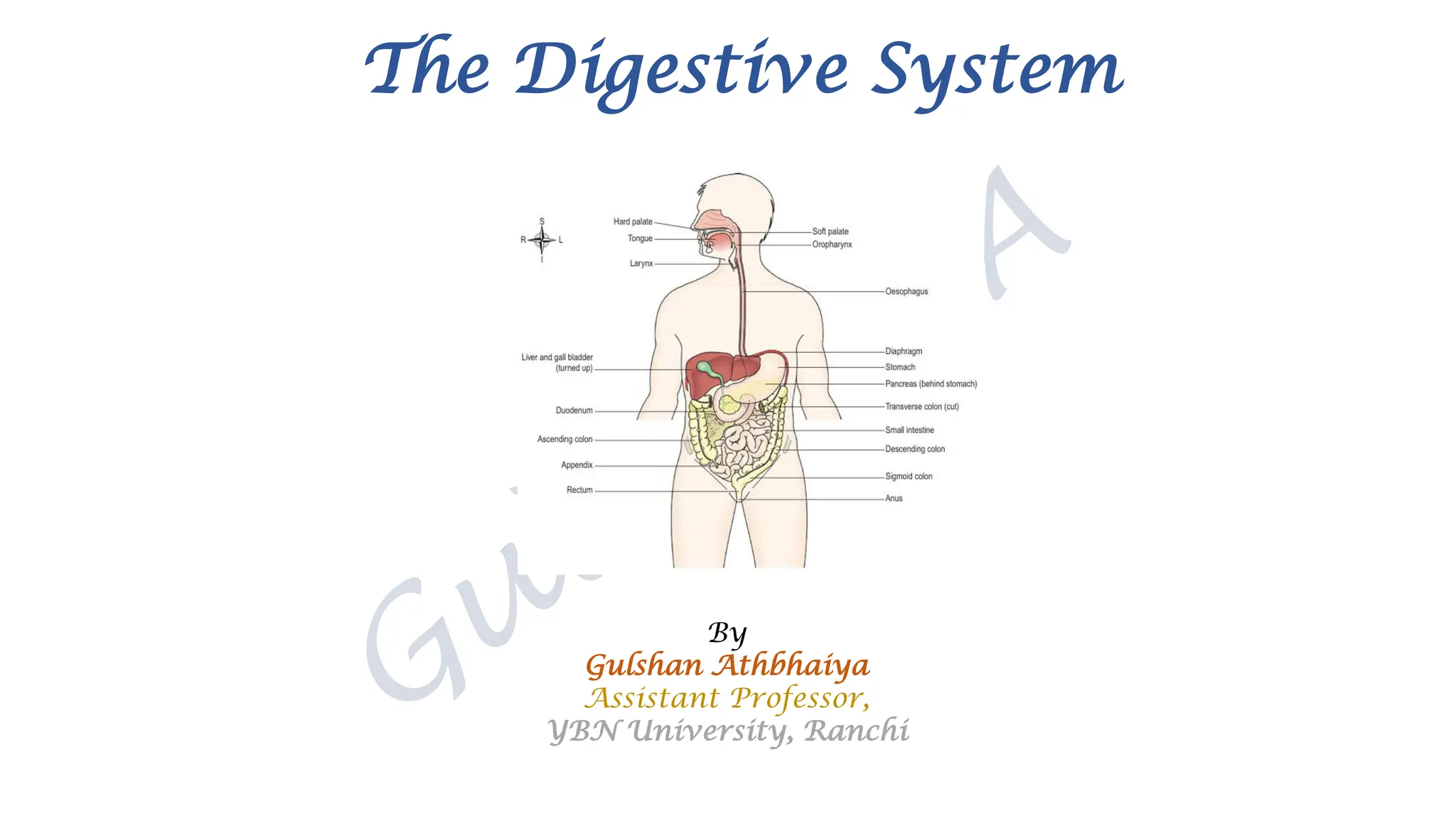
Alimentary canal
Also knownas the gastrointestinal (GI) tract, this is essentially a long tube through which food passes.
It commences at the mouth and terminates at the anus, and the various organs along its length have different functions,
although structurally they are remarkably similar, The parts are:
• mouth
• pharynx
• oesophagus
• stomach
• small intestine
• large intestine
• rectum and anal canal.
Accessory organs
Various secretions are poured into the alimentary tract, some by glands in the lining membrane of the organs, e.g. gastric
juice secreted by glands in the lining the stomach, and some by glands situated outside the tract. The latter are the accessory
organs of digestion and their secretions pass through ducts to enter the tract.
They consist of:
• three pairs of salivary glands
• the pancreas
• the liver and biliary tract.
4.
Layers of thealimentary cannal
Aventitia or serosa:
This is the outermost layer. In the thorax it consists of loose fibrous tissue and in the abdomen the organs are covered by
serous membrane (serosa) called peritoneum.
Peritoneum
The peritoneum is the largest serous membrane of the body.
It provides a physical barrier to local spread of infection, and can isolate an infective focus such as appendicitis, preventing
involvement of other abdominal structures.
It has two layers:
• the parietal peritoneunm, which lines the abdominal wall
• the visceral peritoneum, which covers the organs (viscera) within the abdominal and pelvic cavities.
The parietal peritoneum lines the anterior abdominal wall. The two layers of peritoneum are in close contact, and friction
between them is prevented by the presence of serous fluid secreted by the peritoneal cells, thus the peritoneal cavity is only a
potential.
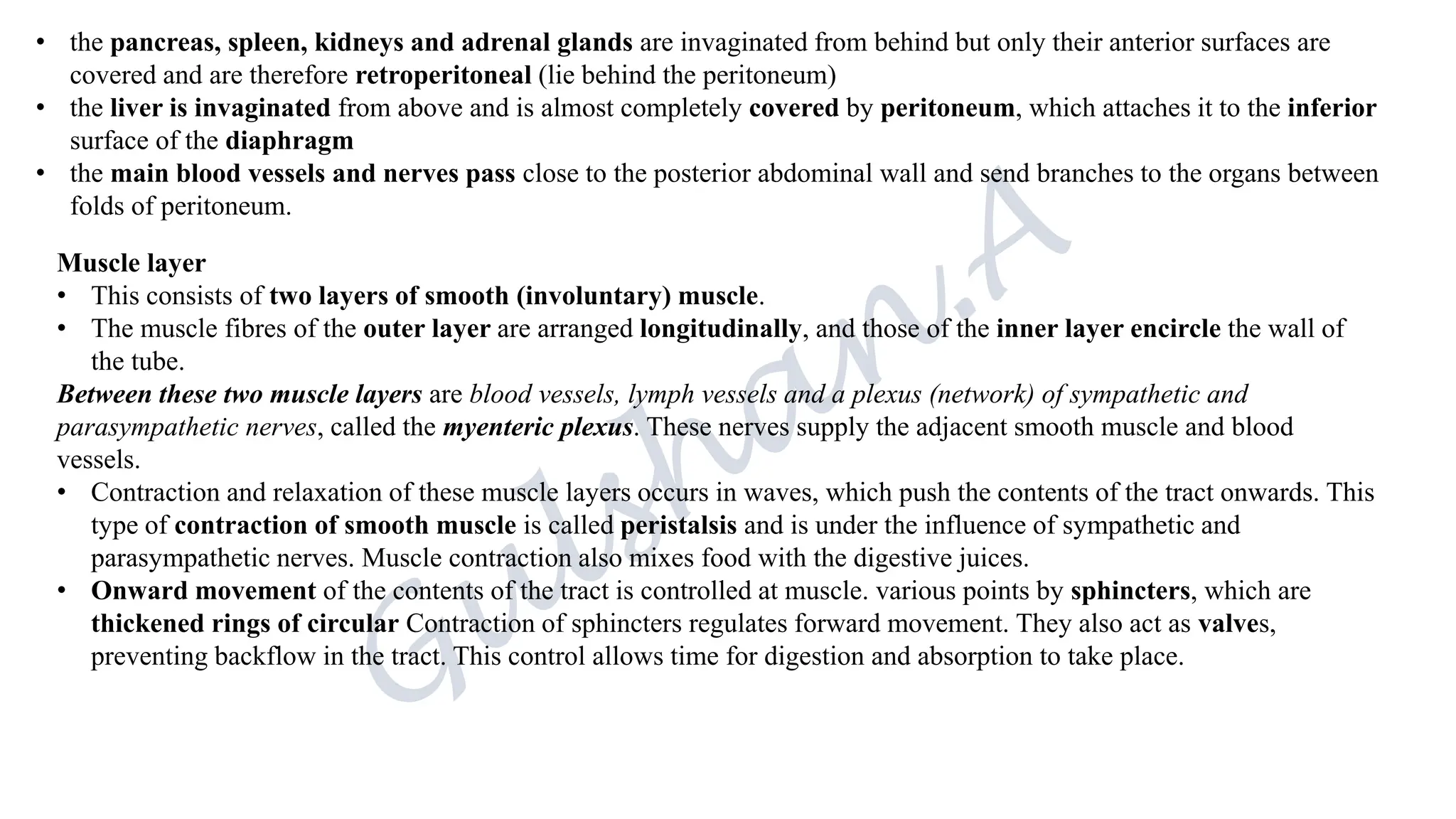
If the organ is completely covered by the visceral peritoneum – intraperitoneal
Organ covered on the anterior side – retroperitoneal.
• pelvic organs are covered only on their superior surface
• the stomach and intestines, deeply invaginated from behind, are almost completely surrounded by peritoneum and
have a double fold (the mesentery) that attaches them to the posterior abdominal wall.
• The fold of peritoneum enclosing the stomach extends beyond the greater curvature of the stomach, and hangs down in
front of the abdominal organs like an apron. This is the greater omentun, which stores fat that provides both insulation
and a long- term energy store.
5.
• the pancreas,spleen, kidneys and adrenal glands are invaginated from behind but only their anterior surfaces are
covered and are therefore retroperitoneal (lie behind the peritoneum)
• the liver is invaginated from above and is almost completely covered by peritoneum, which attaches it to the inferior
surface of the diaphragm
• the main blood vessels and nerves pass close to the posterior abdominal wall and send branches to the organs between
folds of peritoneum.
Muscle layer
• This consists of two layers of smooth (involuntary) muscle.
• The muscle fibres of the outer layer are arranged longitudinally, and those of the inner layer encircle the wall of
the tube.
Between these two muscle layers are blood vessels, lymph vessels and a plexus (network) of sympathetic and
parasympathetic nerves, called the myenteric plexus. These nerves supply the adjacent smooth muscle and blood
vessels.
• Contraction and relaxation of these muscle layers occurs in waves, which push the contents of the tract onwards. This
type of contraction of smooth muscle is called peristalsis and is under the influence of sympathetic and
parasympathetic nerves. Muscle contraction also mixes food with the digestive juices.
• Onward movement of the contents of the tract is controlled at muscle. various points by sphincters, which are
thickened rings of circular Contraction of sphincters regulates forward movement. They also act as valves,
preventing backflow in the tract. This control allows time for digestion and absorption to take place.
6.
Submucosa
• This layerconsists of loose areolar connective tissue containing collagen and some elastic fibres, which binds the
muscle layer to the mucosa.
• Within it are blood vessels and nerves, lymph vessels and varying amounts of lymphoid tissue. The blood vessels are
arterioles, venules and capillaries. The nerve plexus is the submucosal plexus, which contains sympathetic and parasympa-
thetic nerves that supply the mucosal lining.
Mucosa
This consists of three layers of tissue:
1. mucous membrane formed by columnar epithelium is the innermost layer, and has three main functions:
• protection,
• secretion
• and absorption
2. lamina propria consisting of loose connective tissue, which supports the blood vessels that nourish the inner
epithelial layer, and varying amounts of lymphoid tissue that protects against microbial invaders
3. muscularis mucosa, a thin outer layer of smooth muscle that provides involutions of the mucosal layer, e.g. gastric
glands
The secretions include:
• saliva from the salivary glands
• gastric juice from the gastric glands
• intestinal juice from the intestinal glands
• pancreatic juice from the pancreas
• bile from the liver.
These are digestive juices and most contain enzymes that chemically break down food. Under the epithelial lining are varying
amounts of lymphoid tissue that provide protection against ingested microbes.
8.
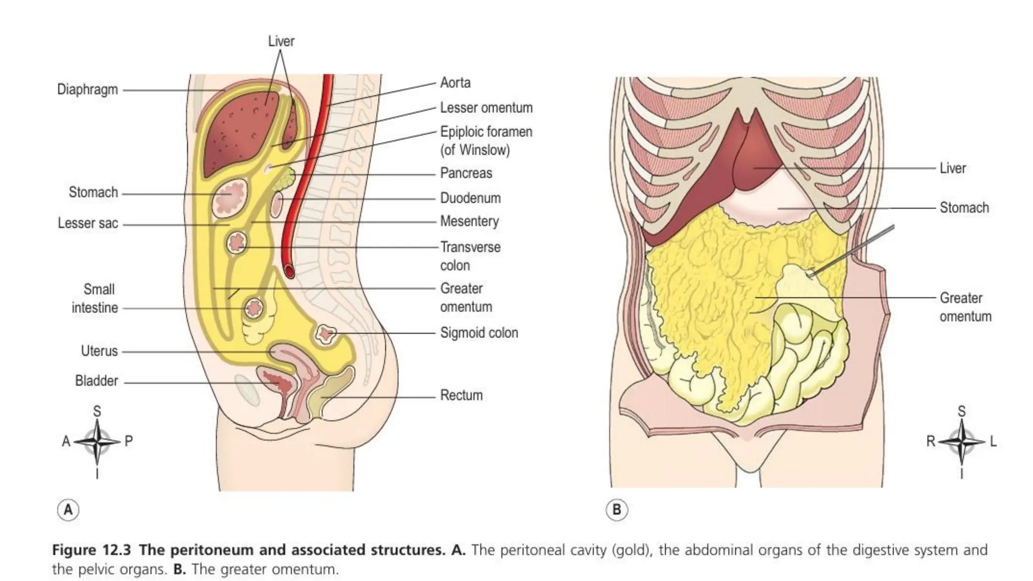
Nerve supply:
Parasymapthetic system:
Sacralnerves supply the most distal part of the GIT.
• increased muscular activity, especially peristalsis, through increased activity of the myenteric plexus
• increased glandular secretion, through increased activity of the submucosal plexus
Sympathetic system
Thoracic and lumbar regions
• decrease muscular activity, especially peristalsis, because there is reduced stimulation of the myenteric plexus
• decrease glandular secretion, as there is less stimulation of the submucosal plexus
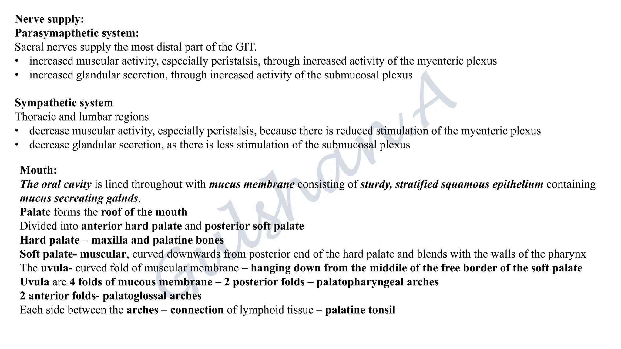
Mouth:
The oral cavity is lined throughout with mucus membrane consisting of sturdy, stratified squamous epithelium containing
mucus secreating galnds.
Palate forms the roof of the mouth
Divided into anterior hard palate and posterior soft palate
Hard palate – maxilla and palatine bones
Soft palate- muscular, curved downwards from posterior end of the hard palate and blends with the walls of the pharynx
The uvula- curved fold of muscular membrane – hanging down from the middile of the free border of the soft palate
Uvula are 4 folds of mucous membrane – 2 posterior folds – palatopharyngeal arches
2 anterior folds- palatoglossal arches
Each side between the arches – connection of lymphoid tissue – palatine tonsil
10.
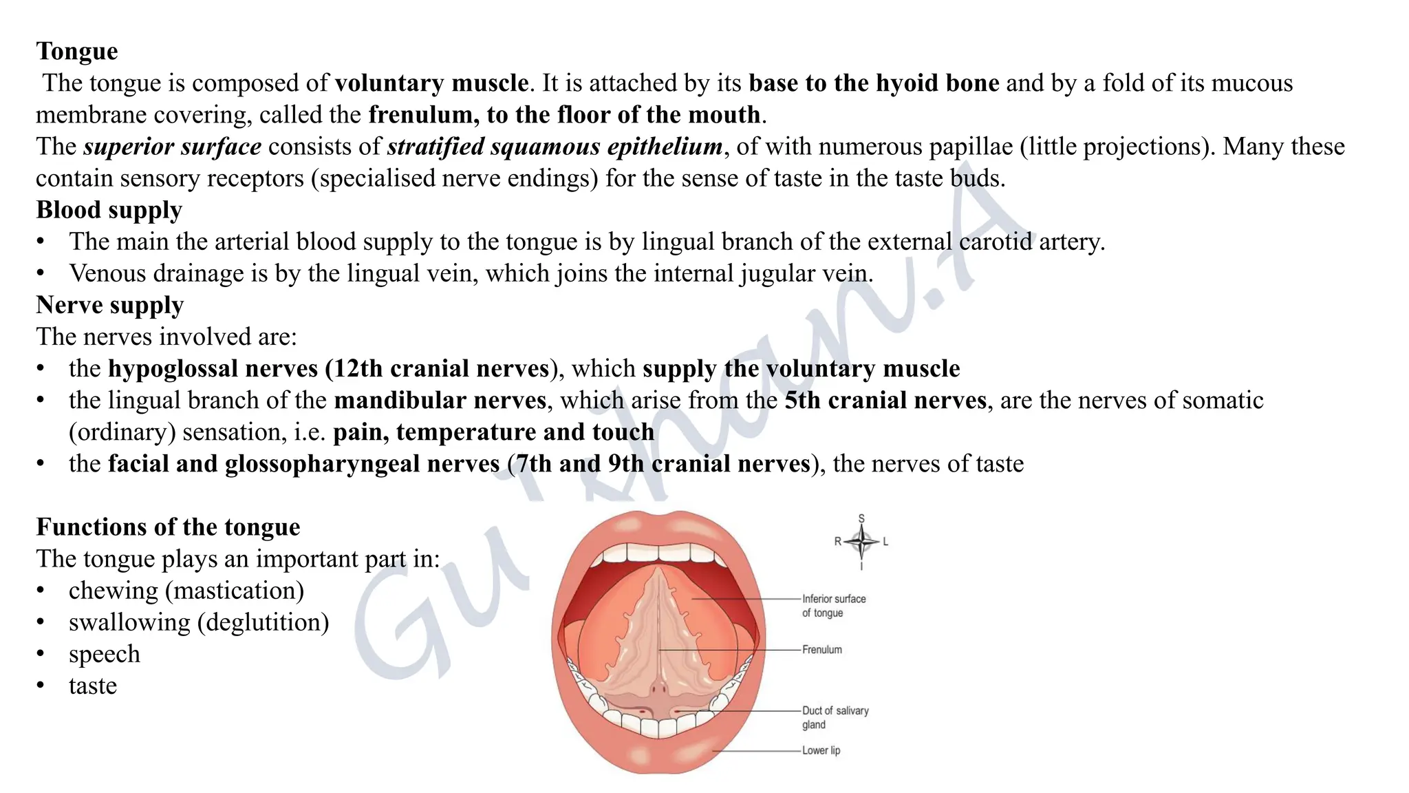
Tongue
The tongue iscomposed of voluntary muscle. It is attached by its base to the hyoid bone and by a fold of its mucous
membrane covering, called the frenulum, to the floor of the mouth.
The superior surface consists of stratified squamous epithelium, of with numerous papillae (little projections). Many these
contain sensory receptors (specialised nerve endings) for the sense of taste in the taste buds.
Blood supply
• The main the arterial blood supply to the tongue is by lingual branch of the external carotid artery.
• Venous drainage is by the lingual vein, which joins the internal jugular vein.
Nerve supply
The nerves involved are:
• the hypoglossal nerves (12th cranial nerves), which supply the voluntary muscle
• the lingual branch of the mandibular nerves, which arise from the 5th cranial nerves, are the nerves of somatic
(ordinary) sensation, i.e. pain, temperature and touch
• the facial and glossopharyngeal nerves (7th and 9th cranial nerves), the nerves of taste
Functions of the tongue
The tongue plays an important part in:
• chewing (mastication)
• swallowing (deglutition)
• speech
• taste
11.
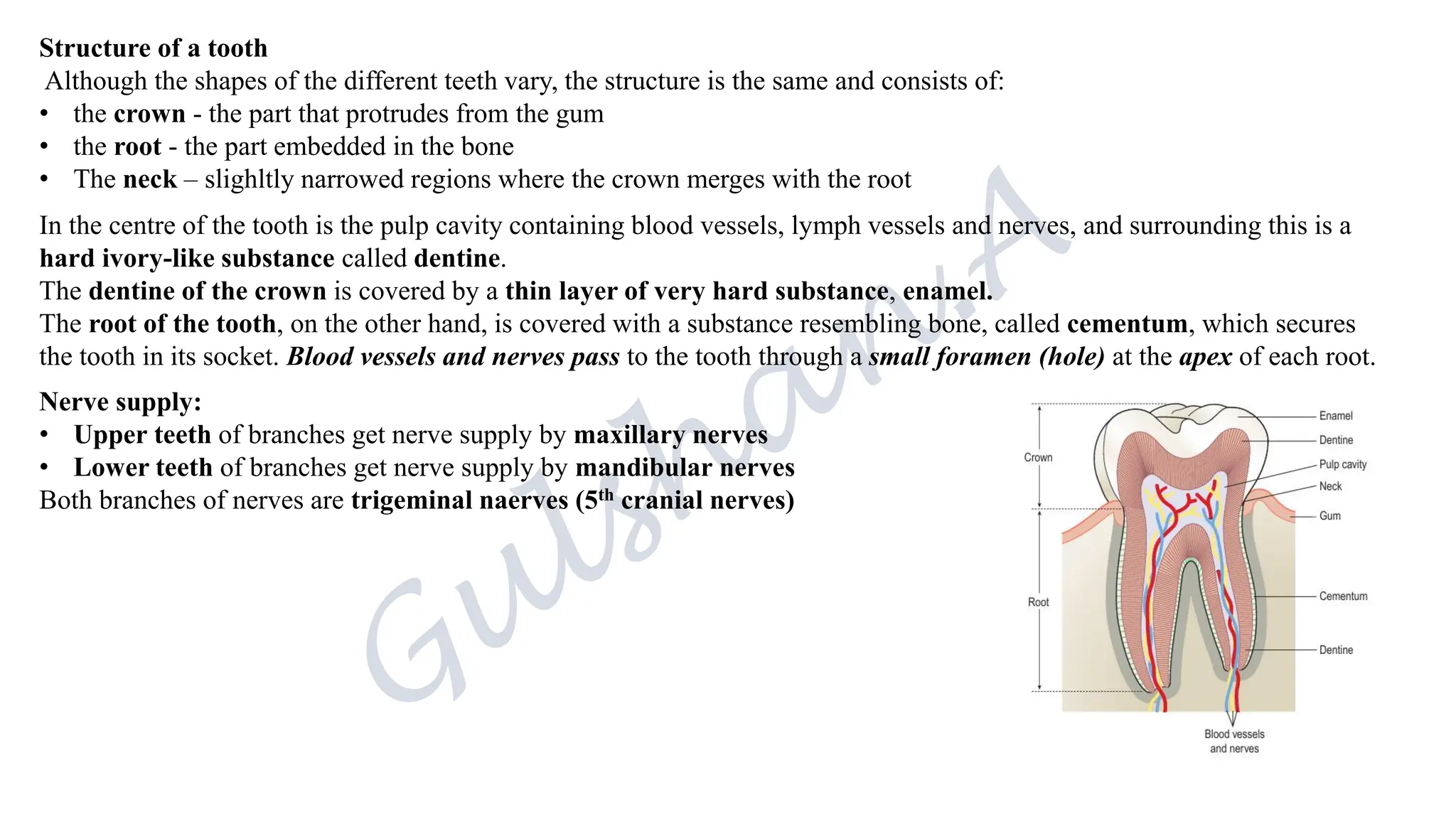
Structure of atooth
Although the shapes of the different teeth vary, the structure is the same and consists of:
• the crown - the part that protrudes from the gum
• the root - the part embedded in the bone
• The neck – slighltly narrowed regions where the crown merges with the root
In the centre of the tooth is the pulp cavity containing blood vessels, lymph vessels and nerves, and surrounding this is a
hard ivory-like substance called dentine.
The dentine of the crown is covered by a thin layer of very hard substance, enamel.
The root of the tooth, on the other hand, is covered with a substance resembling bone, called cementum, which secures
the tooth in its socket. Blood vessels and nerves pass to the tooth through a small foramen (hole) at the apex of each root.
Nerve supply:
• Upper teeth of branches get nerve supply by maxillary nerves
• Lower teeth of branches get nerve supply by mandibular nerves
Both branches of nerves are trigeminal naerves (5th cranial nerves)
12.
Pharynx:
The pharynx isdivided into 3 parts:
Nasopharynx: respiration
Oropharynx & laryngopharynx: both respiration and digestion
The walls of the pharynx consists of :
Mucosa : stratified squamous epithelium, provides thick sturdy lining for wear and tear of swallowing ingested food.
Middile layer consists of connective tissue, contains blood vessels and nerves supply
Outer layer consists of involuntary muscles
Nerves supply:
Parasympathetic system: Glossopharyngeal and vagus nerve
Sympathetic system: Cerviacal ganglia
Oesophagus:
25cm long and 2cm in diameter.
Lies in median plane in thorax, in front of verterbral column, behind. trachea and heart.
Upper and lower ends of the oesophagus are closed by sphincters.
Upper oesophageal sphincter- prevents passage of air during inspiration and aspiration.
Lower oesophageal sphincter- prevents reflux of gastric acid contents.
13.
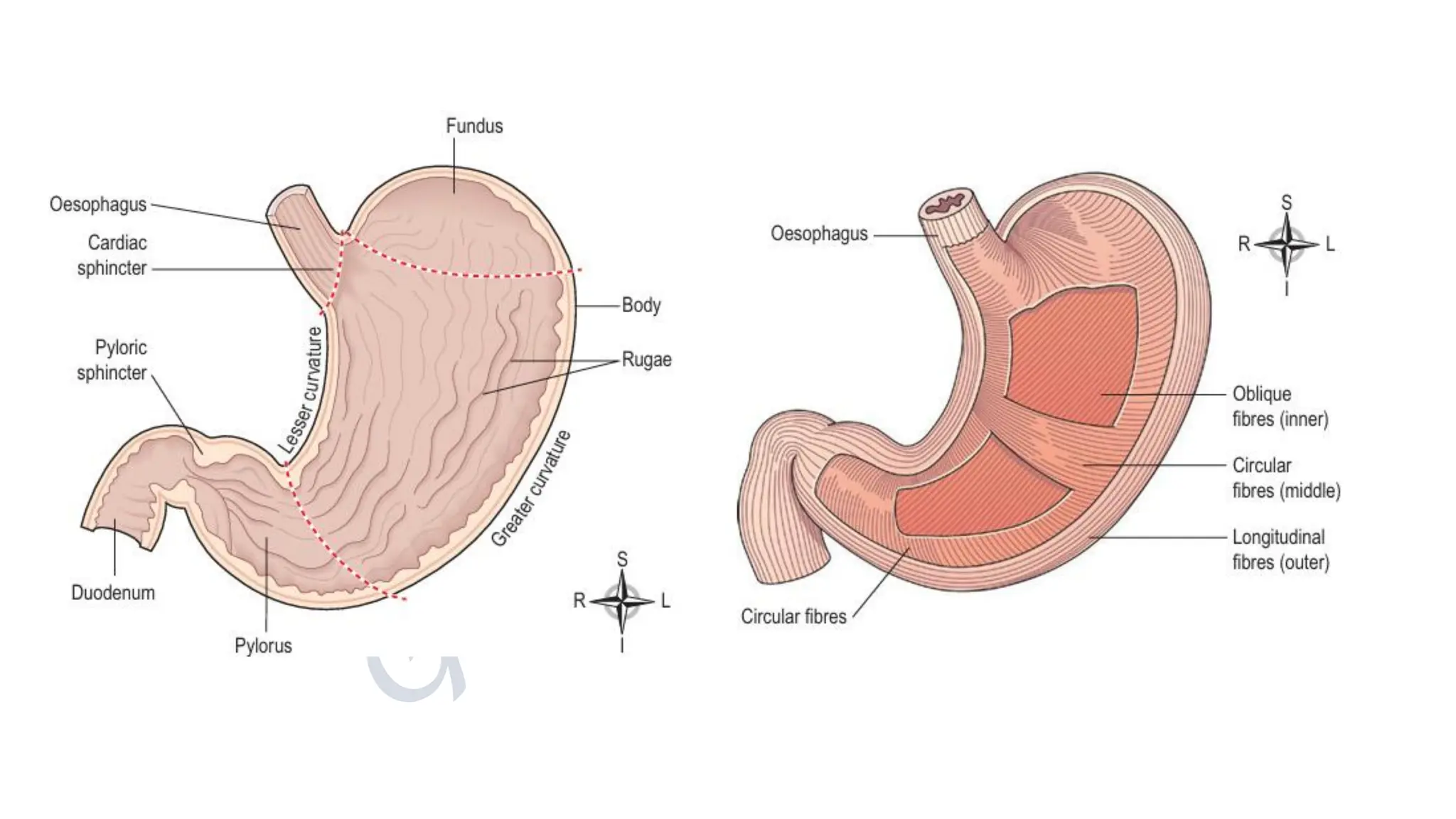
Stomach:
J- shaped dilatedportion of the alimentary tract.
Organs associated with the stomach
Anteriorly - left lobe of liver and anterior abdominal wall
Posteriorly - - abdominal aorta, pancreas, spleen, left kidney and adrenal gland
Superiorly - diaphragm, oesophagus and left lobe of liver
Inferiorly - transverse colon and small intestine
To the left- diaphragm and spleen
To the right - liver and duodenum.
❖ Structure:
Stomach is continuous with oesophagus at lower esophageal sphincter and duodenum at pyloric sphincter
It has 2 curvatures
The stomach is divided into 3 regions:
• Fundus
• Body
• Pylorus
When stomach is inactive pyloric sphincter is relaxed and open, when stomach contains food sphincter closed.
Walls of the stomach:
Same as 4 layers of the GIT, but with some modifications
Muscle layer:
3 layers of smooth muscle fibers:
Outer layer- longitudinal fibers
Middile layer- Circular fibers
14.
Inner layer- Obliquelayers
This arrangement allows for churning and peristaltic activity
Blood supply:
Left gastric artery- coeliac artery
Right gastric artery- Gastroepiploic arteries
The gastric muscles generate churning action to break down bolus mixes with gastric juice.
Peristaltic waves propel contents towards pylorus.
Gastric juices:
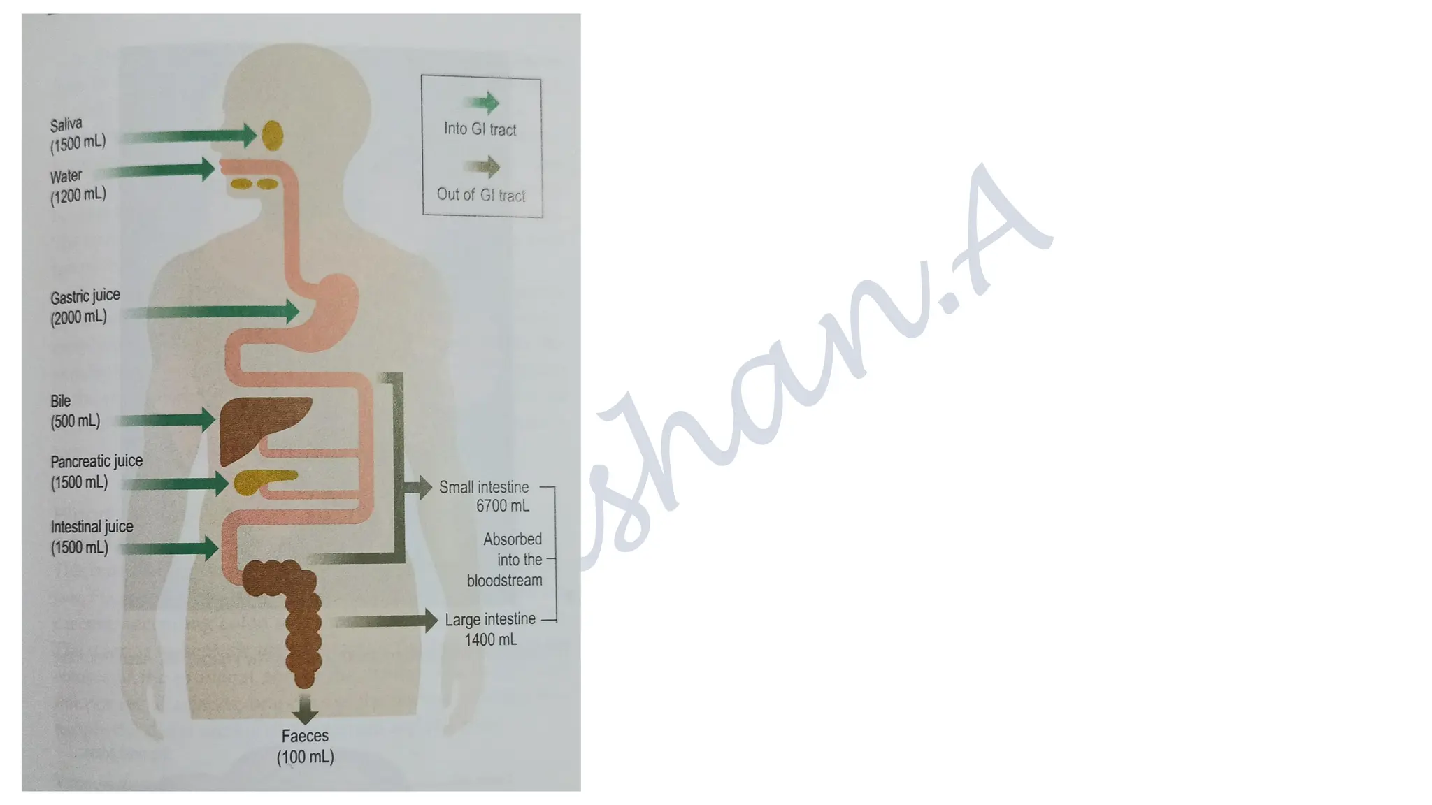
About 2 ltrs of gastric juices secreted daily by specialised secretory glands in the mucosa. It consists of:
• water
• mineral salts
• mucus secreted by mucous neck cells in the glands and surface mucous cells on the stomach surface
• hydrochloric acid
• intrinsic factor secreted by parietal cells in the gastric glands
• inactive enzyme precursors: pepsinogens secreted by chief cells in the glands.
Functions of gastric juice
• Water further liquefies the food swallowed.
• Hydrochloric acid: -
➢ acidifies the food and stops the action of salivary amylase - -
➢ kills ingested microbes
➢ provides the acid environment needed for the action of pepsins.
15.
• Pepsinogens areactivated to pepsins by hydrochloric acid and by pepsins already present in the stomach. These enzymes
begin the digestion of proteins, breaking them into smaller molecules. Pepsins have evolved to act most effectively at a very
low pH, between 1.5 and 3.5.
• Intrinsic factor (a protein) is necessary for the absorption of vitamin B12, from the ileum. (Deficiency leads to pernicious
anemia).
• Mucus prevents mechanical injury to the stomach wall by lubricating the contents. It also prevents chemical injury by
acting as a barrier between the stomach wall and the corrosive gastric juice - hydrochloric acid is present in potentially
damaging concentrations and pepsins would digest the gastric juices.
Secretion of Gastric juice:
Small quantity of gastric juice present in the stomach, even when it contains no food, known as fasting juice
Secretion reaches maximum level at 1 hr after meal, declines to fasting level after 4 hrs.
3 phases of secretion of gastric juice:
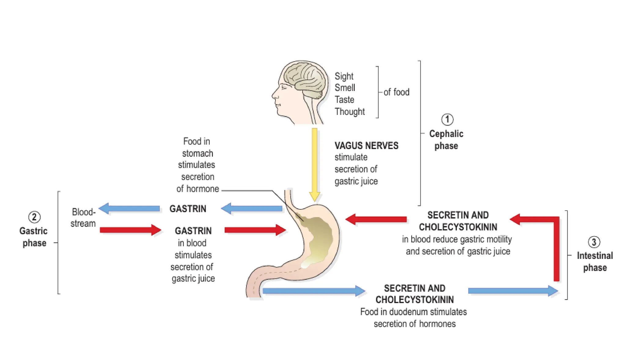
1. Cephalic phase: This flow of juice occurs before food reaches the stomach and is due to reflex stimulation of the vagus
(parasympathetic) nerves initiated by the sight, smell or taste of food. When the vagus nerves have been cut (vagotomy),
this phase of gastric secretion stops. Sympathetic stimulation, e.g. during emotional states, also inhibits gastric activity.
2. Gastric phase: When stimulated by the presence of food the enteroendocrine cells in the pylorus and duodenum secrete
the hormone gastrin, which passes directly into the circulating blood. Gastrin, circulating in the blood which supplies the
stomach, stimulates the gastric glands to produce more gastric juice. In this way secretion of digestive juice is continued
after completion of a meal and the end of the cephalic phase. Gastrin secretion is suppressed when the pH in the pylorus
falls to about 1.5.
16.
3. Intestinal phase:When the partially digested contents of the stomach, reach the small intestine, two hormones, secretin
and cholecystokinin, are produced by endocrine cells in the intestinal mucosa. They slow down the secretion of gastric juice
and reduce gastric motility. By slowing the emptying rate of the stomach, the chyme in the duodenum becomes more
thoroughly mixed with bile and pancreatic juice. This phase of gastric secretion is most marked following a meal with a high
fat content. The rate at which the stomach empties depends largely on the type of food eaten. A carbohydrate meal leaves the
stomach in 2-3 hours, a protein meal remains longer and a fatty meal remains in the stomach longest.
Functions of the stomach
These include:
• temporary storage allowing time for the digestive enzymes, pepsins, to act
• chemical digestion - pepsins break proteins into polypeptides
• mechanical breakdown - the three smooth muscle layers enable the stomach to act as a churn, gastric juice is added and the
contents are liquefied to chyme. Gastric motility and secretion are increased by parasympathetic nerve stimulation
• limited absorption - water, alcohol and some lipid soluble drugs
• non-specific defence against microbes - provided by hydrochloric acid in gastric juice. Vomiting may occur in response to
ingestion of gastric irritants, e.g. microbes or chemicals
• preparation of iron for absorption - the acid environment of the stomach solubilises iron salts, essential for iron absorption
in the small intestine
• production and secretion of intrinsic factor needed for absorption of vitamin B12, in the terminal ileum
• regulation of the passage of gastric contents into the duodenum. When the chyme is sufficiently acidified and liquefied, the
pylorus forces small jets of gastric contents through the pyloric sphincter into the duodenum. This sphincter is normally
closed, preventing backflow of chyme into the stomach
• Secretion of the hormone gastrin
19.
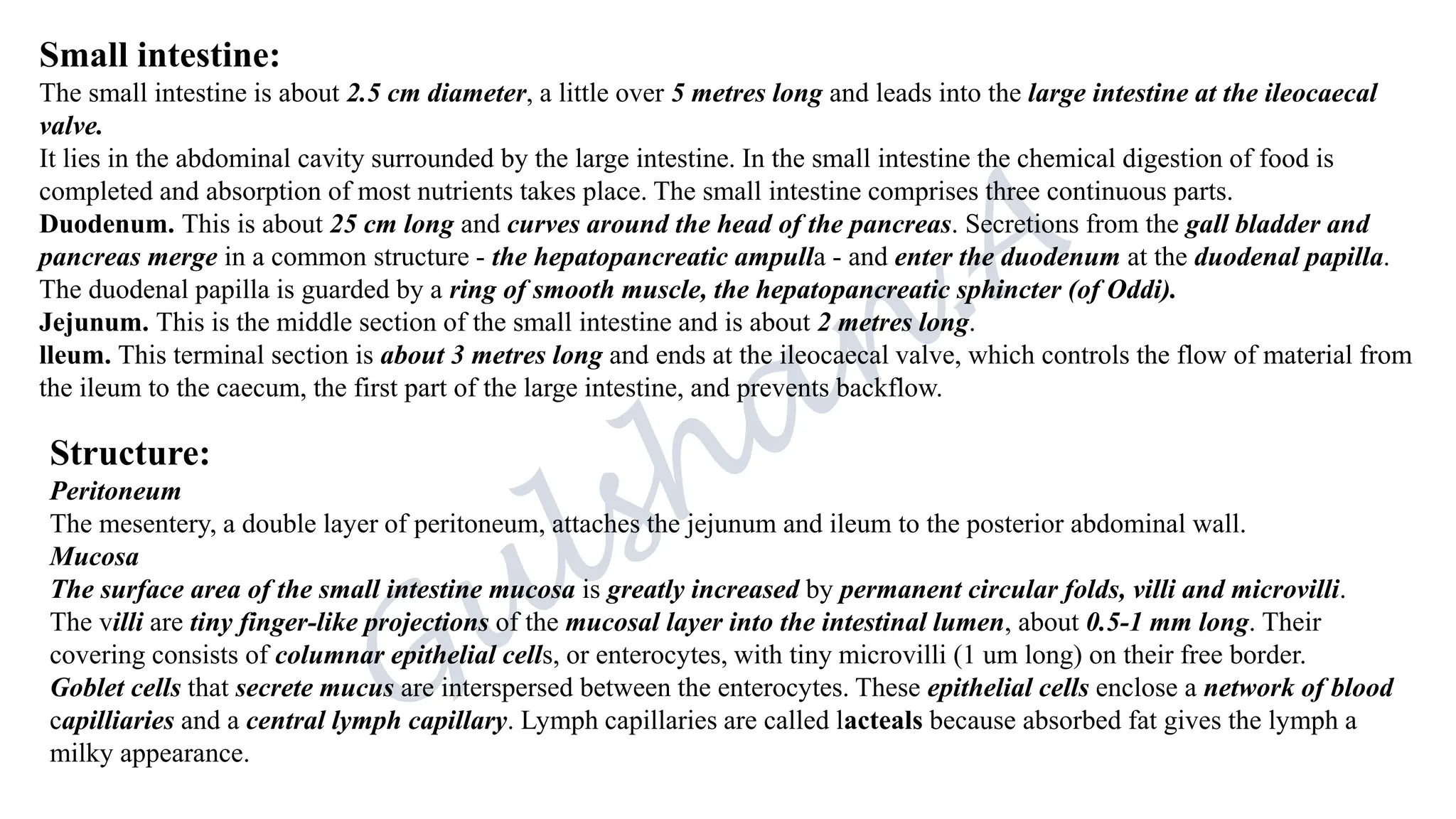
Small intestine:
The smallintestine is about 2.5 cm diameter, a little over 5 metres long and leads into the large intestine at the ileocaecal
valve.
It lies in the abdominal cavity surrounded by the large intestine. In the small intestine the chemical digestion of food is
completed and absorption of most nutrients takes place. The small intestine comprises three continuous parts.
Duodenum. This is about 25 cm long and curves around the head of the pancreas. Secretions from the gall bladder and
pancreas merge in a common structure - the hepatopancreatic ampulla - and enter the duodenum at the duodenal papilla.
The duodenal papilla is guarded by a ring of smooth muscle, the hepatopancreatic sphincter (of Oddi).
Jejunum. This is the middle section of the small intestine and is about 2 metres long.
lleum. This terminal section is about 3 metres long and ends at the ileocaecal valve, which controls the flow of material from
the ileum to the caecum, the first part of the large intestine, and prevents backflow.
Structure:
Peritoneum
The mesentery, a double layer of peritoneum, attaches the jejunum and ileum to the posterior abdominal wall.
Mucosa
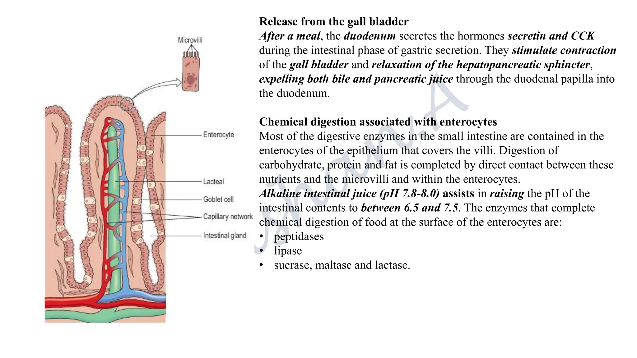
The surface area of the small intestine mucosa is greatly increased by permanent circular folds, villi and microvilli.
The villi are tiny finger-like projections of the mucosal layer into the intestinal lumen, about 0.5-1 mm long. Their
covering consists of columnar epithelial cells, or enterocytes, with tiny microvilli (1 um long) on their free border.
Goblet cells that secrete mucus are interspersed between the enterocytes. These epithelial cells enclose a network of blood
capilliaries and a central lymph capillary. Lymph capillaries are called lacteals because absorbed fat gives the lymph a
milky appearance.
20.
Intestinal juice:
About 1500mL of intestinal juice are secreted daily by the glands of the small intestine. It is slightly basic (alkaline) and
consists of water, mucus and mineral salts.
Functions of the small intestine
The functions are:
• onward movement of its contents by peristalsis, which is increased by parasympathetic stimulation
• secretion of intestinal juice, also increased by parasympathetic stimulation
• completion of chemical digestion of carbohydrates, protein and fats in the enterocytes of the villi
• protection against infection by microbes that have survived the antimicrobial action of the hydrochloric acid in the
stomach, by both solitary and aggregated lymph follicles
• secretion of the hormones cholecystokinin (CCK) and secretin
• absorption of nutrients.
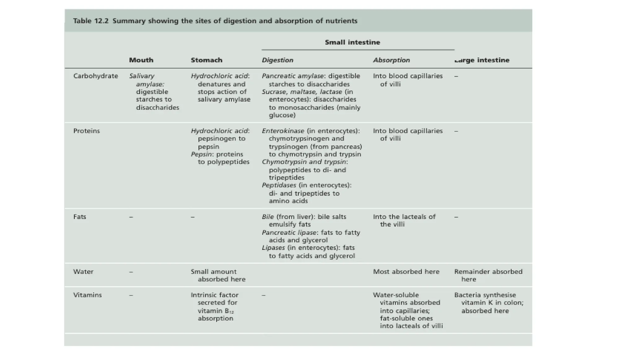
Chemical digestion the small intestine
When acid chyme passes into the small intestine it is mixed with pancreatic juice, bile and intestinal juice, and is in contact
with the enterocytes of the villi. The digestion of all nutrients is completed:
• carbohydrates are broken down to monosaccharides
• proteins are broken down to amino acids
• fats are broken down to fatty acids and glycerol.
21.
Pancreatic juice
Pancreatic juiceis secreted by the exocrine pancreas and enters the duodenum at the duodenal papilla. It consists of:
• water
• mineral salts
• enzymes: -
✓ amylase
✓ lipase
✓ nucleases that digest DNA and RNA
• inactive enzyme precursors including:
✓ trypsinogen
✓ chymotrypsinogen
Pancreatic juice is basic (alkaline, pH 8) because it contains significant quantities of bicarbonate ions, which are basic
(alkaline) in solution. When acid stomach contents enter the duodenum they are mixed with pancreatic juice and bile and
the pH is raised to between 6 and 8. This is the pH at which the pancreatic enzymes, amylase and lipase, act most
effectively.
Functions
Digestion of proteins.
Trypsinogen and chymotrypsinogen are inactive enzyme precursors activated by enterokinase, an enzyme in the
microvilli, which converts them into the active proteolytic enzymes trypsin and chymotrypsin. These enzymes convert
polypeptides to tripeptides, dipeptides and amino acids. It is important that they are produced as inactive precursors and are
activated only upon their arrival in the duodenum, otherwise they would digest the pancreas.
22.
Digestion of carbohydrates.
Pancreaticamylase converts all by digestible polysaccharides (starches) not acted upon salivary amylase to disaccharides.
Digestion of fats.
Lipase converts fats to fatty acids and glycerol. To aid the action of lipase, bile salts emulsify fats, i.e. reduce the size of the
globules, increasing their surface area.
Control of secretion
The secretion of pancreatic juice is stimulated by secretin and CCK, produced by endocrine cells in the walls of the
duodenum. The presence in the duodenum of acid chyme from the stomach stimulates the production of these hormones.
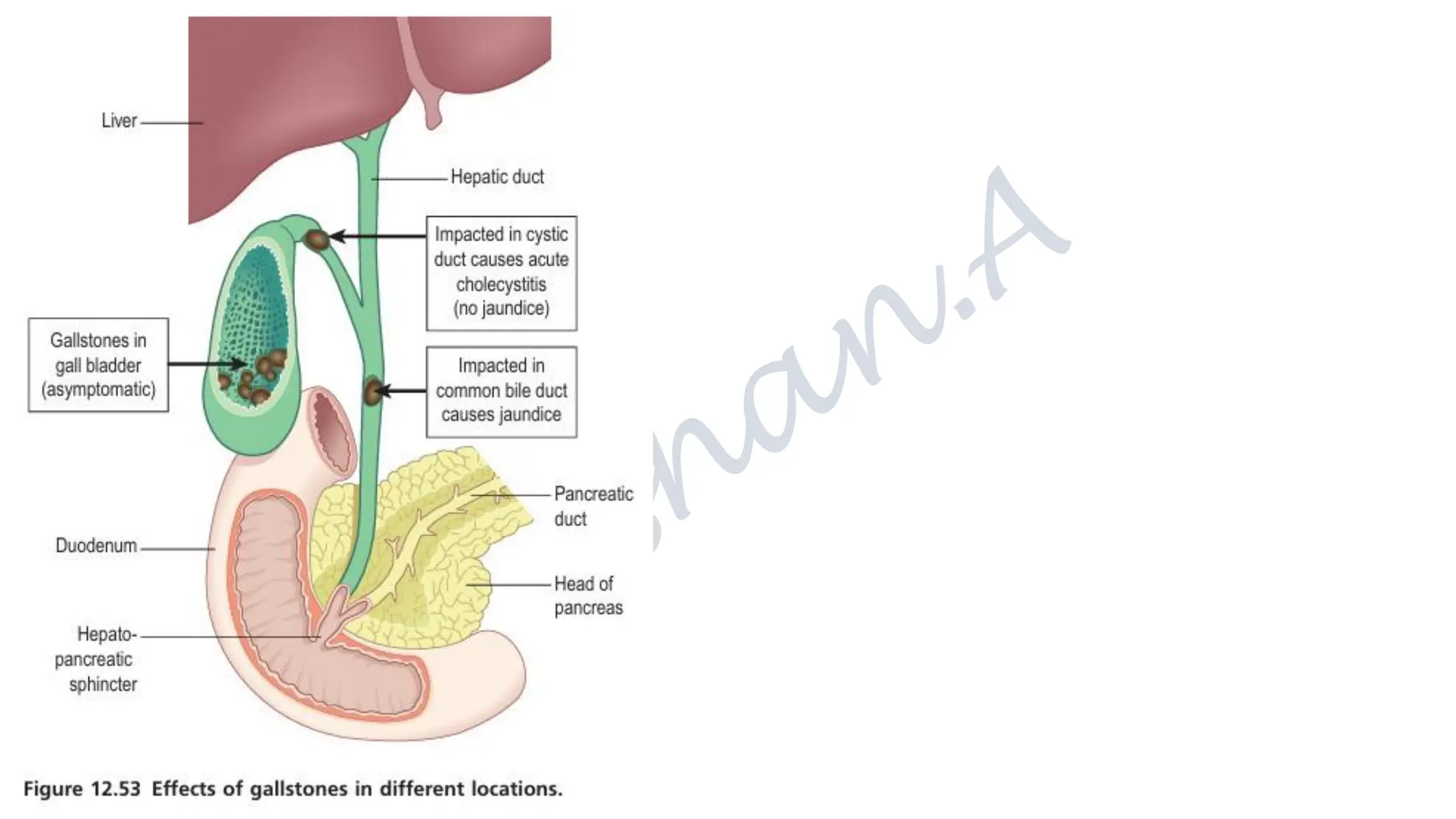
Bile
Bile, secreted by the liver, is unable to enter the duodenum it when the hepatopancreatic sphincter is closed; therefore passes
from the hepatic duct along with the cystic duct to the gall bladder where it is stored. Bile has a pH of around 8 and between
500 and 1000 mL is secreted daily. It consists of:
• water
• mineral salts
• mucus
• bile salts
• bile pigments, mainly bilirubin cholesterol.
The functions of bile are
• emulsification of fats in the small intestine - bile salts
• making cholesterol and fatty acids soluble, enabling their absorption along with the fat-soluble vitamins - bile salts
• Excretion of bilirubin( waste products from the breakdown of red blood cells, most of which is in the form of stercobilin).
23.
Release from thegall bladder
After a meal, the duodenum secretes the hormones secretin and CCK
during the intestinal phase of gastric secretion. They stimulate contraction
of the gall bladder and relaxation of the hepatopancreatic sphincter,
expelling both bile and pancreatic juice through the duodenal papilla into
the duodenum.
Chemical digestion associated with enterocytes
Most of the digestive enzymes in the small intestine are contained in the
enterocytes of the epithelium that covers the villi. Digestion of
carbohydrate, protein and fat is completed by direct contact between these
nutrients and the microvilli and within the enterocytes.
Alkaline intestinal juice (pH 7.8-8.0) assists in raising the pH of the
intestinal contents to between 6.5 and 7.5. The enzymes that complete
chemical digestion of food at the surface of the enterocytes are:
• peptidases
• lipase
• sucrase, maltase and lactase.
24.
Absorption of nutrients
•Absorption of nutrients from the small intestine through the enterocytes occurs by several processes, including diffusion,
osmosis, facilitated diffusion and active transport.
• Water moves by osmosis;
• small fat-soluble substances, e.g. fatty acids and glycerol, are able to diffuse through cell membranes; while others are
generally transported inside the villi by other mechanisms.
• Monosaccharides and amino acids pass into the blood capillaries in the villi.
• Fatty acids and glycerol enter the lacteals and are transported along lymphatic vessels to the thoracic duct where they
enter the circulation.
• A small number of proteins are absorbed unchanged, e.g. antibodies present in breast milk and oral vaccines, such as
poliomyelitis vaccine. Other nutrients such as vitamins, mineral salts and water are also absorbed from the small intestine
into the blood capillaries.
• Fat-soluble vitamins are absorbed into the lacteals along with fatty acids and glvcerol. Vitamin B12 combines with intrinsic
factor in the stomach and is actively absorbed in the terminal ileum.
• The surface area through which absorption takes place in villi the small intestine is greatly increased by the circular folds of
mucous membrane and by the very large number of and microvilli present.
• It has been calculated that the surface area of the small intestine is about five times that of the whole body surface.
• Large amounts of fluid enter the alimentary tract each day. Of this, only about 1500 mL is not absorbed by the small
intestine, and passes into the large intestine.
26.
Large intestine, Rectumand canal:
The large intestine is about 1.5 metres long, beginning at the caecum in the right iliac fossa and terminating at the rectum and
anal canal deep within the pelvis. Its lumen is about 6.5 cm in diameter, larger than that of the small intestine. It forms an
arch round the coiled-up small intestine.
Caecum:
1st part of large intestine
Junction of ileocaecal valve opens from the ileum
The vermiform appendix is a fine tube closed at one end, which leads to form caecum.
8-9cm long, contains more lymphoid tissue.
The appendix has no digestive function, but cause significant problems when inflamed.
The colon
The colon has four parts which have the same structure and functions.
The left ascending colon.
This passes upwards from the caecum to the level of the liver where it curves acutely to the at colon. The hepatic flexure to
become the transverse
transverse colon.
This part extends across the abdominal cavity in front of the duodenum and the stomach to the the area of the spleen where it
forms splenic flexure and curves acutely downwards to become the descending colon.
The descending colon.
This passes down the left side of the abdominal cavity then curves towards the midline. At the level of the iliac crest it is
known as the sigmoid colon.
27.
The sigmoid colonis an S shaped curve in the pelvic cavity continue downwards to become rectum.
The rectum
This is a slightly dilated section of the large intestine about 13 cm long. It leads from the sigmoid colon and terminates in the
anal canal.
The anal canal
This is a short passage about 3.8cm long in the adult and leads from the rectum to the exterior. Two sphincter muscles control
the anus;
the internal sphincter, consisting of smooth muscle, is under the control of the autonomic nervous system and
the external sphincter, formed by skeletal muscle, is under voluntary control
Structure:
Submucosal layer: lymphoid tissue
Mucosal layer: Colon and upper rectum contains large no. of mucus secreting goblet cells
Blood supply:
Superior messentric artery supplies: caecum, ascending colon and most of transverse colon
Inferior messentric artery supplies: remainder of large intestine
Functions:
Absorption
Microbial activity
Mass movement
Defaecation
28.
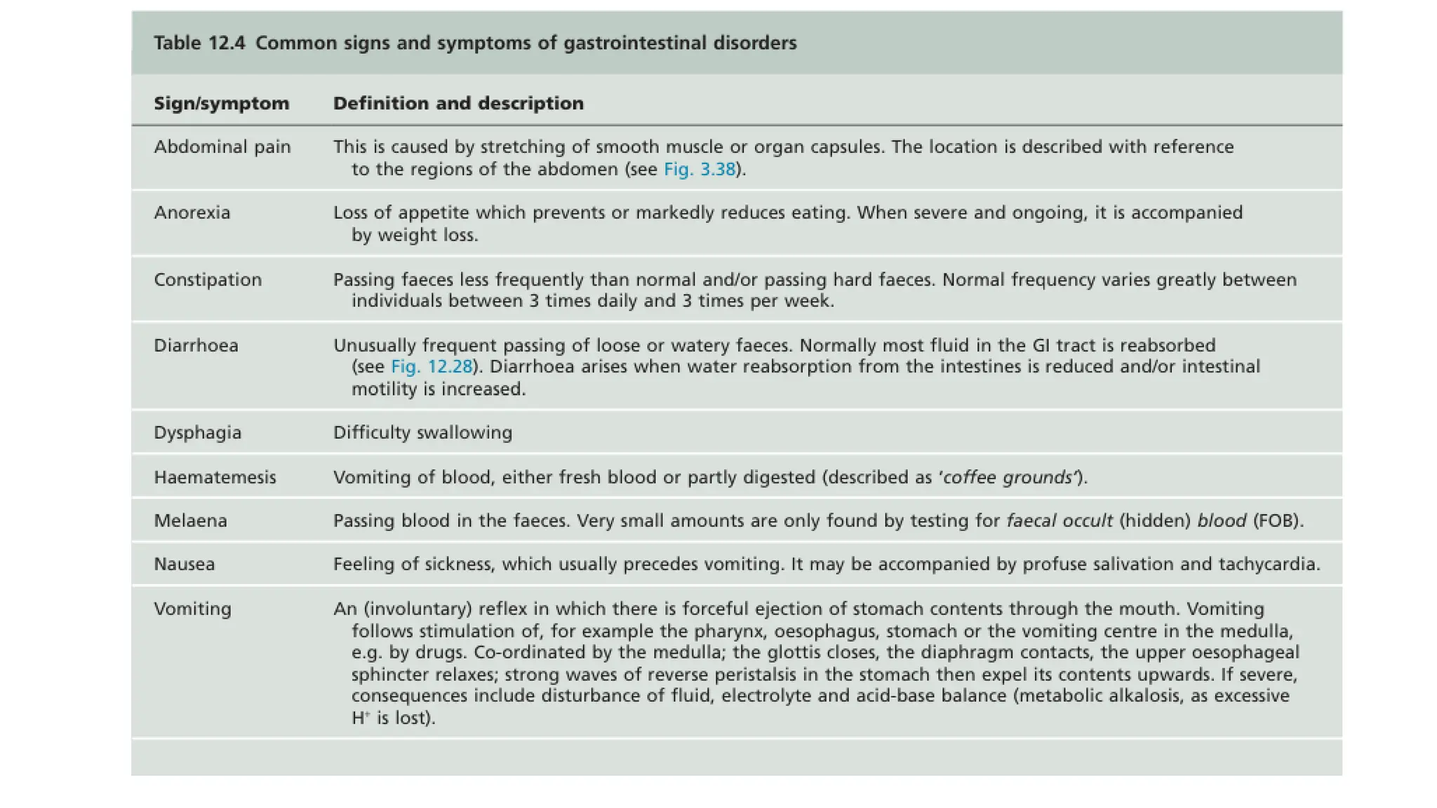
Constituents of faeces.
Thefaeces consist of a semisolid brown mass. The brown colour is due to the presence of stercobilin
Even though absorption of water takes place in the small and large intestines, water still makes up about 60-70% of the weight
of the faeces. The remainder consists of:
• fibre (indigestible cellular plant and animal material)
• dead and live microbes
• epithelial cells shed from the walls of the tract
• fatty acids
• mucus secreted by the epithelial lining of the large intestine.
Mucus helps to lubricate the faeces and an adequate amount of dietary non-starch polysaccharide (NSP, fibre and previously
known as roughage) ensures that the contents of the large intestine are sufficiently bulky to stimulate defaecation.
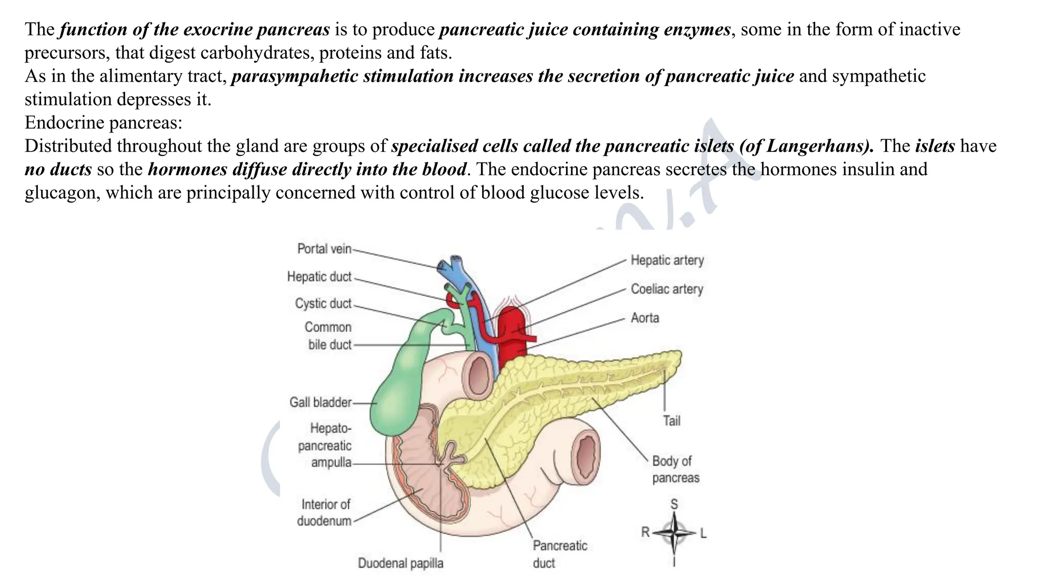
PANCREAS:
The pancreas is a pale grey gland weighing about 60 grams. It is about 12-15 cm long and is situated in the epigastric and left
hypochondriac regions of the abdominal cavity. It consists of a broad head, a body and a narrow tail.
The head lies in the curve of the duodenum, the body behind the stomach and the tail lies in front of the left kidney and just
reaches the spleen. The abdominal aorta and the inferior vena cava lie behind the gland.
The pancreas is both an exocrine and endocrine gland.
Exocrine gland:
This consists of a large number of lobules made up of small acini, the walls of which consist of secretory cells. Each lobule is
drained by a tiny duct and these unite eventually to form the pancreatic duct, which extends along the whole length of the gland
and opens into the duodenum. Just before entering the duodenum the pancreatic duct joins the common bile duct to form the
hepatopancreatic ampulla. The duodenal opening of the ampulla is controlled by the hepatopancreatic sphincter (of Oddi) at
the duodenal papilla.
29.
The function ofthe exocrine pancreas is to produce pancreatic juice containing enzymes, some in the form of inactive
precursors, that digest carbohydrates, proteins and fats.
As in the alimentary tract, parasympahetic stimulation increases the secretion of pancreatic juice and sympathetic
stimulation depresses it.
Endocrine pancreas:
Distributed throughout the gland are groups of specialised cells called the pancreatic islets (of Langerhans). The islets have
no ducts so the hormones diffuse directly into the blood. The endocrine pancreas secretes the hormones insulin and
glucagon, which are principally concerned with control of blood glucose levels.
30.
Liver:
The liver isthe largest gland in the body, weigh between 1 and 2.3 kg. It is situated in the upper part of the weighing
abdominal cavity occupying the greater part of the right hypochondriac region, part of the epigastric region and extending into
the left hypochondriac region. Its upper and anterior surfaces are smooth and curved to fit the under surface of the diaphragm;
its posterior surface is irregular in outline.
Organs associated with the liver
• Superiorly and anteriorly - diaphragm and anterior abdominal wall
• Inferiorly - stomach, bile ducts, duodenum, hepatic flexure of the colon, right kidney and adrenal gland
• Posteriorly - oesophagus, inferior vena cava, aorta, gall bladder, vertebral column and diaphragm
• Laterally - lower ribs and diaphragm.
Structure:
The liver has four lobes.
The two most obvious are the large right lobe and the smaller, wedge-shaped, left lobe.
The other two, the caudate and quadrate lobes, are areas on the posterior surface
The lobes of the liver are made up of tiny functional units, called lobules, which are just visible to the naked eye. Liver lobules
are hexagonal in outline and are formed by cuboidal cells, the hepatocytes, arranged in pairs of columns radiating from a
central vein. Between two pairs of columns of cells are sinusoids (blood vessels with incomplete walls) containing a mixture of
blood from the tiny branches of the portal vein and hepatic artery.
This arrangement allows the arterial blood and portal venous blood (with a high concentration of nutrients) to mix and come
into close contact with the liver cells. Amongst the cells lining the sinusoids are hepatic macrophages (Kupffer cells) whose
function is to ingest and destroy worn out blood cells and any foreign particles present in the blood flowing through from the
liver. Blood drains from the sinusoids into central or centrilobular veins. These then merge with veins from other lobules,
forming larger veins, until eventually they become the hepatic veins, which leave the liver and empty into the inferior vena
cava.
31.
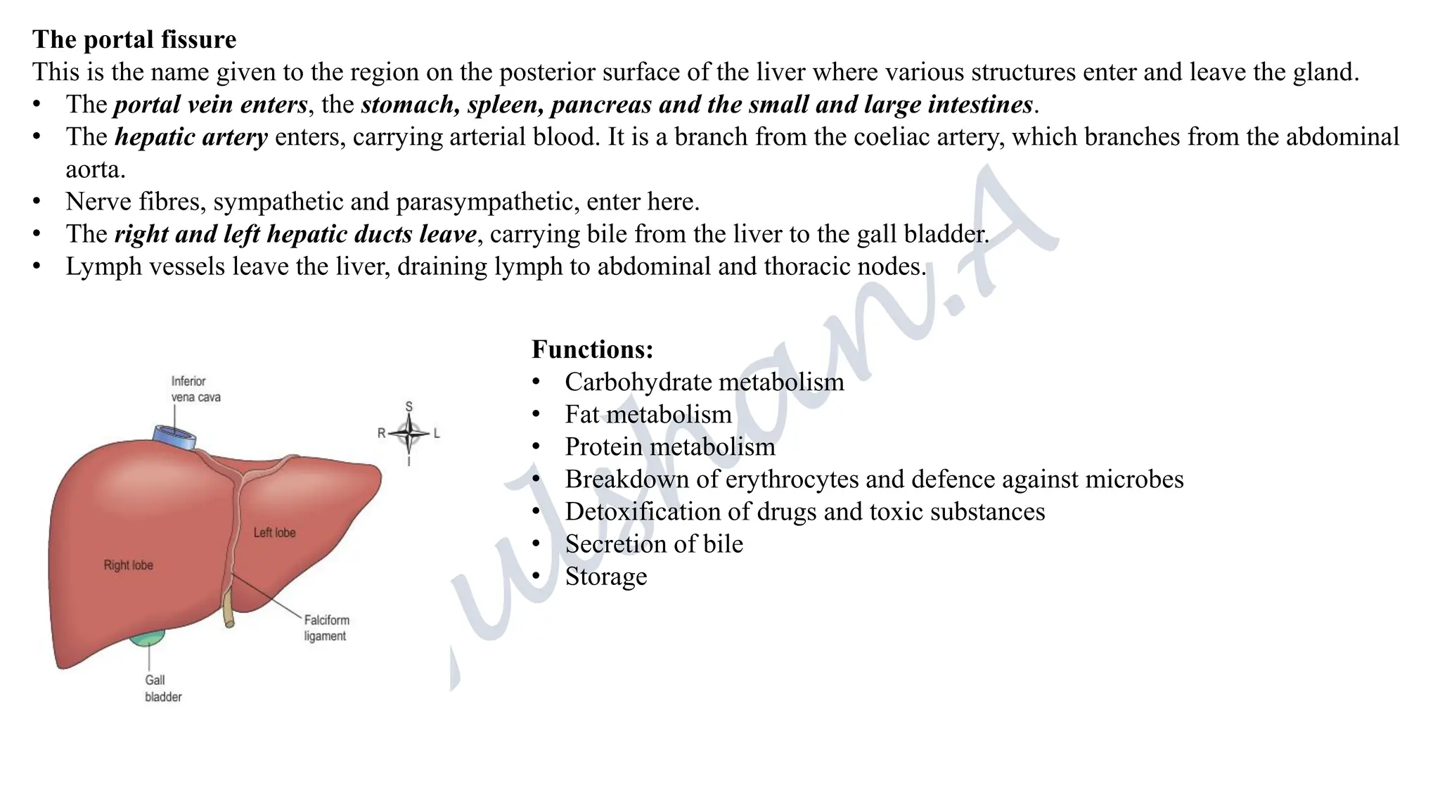
The portal fissure
Thisis the name given to the region on the posterior surface of the liver where various structures enter and leave the gland.
• The portal vein enters, the stomach, spleen, pancreas and the small and large intestines.
• The hepatic artery enters, carrying arterial blood. It is a branch from the coeliac artery, which branches from the abdominal
aorta.
• Nerve fibres, sympathetic and parasympathetic, enter here.
• The right and left hepatic ducts leave, carrying bile from the liver to the gall bladder.
• Lymph vessels leave the liver, draining lymph to abdominal and thoracic nodes.
Functions:
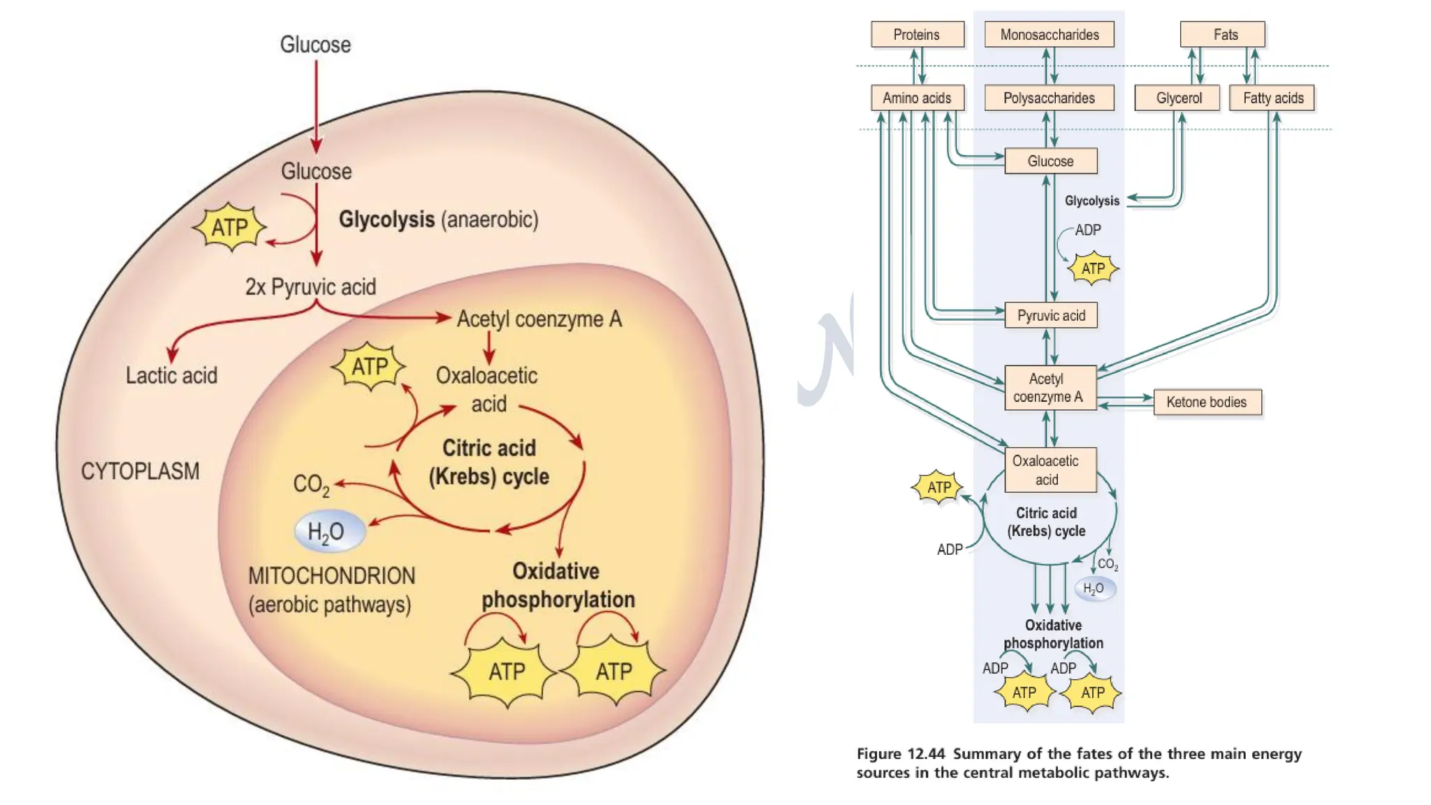
• Carbohydrate metabolism
• Fat metabolism
• Protein metabolism
• Breakdown of erythrocytes and defence against microbes
• Detoxification of drugs and toxic substances
• Secretion of bile
• Storage


![Apporach to lung biopsy [Auto-saved].pptx latest](https://cdn.slidesharecdn.com/ss_thumbnails/apporachtolungbiopsyauto-saved-251211225655-93258539-thumbnail.jpg?width=640&height=640&fit=bounds)


