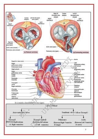
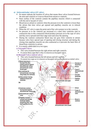
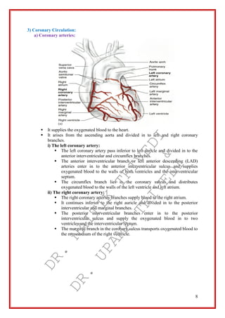
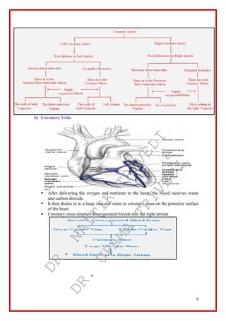
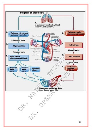
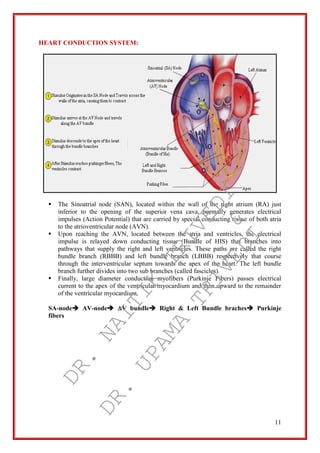
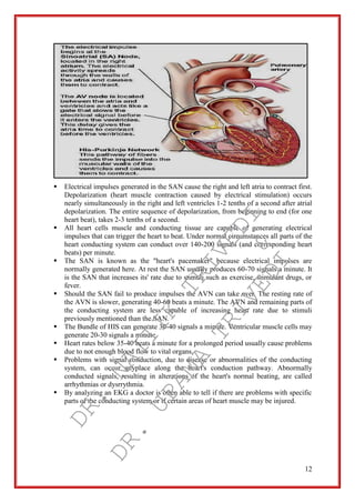
cardiovascular system
blood vessels
biology
b.pharma
Introduction to Heart
Location & position of heart
Anatomy of heart
Heart wall
Valves of heart
Heart – Interior of front
Blood vessels: Arteries, Veins and CapillariesAmir Rifaat
It is one of the circulatory systems. This explains the roles of arteries, veins and capillaries. It also differentiate between the arteries, veins and capillaries. This slide also explained the pulmonary circuit and systemic curcuit. This is an interesting notes and easy to be understand.
The human heart heart length, width, and thickness are 12 cm, 8.5 cm, and 6 cm, respectively. In addition, the mean weight of the heart is 280-340 g in males and 230-280 g in females.
Senses : any of the physical processes by which stimuli are received, transduced, and conducted as impulses to be interpreted in the brain.
The special senses consist of the eyes, ears, nose, throat and skin.
Each of these organs have specialized functions that make if possible for us to experience our environment and to make that experience more pleasant
Unit-III, Chapter-1- Respiratory System.pptAudumbar Mali
B. Pharm. First Year, Sem:II,
Unit III
Respiratory system 10 hours
Anatomy of respiratory system with special reference to anatomy of lungs,
mechanism of respiration, regulation of respiration
Lung Volumes and capacities transport of respiratory gases, artificial respiration,
and resuscitation methods.
Blood is carried through the body via blood vessels. An artery is a blood vessel that carries blood away from the heart, where it branches into ever-smaller vessels.
This PPT includes the heart and it's layers. Heart sounds, coronary circulation, conduction system of heart, cardiac cycle, vascular pathways, Electrocardiogram, applied anatomy of CVS, applied physiology of CVS.
The blood vessels are the components of the circulatory system that transport blood throughout the human body. These vessels transport blood cells, nutrients, and oxygen to the tissues of the body. They also take waste and carbon dioxide away from the tissues.
The circulatory system, also called the cardiovascular system or the vascular system, is an organ system that permits blood to circulate and transport nutrients (such as amino acids and electrolytes), oxygen, carbon dioxide, hormones, and blood cells to and from the cells in the body to provide nourishment and help in fighting diseases, stabilize temperature and pH, and maintain homeostasis.
CVS/Anatomy of heart/ B.pharmacy 2 semesterKondal Reddy
The cardiovascular system is sometimes called the blood-vascular, or simply the circulatory system.
It consists of the heart, which is a muscular pumping device, and a closed system of vessels called arteries, veins, and capillaries.
The main function of the cardiovascular system is therefore to maintain blood flow to all parts of the body, to allow it to survive.
The arteries carry blood away from the heart; the veins carry it back to the heart.
This presentation explores a brief idea about the structural and functional attributes of nucleotides, the structure and function of genetic materials along with the impact of UV rays and pH upon them.
Richard's aventures in two entangled wonderlandsRichard Gill
Since the loophole-free Bell experiments of 2020 and the Nobel prizes in physics of 2022, critics of Bell's work have retreated to the fortress of super-determinism. Now, super-determinism is a derogatory word - it just means "determinism". Palmer, Hance and Hossenfelder argue that quantum mechanics and determinism are not incompatible, using a sophisticated mathematical construction based on a subtle thinning of allowed states and measurements in quantum mechanics, such that what is left appears to make Bell's argument fail, without altering the empirical predictions of quantum mechanics. I think however that it is a smoke screen, and the slogan "lost in math" comes to my mind. I will discuss some other recent disproofs of Bell's theorem using the language of causality based on causal graphs. Causal thinking is also central to law and justice. I will mention surprising connections to my work on serial killer nurse cases, in particular the Dutch case of Lucia de Berk and the current UK case of Lucy Letby.
A brief information about the SCOP protein database used in bioinformatics.
The Structural Classification of Proteins (SCOP) database is a comprehensive and authoritative resource for the structural and evolutionary relationships of proteins. It provides a detailed and curated classification of protein structures, grouping them into families, superfamilies, and folds based on their structural and sequence similarities.
Comparing Evolved Extractive Text Summary Scores of Bidirectional Encoder Rep...University of Maribor
Slides from:
11th International Conference on Electrical, Electronics and Computer Engineering (IcETRAN), Niš, 3-6 June 2024
Track: Artificial Intelligence
https://www.etran.rs/2024/en/home-english/
Richard's entangled aventures in wonderlandRichard Gill
Since the loophole-free Bell experiments of 2020 and the Nobel prizes in physics of 2022, critics of Bell's work have retreated to the fortress of super-determinism. Now, super-determinism is a derogatory word - it just means "determinism". Palmer, Hance and Hossenfelder argue that quantum mechanics and determinism are not incompatible, using a sophisticated mathematical construction based on a subtle thinning of allowed states and measurements in quantum mechanics, such that what is left appears to make Bell's argument fail, without altering the empirical predictions of quantum mechanics. I think however that it is a smoke screen, and the slogan "lost in math" comes to my mind. I will discuss some other recent disproofs of Bell's theorem using the language of causality based on causal graphs. Causal thinking is also central to law and justice. I will mention surprising connections to my work on serial killer nurse cases, in particular the Dutch case of Lucia de Berk and the current UK case of Lucy Letby.
Slide 1: Title Slide
Extrachromosomal Inheritance
Slide 2: Introduction to Extrachromosomal Inheritance
Definition: Extrachromosomal inheritance refers to the transmission of genetic material that is not found within the nucleus.
Key Components: Involves genes located in mitochondria, chloroplasts, and plasmids.
Slide 3: Mitochondrial Inheritance
Mitochondria: Organelles responsible for energy production.
Mitochondrial DNA (mtDNA): Circular DNA molecule found in mitochondria.
Inheritance Pattern: Maternally inherited, meaning it is passed from mothers to all their offspring.
Diseases: Examples include Leber’s hereditary optic neuropathy (LHON) and mitochondrial myopathy.
Slide 4: Chloroplast Inheritance
Chloroplasts: Organelles responsible for photosynthesis in plants.
Chloroplast DNA (cpDNA): Circular DNA molecule found in chloroplasts.
Inheritance Pattern: Often maternally inherited in most plants, but can vary in some species.
Examples: Variegation in plants, where leaf color patterns are determined by chloroplast DNA.
Slide 5: Plasmid Inheritance
Plasmids: Small, circular DNA molecules found in bacteria and some eukaryotes.
Features: Can carry antibiotic resistance genes and can be transferred between cells through processes like conjugation.
Significance: Important in biotechnology for gene cloning and genetic engineering.
Slide 6: Mechanisms of Extrachromosomal Inheritance
Non-Mendelian Patterns: Do not follow Mendel’s laws of inheritance.
Cytoplasmic Segregation: During cell division, organelles like mitochondria and chloroplasts are randomly distributed to daughter cells.
Heteroplasmy: Presence of more than one type of organellar genome within a cell, leading to variation in expression.
Slide 7: Examples of Extrachromosomal Inheritance
Four O’clock Plant (Mirabilis jalapa): Shows variegated leaves due to different cpDNA in leaf cells.
Petite Mutants in Yeast: Result from mutations in mitochondrial DNA affecting respiration.
Slide 8: Importance of Extrachromosomal Inheritance
Evolution: Provides insight into the evolution of eukaryotic cells.
Medicine: Understanding mitochondrial inheritance helps in diagnosing and treating mitochondrial diseases.
Agriculture: Chloroplast inheritance can be used in plant breeding and genetic modification.
Slide 9: Recent Research and Advances
Gene Editing: Techniques like CRISPR-Cas9 are being used to edit mitochondrial and chloroplast DNA.
Therapies: Development of mitochondrial replacement therapy (MRT) for preventing mitochondrial diseases.
Slide 10: Conclusion
Summary: Extrachromosomal inheritance involves the transmission of genetic material outside the nucleus and plays a crucial role in genetics, medicine, and biotechnology.
Future Directions: Continued research and technological advancements hold promise for new treatments and applications.
Slide 11: Questions and Discussion
Invite Audience: Open the floor for any questions or further discussion on the topic.
(May 29th, 2024) Advancements in Intravital Microscopy- Insights for Preclini...Scintica Instrumentation
Intravital microscopy (IVM) is a powerful tool utilized to study cellular behavior over time and space in vivo. Much of our understanding of cell biology has been accomplished using various in vitro and ex vivo methods; however, these studies do not necessarily reflect the natural dynamics of biological processes. Unlike traditional cell culture or fixed tissue imaging, IVM allows for the ultra-fast high-resolution imaging of cellular processes over time and space and were studied in its natural environment. Real-time visualization of biological processes in the context of an intact organism helps maintain physiological relevance and provide insights into the progression of disease, response to treatments or developmental processes.
In this webinar we give an overview of advanced applications of the IVM system in preclinical research. IVIM technology is a provider of all-in-one intravital microscopy systems and solutions optimized for in vivo imaging of live animal models at sub-micron resolution. The system’s unique features and user-friendly software enables researchers to probe fast dynamic biological processes such as immune cell tracking, cell-cell interaction as well as vascularization and tumor metastasis with exceptional detail. This webinar will also give an overview of IVM being utilized in drug development, offering a view into the intricate interaction between drugs/nanoparticles and tissues in vivo and allows for the evaluation of therapeutic intervention in a variety of tissues and organs. This interdisciplinary collaboration continues to drive the advancements of novel therapeutic strategies.
Observation of Io’s Resurfacing via Plume Deposition Using Ground-based Adapt...Sérgio Sacani
Since volcanic activity was first discovered on Io from Voyager images in 1979, changes
on Io’s surface have been monitored from both spacecraft and ground-based telescopes.
Here, we present the highest spatial resolution images of Io ever obtained from a groundbased telescope. These images, acquired by the SHARK-VIS instrument on the Large
Binocular Telescope, show evidence of a major resurfacing event on Io’s trailing hemisphere. When compared to the most recent spacecraft images, the SHARK-VIS images
show that a plume deposit from a powerful eruption at Pillan Patera has covered part
of the long-lived Pele plume deposit. Although this type of resurfacing event may be common on Io, few have been detected due to the rarity of spacecraft visits and the previously low spatial resolution available from Earth-based telescopes. The SHARK-VIS instrument ushers in a new era of high resolution imaging of Io’s surface using adaptive
optics at visible wavelengths.
Deep Behavioral Phenotyping in Systems Neuroscience for Functional Atlasing a...Ana Luísa Pinho
Functional Magnetic Resonance Imaging (fMRI) provides means to characterize brain activations in response to behavior. However, cognitive neuroscience has been limited to group-level effects referring to the performance of specific tasks. To obtain the functional profile of elementary cognitive mechanisms, the combination of brain responses to many tasks is required. Yet, to date, both structural atlases and parcellation-based activations do not fully account for cognitive function and still present several limitations. Further, they do not adapt overall to individual characteristics. In this talk, I will give an account of deep-behavioral phenotyping strategies, namely data-driven methods in large task-fMRI datasets, to optimize functional brain-data collection and improve inference of effects-of-interest related to mental processes. Key to this approach is the employment of fast multi-functional paradigms rich on features that can be well parametrized and, consequently, facilitate the creation of psycho-physiological constructs to be modelled with imaging data. Particular emphasis will be given to music stimuli when studying high-order cognitive mechanisms, due to their ecological nature and quality to enable complex behavior compounded by discrete entities. I will also discuss how deep-behavioral phenotyping and individualized models applied to neuroimaging data can better account for the subject-specific organization of domain-general cognitive systems in the human brain. Finally, the accumulation of functional brain signatures brings the possibility to clarify relationships among tasks and create a univocal link between brain systems and mental functions through: (1) the development of ontologies proposing an organization of cognitive processes; and (2) brain-network taxonomies describing functional specialization. To this end, tools to improve commensurability in cognitive science are necessary, such as public repositories, ontology-based platforms and automated meta-analysis tools. I will thus discuss some brain-atlasing resources currently under development, and their applicability in cognitive as well as clinical neuroscience.
Introduction:
RNA interference (RNAi) or Post-Transcriptional Gene Silencing (PTGS) is an important biological process for modulating eukaryotic gene expression.
It is highly conserved process of posttranscriptional gene silencing by which double stranded RNA (dsRNA) causes sequence-specific degradation of mRNA sequences.
dsRNA-induced gene silencing (RNAi) is reported in a wide range of eukaryotes ranging from worms, insects, mammals and plants.
This process mediates resistance to both endogenous parasitic and exogenous pathogenic nucleic acids, and regulates the expression of protein-coding genes.
What are small ncRNAs?
micro RNA (miRNA)
short interfering RNA (siRNA)
Properties of small non-coding RNA:
Involved in silencing mRNA transcripts.
Called “small” because they are usually only about 21-24 nucleotides long.
Synthesized by first cutting up longer precursor sequences (like the 61nt one that Lee discovered).
Silence an mRNA by base pairing with some sequence on the mRNA.
Discovery of siRNA?
The first small RNA:
In 1993 Rosalind Lee (Victor Ambros lab) was studying a non- coding gene in C. elegans, lin-4, that was involved in silencing of another gene, lin-14, at the appropriate time in the
development of the worm C. elegans.
Two small transcripts of lin-4 (22nt and 61nt) were found to be complementary to a sequence in the 3' UTR of lin-14.
Because lin-4 encoded no protein, she deduced that it must be these transcripts that are causing the silencing by RNA-RNA interactions.
Types of RNAi ( non coding RNA)
MiRNA
Length (23-25 nt)
Trans acting
Binds with target MRNA in mismatch
Translation inhibition
Si RNA
Length 21 nt.
Cis acting
Bind with target Mrna in perfect complementary sequence
Piwi-RNA
Length ; 25 to 36 nt.
Expressed in Germ Cells
Regulates trnasposomes activity
MECHANISM OF RNAI:
First the double-stranded RNA teams up with a protein complex named Dicer, which cuts the long RNA into short pieces.
Then another protein complex called RISC (RNA-induced silencing complex) discards one of the two RNA strands.
The RISC-docked, single-stranded RNA then pairs with the homologous mRNA and destroys it.
THE RISC COMPLEX:
RISC is large(>500kD) RNA multi- protein Binding complex which triggers MRNA degradation in response to MRNA
Unwinding of double stranded Si RNA by ATP independent Helicase
Active component of RISC is Ago proteins( ENDONUCLEASE) which cleave target MRNA.
DICER: endonuclease (RNase Family III)
Argonaute: Central Component of the RNA-Induced Silencing Complex (RISC)
One strand of the dsRNA produced by Dicer is retained in the RISC complex in association with Argonaute
ARGONAUTE PROTEIN :
1.PAZ(PIWI/Argonaute/ Zwille)- Recognition of target MRNA
2.PIWI (p-element induced wimpy Testis)- breaks Phosphodiester bond of mRNA.)RNAse H activity.
MiRNA:
The Double-stranded RNAs are naturally produced in eukaryotic cells during development, and they have a key role in regulating gene expression .
Professional air quality monitoring systems provide immediate, on-site data for analysis, compliance, and decision-making.
Monitor common gases, weather parameters, particulates.
1. 1
!!JAY AMBE!!
CARDIOVASCULAR SYSTEM
PREPARED BY
DR. NAITIK D. TRIVEDI,
M.PHARM, PH.D
LECTURER,
A. R. COLLEGE OF PHARMACY &
G. H. PATEL INSTITUTE OF PHARMACY,
VALLABH VIDYANAGAR, ANAND, GUJARAT.
Mobile: 91 – 9924567864
E-mail: mastermindnaitik@gmail.com
DR. UPAMA N. TRIVEDI,
M. PHARM, PH. D
ASSOCIATE PROFESSOR,
INDUBHAI PATEL COLLEGE OF PHARMACY AND RESEARCH CENTRE,
DHARMAJ.
E-mail: ups.aasthu@gmail.com
DR.
NAITIK
TRIVEDI&
DR.
UPAMA
TRIVEDI
2. 2
CARDIOVASCULAR SYSTEM
INTRODUCTION:
Cardiovascular is the system which includes the study of the heart, blood vessels and blood.
These system is some time known as circularly system because it circulate or transport the
nutrients, oxygen, carbon dioxide and essential molecules from environment to cells and
cells to environment. It is involuntary in nature and gives continuous work. The heart
propelling or impelling blood around 100,000 km of blood vessels and it pumps 14000 liters
blood in day and 10 million liters in year.
LOCATION OF HEART:
Cone shaped heart is relatively small, about the same size of closed fist of person.
It is 12 cm (5 in.) long, 9 cm (3.5 in.) wide and 6 cm (2.5 in.) thick.
In an adult, average weight of heart is 300gm.
The heart consist four chambers:
a) Two atria or atrium
b) Two ventricles
It is located near to the middle of thoracic cavity in the mediastinum (the space
between the lungs) and it rest on to the diaphragm.
About two third of the mass of the heart lies to the left of the body’s midline.
Pointed end portion which is formed by the tip of left ventricle is known as apex and
opposite to apex the wide superior and posterior margin is known as base.
LAYER OF THE HEART:
Heart layer is formed by pericardium ("around the heart") which is triple-layered
fluid-filled sac that surrounds and protects the heart.
The pericardium consist of two principle portion
Layer of Heart (Pericardium)
Fibrous Pericardium Serous Pericardium
Parietal Layer (Outer Layer) Visceral Layer (Inner Layer)
(Epicardium)
DR.
NAITIK
TRIVEDI&
DR.
UPAMA
TRIVEDI
3. 3
a) Fibrous pericardium:
It is a strong layer of dense connective tissue.
It adheres to the diaphragm inferiorly, and superiorly it is fused to the roots of
the great vessels that leave and enter the heart.
The fibrous pericardium acts as a tough outer coat that holds the heart in place
and keeps it from overfilling with blood.
It prevents overstretching of the heart, provides protection and fix the heart in
mediastinum.
b) Serous pericardium:
It is a Deep to the fibrous pericardium and form double layer around the heart.
The outer layer is known as parietal layer which is fused to the fibrous
pericardium.
The inner layer is known as visceral layer also known as epicardium.
Between the outer layer (parietal layer) and inner layer (visceral layer) is gap
known as pericardial cavity filled by fluid is known as pericardial fluid.
This fluid ac as lubricant and reduces friction between the layer during
contraction and relaxation.
WALL OF THE HEART:
The wall of the heart is formed by three layers:
a) Epicardium (External layer):
It is also known as the visceral layer of the serous pericardium.
It is composed of mesothelium and delicated connective tissue.
It is the outer or external wall of the heart.
b) Myocardium (Middle layer):
Myo means muscles so it is made up by cardiac muscles tissue.
It provides the bulkiness to the heart and it is responsible for the pumping
action of heart.
c) Endocardium (Inner layer):
The endocardium is an innermost, thin, smooth layer of epithelial tissue that
lines the inner surface of the heart chambers and valves.
DR.
NAITIK
TRIVEDI&
DR.
UPAMA
TRIVEDI
4. 4
INTERIOR OF THE HEART:
The heart is divided in to right and left side by partition known as the septum which is
made up by myocardium covered by epithelium
a) Chamber of heart:
Heart consists of four chambers;
i) Two atria:
The two superior chambers are known as right atrium and left atrium.
The posterior wall of the atrium is smooth surface while the anterior wall of
the atrium is rough surface.
On the surface of the atrium is wrinkle pouch like structure is known as auricle
because it resemble like dog ear.
ii) Two ventricles
The two inferior are known as right ventricle and left ventricles.
On the surface of heart grooves like structures known as coronary sulcus which
separate the atria to ventricle.
The thickness of the wall of the four chambers varies according to their function.
Example: The wall of the atria is thin and it pumps blood in to ventricles & the
ventricle wall is thick in which right ventricle pumps blood in to lungs and
left ventricle pumps blood in to aorta so the work load on left ventricle is
high because of that the wall of left ventricle is two to four times thicker
than right ventricle.
b) Valve of the heart:
In the heart blood passes from various compartment and finally it enter in to the
artery.
During these circulations for the prevention of back flow of blood there is special type
of structure in heart which is known as valve.
These special types of structures or valves are composed by dense connective tissue
which is covered by endocardium.
Valves are opened and closed in response to the contraction or relaxation of heart.
DR.
NAITIK
TRIVEDI&
DR.
UPAMA
TRIVEDI
5. 5
It is mainly classified in to two types:
DR.
NAITIK
TRIVEDI&
DR.
UPAMA
TRIVEDI
6. 6
a) Atrioventricular valves (AV valve):
Its names indicate the location of valves that means these valves located between
the atria and ventricle or it moves blood from atrium to ventricle..
Inner surface of the ventricle contain the papillary muscles which is connected
with the end or tip part of valve.
Blood moves atrium to ventricle when the pressure in to the ventricle is lower than
the atrium that time valves get opened and papillary muscles are in relaxed
condition.
When the AV valve is open the point end of the valve project in to the ventricle.
So pressure in to the ventricle get increased at a same time ventricles start to
contraction due to this contraction blood produce pressure on to the cups of valve
and they move upward until their edge meet and close the opening.
During the ventricle contraction blood may not goes from ventricle to atrium
because valve have special type of opening and closing structure and during the
closing time papillary muscles are also contracted so it prevents the back flow of
blood from ventricles to atrium.
It is mainly subdivided in to two types:
i) Tricuspid Valves:
This valve located between the right atrium and right ventricle.
It consist three cups that’s why it is known as tricuspid valves.
ii) Bicuspid Valves (mitral valve):
This valve located between the left atrium and left ventricle.
It consist two cups so it is known as bicuspid valves also known as mitral valve.
DR.
NAITIK
TRIVEDI&
DR.
UPAMA
TRIVEDI
7. 7
b) Semilunar Valves:
These valves consist three Semilunar (half moon like) cups.
It is further subdivided in to:
i) Pulmonary Semilunar valve:
It passes the blood from right ventricle to lungs during opening stage.
ii) Aortic Semilunar valve:
It passes the blood from left ventricle to aorta during opening condition.
CIRCULATRY SYSTEM OF BLOOD THROUGH HEART:
1) Pulmonary circulation:
In the body system cells receive O2 from blood and give CO2 in to blood so blood
become deoxygenate.
These Deoxygenated blood by the help of Superior venacava, Inferior venacava and
coronary sinus transfer in to the right atrium.
Right atrium flow this deoxygenated blood in to right ventricle with the help of
tricuspid valves.
Right ventricle pumps blood in to pulmonary trunk via pulmonary Semilunar valve.
The pulmonary trunk divided in to right pulmonary artery and left pulmonary artery,
the right pulmonary artery gives blood to right lungs and left pulmonary artery gives
blood to left lungs.
2) Systemic Circulation:
In the lungs blood become oxygenated means it gain O2 and loss CO2.
Now, the oxygenated blood returns in to heart through pulmonary veins.
Then pulmonary veins pass blood in to left atrium which pumps blood in to left
ventricle with the help of bicuspid valve.
Then, left ventricle through blood enters in to aorta with the help aortic valve.
Finally, bloods flows in systemic circulation from aorta and reach near to each and
every cells of the body and gives O2 and take CO2.
Again the deoxygenated blood flow trough pulmonary circulation.
DR.
NAITIK
TRIVEDI&
DR.
UPAMA
TRIVEDI
8. 8
3) Coronary Circulation:
a) Coronary arteries:
It supplies the oxygenated blood to the heart.
It arises from the ascending aorta and divided in to left and right coronary
branches.
i) The left coronary artery:
The left coronary artery pass inferior to left auricle and divided in to the
anterior interventricular and circumflex branches.
The anterior interventricular branch or left anterior descending (LAD)
arteries enter in to the anterior interventricular sulcus and supplies
oxygenated blood to the walls of both ventricles and the interventricular
septum.
The circumflex branch lies in the coronary sulcus and distributes
oxygenated blood to the walls of the left ventricle and left atrium.
ii) The right coronary artery:
The right coronary arteries branches supply blood to the right atrium.
It continues inferior to the right auricle and divided in to the posterior
interventricular and marginal branches.
The posterior interventricular branches enter in to the posterior
interventricular sulcus and supply the oxygenated blood in to two
ventricles and the interventricular septum.
The marginal branch in the coronary sulcus transports oxygenated blood to
the myocardium of the right ventricle.
DR.
NAITIK
TRIVEDI&
DR.
UPAMA
TRIVEDI
9. 9
b) Coronary Vein:
After delivering the oxygen and nutrients to the heart, the blood receives waste
and carbon dioxide.
It then drains in to a large vascular sinus or coronary sinus on the posterior surface
of the heart.
Coronary sinus empties deoxygenated bloods into the right atrium.
DR.
NAITIK
TRIVEDI&
DR.
UPAMA
TRIVEDI
11. 11
HEART CONDUCTION SYSTEM:
The Sinoatrial node (SAN), located within the wall of the right atrium (RA) just
inferior to the opening of the superior vena cava, normally generates electrical
impulses (Action Potential) that are carried by special conducting tissue of both atria
to the atrioventricular node (AVN).
Upon reaching the AVN, located between the atria and ventricles, the electrical
impulse is relayed down conducting tissue (Bundle of HIS) that branches into
pathways that supply the right and left ventricles. These paths are called the right
bundle branch (RBBB) and left bundle branch (LBBB) respectively that course
through the interventricular septum towards the apex of the heart. The left bundle
branch further divides into two sub branches (called fascicles).
Finally, large diameter conduction myofibers (Purkinje Fibers) passes electrical
current to the apex of the ventricular myocardium and then upward to the remainder
of the ventricular myocardium.
SA-node AV-node AV bundle Right & Left Bundle braches Purkinje
fibers
DR.
NAITIK
TRIVEDI&
DR.
UPAMA
TRIVEDI
12. 12
Electrical impulses generated in the SAN cause the right and left atria to contract first.
Depolarization (heart muscle contraction caused by electrical stimulation) occurs
nearly simultaneously in the right and left ventricles 1-2 tenths of a second after atrial
depolarization. The entire sequence of depolarization, from beginning to end (for one
heart beat), takes 2-3 tenths of a second.
All heart cells muscle and conducting tissue are capable of generating electrical
impulses that can trigger the heart to beat. Under normal circumstances all parts of the
heart conducting system can conduct over 140-200 signals (and corresponding heart
beats) per minute.
The SAN is known as the "heart's pacemaker" because electrical impulses are
normally generated here. At rest the SAN usually produces 60-70 signals a minute. It
is the SAN that increases its' rate due to stimuli such as exercise, stimulant drugs, or
fever.
Should the SAN fail to produce impulses the AVN can take over. The resting rate of
the AVN is slower, generating 40-60 beats a minute. The AVN and remaining parts of
the conducting system are less capable of increasing heart rate due to stimuli
previously mentioned than the SAN.
The Bundle of HIS can generate 30-40 signals a minute. Ventricular muscle cells may
generate 20-30 signals a minute.
Heart rates below 35-40 beats a minute for a prolonged period usually cause problems
due to not enough blood flow to vital organs.
Problems with signal conduction, due to disease or abnormalities of the conducting
system, can occur anyplace along the heart's conduction pathway. Abnormally
conducted signals, resulting in alterations of the heart's normal beating, are called
arrhythmias or dysrrythmia.
By analyzing an EKG a doctor is often able to tell if there are problems with specific
parts of the conducting system or if certain areas of heart muscle may be injured.
DR.
NAITIK
TRIVEDI&
DR.
UPAMA
TRIVEDI
13. 13
ELECTROCARDIOGRAPHY:
Electrocardiography (ECG or EKG from the German Elektrokardiogramm) is a
transthoracic interpretation of the electrical activity of the heart over time captured
and externally recorded by skin electrodes.
It is a noninvasive recording produced by an electrocardiographic device.
The ECG works mostly by detecting and amplifying the tiny electrical changes on the
skin that are caused when the heart muscle "depolarizes" during each heart beat.
At rest, each heart muscle cell has a charge across its outer wall, or cell membrane.
Reducing this charge towards zero is called de-polarization, which activates the
mechanisms in the cell that cause it to contract.
During each heartbeat a healthy heart will have an orderly progression of a wave of
depolarization that is triggered by the cells in the sinoatrial node, spreads out through
the atrium, passes through "intrinsic conduction pathways" and then spreads all over
the ventricles. This is detected as tiny rises and falls in the voltage between two
electrodes placed either side of the heart which is displayed as a wavy line either on a
screen or on paper. This display indicates the overall rhythm of the heart and
weaknesses in different parts of the heart muscle.
Usually more than 2 electrodes are used and they can be combined into a number of
pairs. (For example: Left arm (LA), right arm (RA) and left leg (LL) electrodes form
the pairs: LA+RA, LA+LL, RA+LL) The output from each pair is known as a lead.
Each lead is said to look at the heart from a different angle.
Different types of ECGs can be referred to by the number of leads that are recorded,
for example 3-lead, 5-lead or 12-lead ECGs (sometimes simply "a 12-lead").
A 12-lead ECG is one in which 12 different electrical signals are recorded at
approximately the same time and will often be used as a one-off recording of an ECG,
typically printed out as a paper copy. 3- and 5-lead ECGs tend to be monitored
continuously and viewed only on the screen of an appropriate monitoring device, for
example during an operation or whilst being transported in an ambulance.
There may, or may not be any permanent record of a 3- or 5-lead ECG depending on
the equipment used.
Placement of electrodes:
Ten electrodes are used for a 12-lead ECG. The electrodes usually consist of a
conducting gel, embedded in the middle of a self-adhesive pad onto which cables
clip. Sometimes the gel also forms the adhesive. They are labeled and placed on
the patient's body as follows:
DR.
NAITIK
TRIVEDI&
DR.
UPAMA
TRIVEDI
14. 14
Electrode
label (in
the USA)
Electrode placement
RA On the right arm, avoiding bony prominences.
LA In the same location that RA was placed, but on the left arm this time.
RL On the right leg, avoiding bony prominences.
LL In the same location that RL was placed, but on the left leg this time.
V1 In the fourth intercostal space (between ribs 4 & 5) just to the right of
the sternum (breastbone).
V2 In the fourth intercostal space (between ribs 4 & 5) just to the left of the
sternum.
V3 Between leads V2 and V4.
V4 In the fifth intercostal space (between ribs 5 & 6) in the mid-clavicular
line (the imaginary line that extends down from the midpoint of the
clavicle (collarbone)).
V5 Horizontally even with V4, but in the anterior axillary line. (The anterior
axillary line is the imaginary line that runs down from the point midway
between the middle of the clavicle and the lateral end of the clavicle; the
lateral end of the collarbone is the end closer to the arm.)
V6 Horizontally even with V4 and V5 in the midaxillary line. (The
midaxillary line is the imaginary line that extends down from the middle
of the patient's armpit.)
Waves and intervals:
A typical ECG tracing of the cardiac cycle (heartbeat) consists of a P wave, a
QRS complex, a T wave, and a U wave which is normally visible in 50 to 75%
of ECGs. The baseline voltage of the electrocardiogram is known as the
isoelectric line. Typically the isoelectric line is measured as the portion of the
tracing following the T wave and preceding the next P wave.
DR.
NAITIK
TRIVEDI&
DR.
UPAMA
TRIVEDI
15. 15
Feature Description Duration
RR interval The interval between an R wave and the next R wave is the
inverse of the heart rate. Normal resting heart rate is between 50
and 100 bpm
0.6 to
1.2s
P wave During normal atrial depolarization, the main electrical vector is
directed from the SA node towards the AV node, and spreads
from the right atrium to the left atrium. This turns into the P wave
on the ECG.
80ms
PR interval The PR interval is measured from the beginning of the P wave to
the beginning of the QRS complex. The PR interval reflects the
time the electrical impulse takes to travel from the sinus node
through the AV node and entering the ventricles. The PR interval
is therefore a good estimate of AV node function.
120 to
200ms
PR segment The PR segment connects the P wave and the QRS complex. This
coincides with the electrical conduction from the AV node to the
bundle of His to the bundle branches and then to the Purkinje
Fibers. This electrical activity does not produce a contraction
directly and is merely traveling down towards the ventricles and
this shows up flat on the ECG. The PR interval is more clinically
relevant.
50 to
120ms
QRS
complex
The QRS complex reflects the rapid depolarization of the right
and left ventricles. They have a large muscle mass compared to
the atria and so the QRS complex usually has a much larger
amplitude than the P-wave.
80 to
120ms
J-point The point at which the QRS complex finishes and the ST
segment begins. Used to measure the degree of ST elevation or
depression present.
N/A
ST segment The ST segment connects the QRS complex and the T wave. The
ST segment represents the period when the ventricles are
depolarized. It is isoelectric.
80 to
120ms
T wave The T wave represents the repolarization (or recovery) of the
ventricles. The interval from the beginning of the QRS complex
to the apex of the T wave is referred to as the absolute refractory
period. The last half of the T wave is referred to as the relative
refractory period (or vulnerable period).
160ms
ST interval The ST interval is measured from the J point to the end of the T
wave.
320ms
QT interval The QT interval is measured from the beginning of the QRS
complex to the end of the T wave. A prolonged QT interval is a
risk factor for ventricular tachyarrhythmias and sudden death. It
varies with heart rate and for clinical relevance requires a
correction for this, giving the QTc.
300 to
430ms
U wave The U wave is not always seen. It is typically low amplitude, and,
by definition, follows the T wave.
J wave The J wave, elevated J-Point or Osborn Wave appears as a late
delta wave following the QRS or as a small secondary R wave. It
is considered pathognomic of hypothermia or hypocalcemia.
DR.
NAITIK
TRIVEDI&
DR.
UPAMA
TRIVEDI
16. 16
Some pathological entities which can be seen on the ECG:
Shortened QT
interval
Hypercalcemia, some drugs, certain genetic abnormalities.
Prolonged QT
interval
Hypocalcemia, some drugs, certain genetic abnormalities.
Elevated ST
segment
Myocardial infraction
Depressed ST
segment
The heart muscles receive insufficient oxygen
Prolonged PQ
intervals
Coronary artery disease, Rheumatic fever and Scar tissue may
be form in the heart.
Flattened or
inverted T Waves
Coronary ischemia, left ventricular hypertrophy, digoxin effect,
some drugs.
Hyper acute T
Waves
Possibly the first manifestation of acute myocardial infarction.
Prominent U Waves Hypokalemia.
Larger P Wave Indicate enlargement of an Atrium, as may occur in mitral
stenosis in which blood back up in to the left atrium.
Enlarge Q Wave Indicates myocardium infrection.
Enlarge R Wave Indicates enlarged ventricles
The ECG cannot reliably measure the pumping ability of the heart, for which
ultrasound-based (echocardiography) or nuclear medicine tests are used. It is possible
to be in cardiac arrest with a normal ECG signal (a condition known as pulseless
electrical activity).
CARDIAC CYCLES:
“A cardiac cycle include all the events associated within one heart beat”
The normal heart beats in healthy adult is 75 beats/min and cardiac cycle last for 0.8
sec.
In the cardiac cycle due to the pressure changes atria and ventricles alternately
contract and relax, and blood flows from areas of higher blood pressure to areas of
lower blood pressure.
The term systole is used for the contraction and diastole used for the relaxation.
In a normal cardiac cycle, the two atria contract while the two ventricles relax. Then,
while the two ventricle contract, the two atria relax.
Cardiac cycle is described by the following phase:
1) Atrial systole
In this phase Atrial contraction is begins which is last about 0.1 sec at same time
ventricle are relaxed.
So, the Right and Left AV valves are open and Atria send blood into the relaxed
ventricles.
Atrial systole pushes 25 ml of blood in to ventricles which already contain 105 ml
blood so at the end of atrial systole means end of ventricle diastole Ventricles
contain maximum blood volume which is 130 ml known as end-diastolic volume
(EDV).
In the ECG or EKG it is noted as P wave.
DR.
NAITIK
TRIVEDI&
DR.
UPAMA
TRIVEDI
17. 17
2. Ventricular systole:
In this phase ventricles begin contraction which is last for 0.3 sec.
Pressure in ventricles rises due to contraction and shut the AV valves which is
heard by “Lubb” Sound.
For about 0.05 sec all four valve are closed which is known as isovolumetric
contraction.
Ventricular contraction pushes blood out of the ventricles and opens the both
Semilunar valve and ejected 70 ml blood in to aorta and same amount of blood in
to pulmonary trunk by respectively left and right ventricles so the 60 ml of blood
remains in the each ventricle out of 130 ml.
This is known as ventricular ejection which last for 0.25 sec
Ventricular systole = isovolumetric contraction + ventricular ejection
= (0.05) + (0.25)
= 0.3 sec.
The blood volume in the Ventricles at the end of ventricle systole is 60 ml known
as end-systolic volume (ESV).
The blood ejection per beat from each ventricle is known as stroke volume.
Stroke volume = EDV – ESV
= 130 ml – 60 ml
= 70 ml
It is noted as QRS wave in ECG or EKG.
3. Ventricular Diastole or Relaxation period:
In this phase both ventricles and atria are in relaxation.
Relaxation which is last for 0.4 sec.
Pressure in the ventricles drops so blood in the Pulmonary Trunk and aorta
backflows closing the Semilunar Valves.
This is heard by “Dubb” sound.
In this period, ventricular pressure is higher than atrial pressure hence all heart
valves are closed again and this period is known as isovolumetric.
It is noted as T wave in ECG or EKG.
Again the AV valves open and fill up the ventricle and complete the one Cardiac
cycle.
DR.
NAITIK
TRIVEDI&
DR.
UPAMA
TRIVEDI
18. 18
CARDIAC OUTPUT:
Cardiac output is the amount of blood ejected from the left ventricle (or the right
ventricle) in to the aorta (or pulmonary trunk) each minute and it is equal to the
product of stroke volume and heart rate.
Thus,
Cardiac Output = Stroke volume (ml/beat) * Heart rate (beats/min)
= 70 ml/beat * 75 beats/min
= 5250 ml/min or 5.25 liters/min.
Note: Stroke volume = EDV – ESV = 130 ml/beat – 60 ml/beat = 70 ml/beat
DR.
NAITIK
TRIVEDI&
DR.
UPAMA
TRIVEDI
19. 19
Means the cardiac out put volume is close to the total blood volume, which is about 5
liters in the typical adult male. The entire blood volume thus flows through the
pulmonary and systemic circulations about once a minute.
When the demand of oxygen is increase or decrease, cardiac output changes to meet
the need by increasing or decreasing the stroke volume and heart rate.
For example during the mild exercise demand of oxygen is increase so the stroke
volume may increase to 100 ml/beat and heart rate to 100 beats/min. cardiac output
then would be 10 liters/min.
Factor affecting on Stroke volume:
Three important factors regulate the stroke volume in different circumstances and
ensure that the left and right ventricles pump equal volume of blood.
i) Preload:
The blood supply to the ventricle is often referred to as preload. Technically, the
definition of preload is the volume or pressure in the ventricle at the end of diastole or
refers as end-diastolic volume.
According to Frank-Starling law of the heart, more the heart is filling during diastole,
the greater the force of contraction during systole.
The duration of ventricular diastole and venous pressure are the two key factors that
determine EDV.
When the heart rate is increase, the duration of diastole is shorter so it takes smaller
filling times means smaller EDV.
On the other hand, when venous pressure increase, a greater volume of blood is forced
into the ventricles and the EDV is increased.
For example:
When heart rate exceeds about 160 beats/min, stroke volumes usually decrease. At
such rapid heart rate decrease the ventricular filling time so decrease the EDV and
thus the preload is lower.
People, who have slow resting heart rate, usually have large stroke volumes because
filling time is prolonged and preload thus is larger.
ii) Contractility:
The second factor that influences stroke volume is myocardial contractility, which is
the strength of contraction at any given preload.
Increases the contractility are called positive inotropic agents and decreases the
contractility are called negative inotropic agents.
Thus, for a constant preload, stroke volume is larger when positive inotropic agent is
present and it promotes the forceful contraction.
iii) Afterload:
The resistance to the ejection of blood by the ventricle is called afterload.
For example:
In the atherosclerosis condition arteries are narrowed so they resist the blood flow
from ventricles to out of heart and it decrease the stroke volume, and more blood
remains in the ventricles at the end of systole.
DR.
NAITIK
TRIVEDI&
DR.
UPAMA
TRIVEDI
20. 20
Factor affecting on stroke volume
iv) Other factors are:
Sympathetic stimulation
Increase the Epinephrine (E) and Non Epinephrine (NE) level
Causes ventricles to contract with more force and increases ejection fraction and
decreases ESV and increase the stroke volume.
Parasympathetic activity
Increases the Acetylcholine (Ach) released by vagus nerves
Reduces force of cardiac contractions
IMPORTANT QUESTIONS:
1. Draw the neat and labeled diagram of heart.
2. Write short note on layer and wall of heart.
3. Write short note on cardiac cycle.
4. Explain the blood circulation through the heart/Circulatory systems.
5. Write short note on ECG.
6. Write short note on cardiac output.
7. Explain the valve of heart.
A serious person can never be innocent, and one
who is innocent can never be serious.
DR.
NAITIK
TRIVEDI&
DR.
UPAMA
TRIVEDI