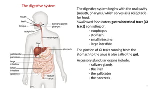
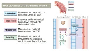
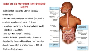
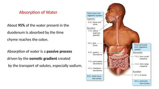
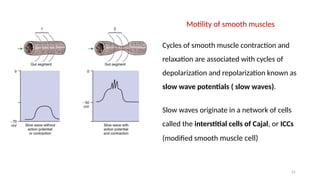
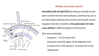
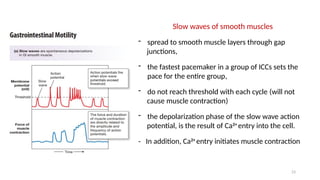
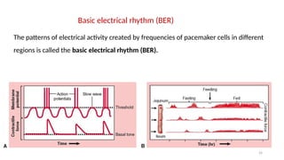
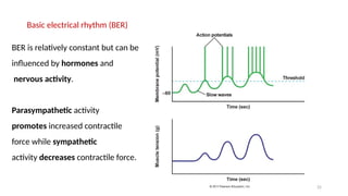
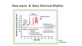
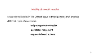
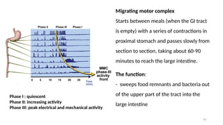
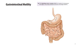
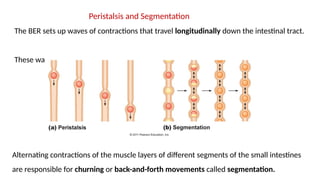
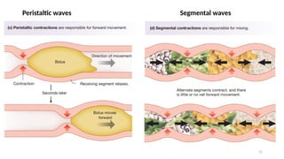
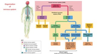
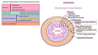
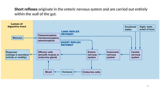
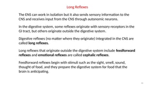
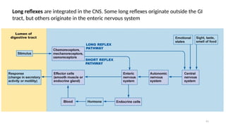
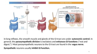
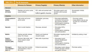
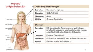
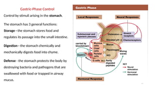
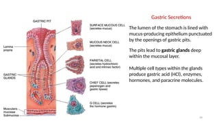
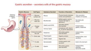
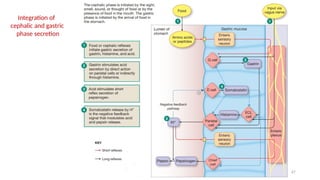
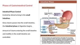
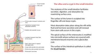
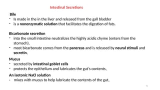
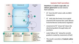
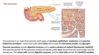
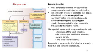
Digestive system
2
The digestive system begins with the oral cavity
(mouth, pharynx), which serves as a receptacle
for food.
Swallowed food enters gastrointestinal tract (GI
tract) consisting of:
- esophagus
- stomach
- small intestine
- large intestine
The portion of GI tract running from the
stomach to the anus is also called the gut.
Accessory glandular organs include:
- salivary glands
- the liver
- the gallblader
- the pancreas
The digestive system
3
Salivary glands
The oral cavity, the first stages of digestion begin
with chewing and the secretion of saliva by 3
Pairs of salivary galnds:
- sublingual glands
- submandibular gland
- parotid gland
4
Stomach
The stomach has 3 sections:
- the upper fundus
- the central body
- the lower antrum
The stomach continues digestion by mixing food
with acid and enzymes to create chyme.
Chyme – is the soupy mixture of food and secretions
The pylorus (gatekeeper) opening between the
stomach and the small intestine is guarded by the
pylori valve.
5
Small intestine
Most digestion takes place in the small intestine,
Sections of the small intestine:
- duodenum (25 cm)
- jejunum
- ileum
Digestion finishes in the small intestine, almost all
digested nutrients and secreted fluids are absorbed
there, leaving about 1.5l of chyme per day to pass
into the large intestine.
260 cm
6
Large intestine
In the colon – the proximal section of the
large intestine, watery chyme becomes
semisolid feces as water and electrolytes
are absorbed out of chyme into ECF.
7
The wall of the GI tract
The basic structure of the GI wall is similar in stomach and intestines.
The gut wall consists of four layers
- an inner mucosa facing the lumen
- submucosa layer
- layers of smooth muscles - muscularis externa
- a covering of connective tissue called the serosa
8
9
10
Other tasks of the digestive system
1. Avoiding autodigestion
- digestive systems secrete powerful enzymes to digest food into molecules,
- these enzymes must not digest the cells of the GI tract itself (autodigestion).
- If protective mechanisms against autodigestion fail, peptic ulcers develop on the
walls of the GI tract.
2. Mass balance
- maintaining mass balance by matching fluid input with output
11
Other tasks of the digestive system
3. Fluid secretion and reabsorption disorders
Normally intestinal reabsorption is very efficient (only ≈100 mL of fluid is lost in the feces).
Vomiting and diarrhea - excessively lost of water can become an emergency when the
secretion will be greater than reabsorption. In severe cases, this fluid loss can deplete
ECF volume to the point that the circulatory system is unable to maintain adequate blood pressure.
4. Defense
- protecting the body from foreign invaders (lumen of the digestive system is the largest area of
contact between the body’s internal environment and the outside world)
- physiological defense mechanisms: mucus, digestive enzymes, acid, the largest collection of
lymphoid tissue in the body, the gut-associated lymphoid tissue (GALT).
12
Mass balance in the digestive system
To maintain homeostasis, the volume of
fluid entering the gastrointestinal tract
by intake or secretion must equal the
volume leaving the lumen.
13
Rates of Fluid Movement in the Digestive
System
The fluid that enters the GI tract each day
comes from:
- the liver and pancreatic secretions (~ 2,5 liters)
- salivary gland secretions (~ 1.5 liters),
- secretions by glands of the stomach and small
intestines (~ 3.5 liters)
- and ingested water (~ 2 liters).
Most of this total (approximately 7,5 liters) is
absorbed by the small intestines, the colon also
absorbs some. Only a small amount (~ 100 ml) is
eliminated in the feces.
14
Absorption of Water
About 95% of the water present in the
duodenum is absorbed by the time
chyme reaches the colon.
Absorption of water is a passive process
driven by the osmotic gradient created
by the transport of solutes, especially sodium.
15
Digestive Enzymes
- are secreted either by exocrine glands (salivary glands and the pancreas) or by
epithelial cells in the stomach and small intestine,
- are proteins synthesized on the rough endoplasmic reticulum, packaged by the Golgi
complex into secretory vesicles, and then stored in the cell and released on demand by
exocytosis,
- many intestinal enzymes remain bound to the apical membranes of intestinal cells, anchored
by transmembrane protein “stalks” or lipid anchors,
16
Digestive Enzymes
- some digestive enzymes are secreted in an inactive proenzyme form (zmogens).
Zymogens must be activated in the GI lumen before they can carry out digestion.
- a nonfunctional form of enzymes protects cells from damage,
- zymogen names often have the suffix –ogen added to the enzyme name, such as pepsinogen
17
Mucus
- mucus is a viscous secretion composed primarily of glycoproteins called mucins,
- functions of mucus are to form a protective coating over the GI mucosa and to
lubricate the contents of the gut,
- mucus is made in specialized exocrine cell:
- in the stomach and salivary glands - mucous cells,
- in the intestine - goblet cells
18
Mucus
- goblet cells make up between 10% and 24% of the intestinal cell population,
- the signals for mucus release include:
- parasympathetic innervation,
- a variety of neuropeptides found in the enteric nervous system,
- cytokines from immunocytes
- parasitic infections
- inflammatory processes in the gut
the role of a protective barrier
19
Motility of smooth muscles
Role of motility in the GI tract:
- moving food from the mouth to the anus
- mechanically mixing food to break it into uniformly small particles
This mixing maximizes exposure of the particles to digestive enzymes by increasing
particle surface area.
Gastrointestinal motility is determined by:
- the properties of the GI smooth muscle
- and is modified by chemical input from nerves, hormones, and
paracrine signals
20
Motility of smooth muscles
Most of the gastrointestinal tract is composed of
single-unit smooth muscle, with groups of cells
electrically connected by gap junctions to create
contracting segments.
Different regions exhibit different types of
contraction:
- tonic contractions - are sustained for
minutes
or hours. They occur in some sphincters and in
the anterior portion of the stomach.
- phasic contractions - with contraction-
relaxation cycles lasting a few seconds,
occur in the posterior region of the stomach
21
Motility of smooth muscles
Cycles of smooth muscle contraction and
relaxation are associated with cycles of
depolarization and repolarization known as
slow wave potentials ( slow waves).
Slow waves originate in a network of cells
called the interstitial cells of Cajal, or ICCs
(modified smooth muscle cell)
22
Motility of smooth muscles
Interstitial cells of Cajal (ICCs) lie between smooth muscle
layers and the intrinsic nerve plexuses, and they may act as
an intermediary between the neurons and smooth muscle.
It appears that ICCs function as the pacemakers for slow
wave activity in different regions of the GI tract.
Slow wave potentials:
- frequency - (3–12 waves/min)
- frequency varies by region of the digestive tract,
(3 waves/min in the stomach, 12 waves/min in the
duodenum).
23
Slow waves of smooth muscles
- spread to smooth muscle layers through gap
junctions,
- the fastest pacemaker in a group of ICCs sets the
pace for the entire group,
- do not reach threshold with each cycle (will not
cause muscle contraction)
- the depolarization phase of the slow wave action
potential, is the result of Ca2+
entry into the cell.
- In addition, Ca2+
entry initiates muscle contraction
24
The patterns of electrical activity created by frequencies of pacemaker cells in different
regions is called the basic electrical rhythm (BER).
Basic electrical rhythm (BER)
25
BER is relatively constant but can be
influenced by hormones and
nervous activity.
Parasympathetic activity
promotes increased contractile
force while sympathetic
activity decreases contractile force.
Basic electrical rhythm (BER)
26
27
Motility of smooth muscles
Muscle contractions in the GI tract occur in three patterns that produce
different types of movement:
- migrating motor complex
- peristalsis movement
- segmental contractions
28
Migrating motor complex
Starts between meals (when the GI tract
is empty) with a series of contractions in
proximal stomach and passes slowly from
section to section, taking about 60-90
minutes to reach the large intestine.
The function:
- sweeps food remnants and bacteria out
of the upper part of the tract into the
large intestine
Phase I : quiescent
Phase II: increasing activity
Phase III: peak electrical and mechanical activity
29
Peristalsis and Segmentation
The BER sets up waves of contractions that travel longitudinally down the intestinal tract.
These waves are called peristalsis.
Alternating contractions of the muscle layers of different segments of the small intestines
are responsible for churning or back-and-forth movements called segmentation.
31
Peristaltic waves Segmental waves
32
Organization
of
nervous system
33
The enteric nervous system (ENS) controls:
- motility,
- secretion,
- and growth of the digestive tract.
The Enteric Nervous System
34
Characteristics
of the
Enteric Nervous System
• Intrinsic neurons
The intrinsic neurons of the two nerve plexuses of
the GI tract are lie completely within the wall of the
gut.
Autonomic neurons that bring signals from the CNS
to the digestive system are called extrinsic neurons.
• Neurotransmitters and neuromodulators
ENS neurons release more than 30
neurotransmitters and neuromodulators,
most of which are identical to molecules
found in the brain.
These neurotransmitters are sometimes
called NANC (nonadrenergic, noncholinergic).
The best known GI neurotransmitters and
neuromodulators are serotonin,
vasoactive intestinal peptide (VIP),
nitric oxid (NO).
35
Characteristics of the Enteric Nervous System
• Glial support cells
The glial cells of neurons within the ENS are more similar to astroglia of the
brain than to Schwann cells of the peripheral nervous system.
• Diffusion barrier
The capillaries that surround ganglia in the ENS are not very permeable and
create a diffusion barrier that is similar to the blood-brain barrier of cerebral
blood vessels.
• Integrating center
reflexes that originate in the GI tract can be integrated and acted on without
neural signals leaving the ENS. The neuron network of the ENS is its own
integrating center, much like the brain and spinal cord
36
Short Reflexes in the Enteric Nervous ­
System
The enteric nerve plexuses in the gut wall act as a “little brain,” allowing local reflexes to
begin, be integrated, and end completely in the GI tract
Reflexes that originate within the enteric nervous system and are integrated there without
outside input are called short reflexes.
The submucosal plexus
- contains sensory neurons that receive signals from the lumen of the gut,
- the ENS network integrates this sensory information, then initiates responses,
- neurons of this plexus controls secretion by GI epithelial cells,
The myenteric plexus
- neurons are located in the muscularis externa and influence motility
37
Enteric Plexus of the
Enteric Nervous System
Mesentery
38
39
Short reflexes originate in the enteric nervous system and are carried out entirely
within the wall of the gut.
40
Long Reflexes
The ENS can work in isolation but it also sends sensory information to the
CNS and receives input from the CNS through autonomic neurons.
In the digestive system, some reflexes originate with sensory receptors in the
GI tract, but others originate outside the digestive system.
Digestive reflexes (no matter where they originate) integrated in the CNS are
called long reflexes.
Long reflexes that originate outside the digestive system include feedforward
reflexes and emotional reflexes are called cephalic reflexes.
Feedforward reflexes begin with stimuli such as the sight, smell, sound,
thought of food, and they prepare the digestive system for food that the
brain is anticipating.
41
Long reflexes are integrated in the CNS. Some long reflexes originate outside the GI
tract, but others originate in the enteric nervous system
42
In long reflexes, the smooth muscle and glands of the GI tract are under autonomic control. In
general, the parasympathetic division is excitatory and enhances GI functions (“rest and
digest.”) Most parasympathetic neurons to the GI tract are found in the vagus nerve.
Sympathetic neurons usually inhibit GI function.
43
Peptides of the digestive tract
Peptides secreted by cells of the digestive tract may act as hormones or paracrine signals.
In the digestive system, GI peptides excite or inhibit motility and secretion.
Some paracrine peptides are secreted into the lumen, where they combine with receptors on
the apical membrane of the luminal epithelium to elicit a response.
Others are secreted into the extracellular fluid where they diffuse and act on neighboring
cells.
GI peptides also act outside the GI tract.
- GI hormone cholecystokinin (CCK) - enhances satiety and CCK is also release by
neurons and functions as a neurotransmitter in the brain.
- Ghrelin (peptide) is secreted by the stomach and acts on the brain to increase
food intake.
There are over 30 peptides from the GI mucosa, but only some of them are hormones.
44
GI Hormones
- are secreted into the blood and transported throughout the body,
- they act on the GI tract, on accessory organs (the pancreas or on distant organs such as the
brain)
The gastrointestinal hormones are divided into three families.
(all hormones of a family have similar amino acid sequences, and in some cases there is
overlap in their ability to bind to receptors).
GI Hormone Families
1. The gastrin family
2. Secretin family
3. The third family of peptides - contains those
that do not fit into the other two families.
45
GI Hormone Families
1. The gastrin family
- includes the hormones gastrin and cholecystokinin (CCK) and several variants of each.
- gastrin and CCK can bind to and activate the same CCKB receptor.
2. The secretin family
- includes: - secretin
- vasoactive intestinal peptide (VIP) - a nonadrenergic-noncholinergic
neurotransmiter (NANC),
- GIP – (old name gastric inhibitory peptide, a new name: glucose-dependent
insulinotropic peptide - it stimulates insulin release in response to glucose in
the intestinal lumen)
- glucagon-like peptide-1 (GLP-1)
3. The third family of peptides
- the primary member of this group is the motilin
- increases in motilin secretion are associated with the migrating motor complex
46
Cells in gastric mucosa
47
Overview
of digestive function
48
Food processing is divided into three phases:
- a cephalic phase,
- a gastric phase,
- an intestinal phase
Phases of Gastrointestinal Control
49
Cephalic-Phase Control
Control of gastrointestinal function by stimuli arising
in the head (smell, taste, thought of food) and can
make our mouths water and our stomachs rumble.
These long reflexes that begin in the brain create a
feedforward response known as the cephalic
phase of digestion.
50
Cephalic-Phase Control
Anticipatory stimuli and the stimulus of food in the
oral cavity activate neurons in the medulla oblongata.
The medulla in turn sends an efferent signal through
autonomic neurons to the salivary glands, and
through the vagus nerve to the enteric nervous
system.
In response to these signals, the stomach, intestine,
and accessory glandular organs begin secretion and
increase motility in anticipation of the food to come.
51
Chewing and Swallowing
The upper part of the GI tract, mouth, pharynx and upper esophagus is controlled by
skeletal muscle and control comes directly from the CNS.
Chewing is under both conscious and unconscious control.
Unconscious chewing is controlled by the chewing reflex. The presence of food in the
mouth stimulates pressure receptors that reflexively inhibit jaw-closing muscles allowing
the jaw to drop.
When the jaw drops the pressure on the pressure receptors is relieved and the jaw-closing
muscles become dis-inhibited. The alternate opening and closing of the jaw is thus
maintained by this reflex.
52
The Swallowing reflex
53
The Swallowing reflex
54
The Swallowing reflex
55
Gastric-Phase Control
Control by stimuli arising in the stomach.
The stomach has 3 general functions:
Storage - the stomach stores food and
regulates its passage into the small intestine.
Digestion - the stomach chemically and
mechanically digests food into chyme.
Defense - the stomach protects the body by
destroying bacteria and pathogens that are
swallowed with food or trapped in airway
mucus.
56
Gastric Motility
Stomach motility performs two tasks:
1. Mixing of food and gastric juice to form a chyme.
2. Regulated emptying of the chyme into the small intestines.
Gastric Motility Patterns
About three times a minute a peristaltic wave travels from the upper body of the
stomach toward the pylorus which mixes the stomach contents.
Peristaltic waves also propel some chyme forward into the duodenum.
The rate of gastric emptying depends on:
1. Composition of chyme
2. Volume of chyme
3. Force of gastric contractions
57
Regulation of Gastric Motility
The force of gastric contraction increases in response to gastrin and decreases in
response to CCK, secretin and GIP (gastric inhibitory peptide).
Gastric motility is controlled by:
- Cephalic-phase stimuli
- Pain, fear, depression inhibit gastric motility
- Anger and aggression stimulate gastric motility.
58
Gastric Secretions
The lumen of the stomach is lined with
mucus-producing epithelium punctuated
by the openings of gastric pits.
The pits lead to gastric glands deep
within the mucosal layer.
Multiple cell types within the glands
produce gastric acid (HCl), enzymes,
hormones, and paracrine molecules.
59
Gastric secretion – secretory cells of the gastric mucosa
60
Gastrin Secretion
G cells - found deep in the gastric glands,
secrete the hormone gastrin into the blood.
In short reflexes (mediated by ENS
neurotransmiter gastric-releasing peptide GRP),
gastrin release is stimulated by the presence of
amino acids and peptides in the stomach and by
distension of the stomach.
Coffee also stimulates gastrin release.
Gastrin release is also triggered by neural
reflexes (long reflex) - parasympathetic neurons
from the Xn. stimulate G cells to release gastrin
into the blood.
Gastrin’s primary action is to promote acid
release. It does this directly by acting on parietal
cells and indirectly by stimulating histamine
release
61
Gastric acid (HCl)
- parietal cells in the gastric glands secrete gasstric acid
into the lumen of the stomach
- 1-3 liters per day, acid create luminar pH as low as 1
Role of gastric acid:
- in the stomach causes release and activation of
pepsin an enzyme that digest proteins,
- denaturates proteins – help prepare proteins to
digestion by pepsin,
- helps kill bacteria and other ingested microorganisms,
- inactivates salivary amylase, stopping carbohydrate
digestion that began in the mouth.
62
Gastric acid (HCl)
The parietal cell secrete acid in the stomach.
↓
the H+ from water inside the parietal cell is pumped into the
stomach lumen by an H+-K+-ATPase in exchange for K+
entering the cell.
↓
Cl- then follows the electrical gradient created by H+ by
moving through open chloride channels
↓
The net result is secretion of HCl by the cell.
↓
While acid is being secreted into the lumen, bicarbonate
made from CO2 and the OH– from water is absorbed into
the blood.
↓
The buffering action of HCO3 – makes blood leaving the
stomach less acidic, creating an alkaline tide that can be
measured as a meal is being digested.
63
Mucus –Bicarbonate Barrier
The gastric mucosa protects itself from
autodigestion by acid and enzymes with a
mucus-bicarbonate barrier.
Mucous cells on the luminal surface and in
the neck of gastric glands secrete both
substances.
The mucus forms a physical barrier, and
the bicarbonate creates a chemical buffer
barrier underlying the mucus.
Mucus secretion is increased by irritants
such as aspirin (acetylsalicylic acid) or
alcohol.
64
Enzyme Secretion
The stomach produces two enzymes pepsin and a
gastric lipase.
Pepsin
- digests proteins into smaller peptides (cannot
break them down into amino acids),
- is effective in digesting collagen (plays an
important role in digesting meat),
- is secreted as the inactive enzyme pepsinogen by
chief cells in the gastric glands.
- acid stimulates pepsinogen release from chief cells
through a short reflex mediated in the ENS
Gastric lipase
- is co-secreted with pepsin,
- lipases are enzymes that break down triglycerides,
- less than 1/3 of fat digestion takes place in the
stomach
65
Paracrine secretions from the gastric mucosa
include:
- histamine,
- somatostatin,
- intrinsic factor
Histamine
- is a paracrine signal secreted by
enterochromaffinlike cells (ECL cells) in
response to gastrin or acetylcholine
stimulation,
- it diffuses to the parietal cells, and
stimulates acid secretion by combining with
H2 receptors on parietal cells,
- H2 receptor antagonists (cimetidine and
ranitidine, for example) that block histamine
action are a second class of drugs used to
treat acid hypersecretion.
Paracrine Secretions
66
Intrinsic factor
- is a protein secreted by the same gastric
parietal cells that secrete acid,
- in the lumen of the stomach intrinsic factor
complexes with vitamin B12, a step that is
needed for the vitamin’s absorption in the
intestine,
Somatostatin (SST)
- also known as hypothalamic growth
hormone-inhibiting hormone,
- is secreted by D cells in the stomach.
- somatostatin is the primary negative
feedback signal for gastric phase secretion,
- it shuts down acid secretion directly and
indirectly by decreasing gastrin and
histamine secretion,
- somatostatin also inhibits pepsinogen
secretion
Paracrine Secretions
67
Integration of
cephalic and gastric
phase secretion
68
Phases of Gastrointestinal Control
Intestinal-Phase Control
Control by stimuli arising in the small
intestines.
Once chyme passes into the small intestine,
the intestinal phase of digestion begins.
Amount of chyme entering the small intestine
and motility in the small intestine are
controlled.
69
Motility of the Small Intestines
Motility Patterns in the Small Intestines
The presence of chyme in the small intestines causes brief periods of peristaltic
contraction that propels the chyme forward and longer periods of segmentation during
which neighboring regions of the small intestines alternately relax and contract.
Segmentation facilitates the mixing of the chyme with bicarbonate, digestive enzymes
and bile. The stirring also aids absorption by bringing the chyme into contact with the
mucosa.
During fasting peristalsis and segmentation are replaced by migrating motility
complexes which are a series of intense contractions that periodically move through the
intestines and clean it of its contents.
70
The anatomy of the small intestine facilitates
secretion, digestion, and absorption by
maximizing surface area.
The surface of the lumen is sculpted into
fingerlike villi and deep crypts.
Most absorption takes place along the villi while
fluid and hormone secretion and cell renewal
from stem cells occurs in the crypts.
The apical surface of the enterocytes is modified
into microvilli whose surfaces are covered with
membrane-bound enzymes and a glycocalyx
coat.
The surface of the intestinal epithelium is called
the brush border.
The villus and a crypt in the small intestine
71
Most nutrients absorbed across the
intestinal epithelium move into
capillaries in the villi for distribution
through the circulatory system.
The exception is digested fats, most of
which pass into lacteals of the lymphatic
system.
The intestinal phase
72
Venous blood from the digestive tract passes
into the hepatic portal system.
This specialized region of the circulation has
two sets of capillary beds: one that picks up
absorbed nutrients at the intestine, and
another that delivers the nutrients directly
to the liver.
The intestinal phase
73
Intestinal Secretions
Intestinal secretions are produced by the liver, pancreas, and intestine
(3 liters/day).
The added secretions include:
- digestive enzymes,
- bile,
- bicarbonate,
- mucus,
- isotonic NaCl solution.
74
Intestinal Secretions
Digestive enzymes
- are produced by the intestinal epithelium and the exocrine pancreas,
- intestinal brush border enzymes are anchored to the luminal cell membranes
and are not swept out of the small intestine as chyme is propelled forward,
- the control pathways for enzyme release include neural, hormonal, and
paracrine signals,
- usually, stimulation of parasympathetic neurons (n X) enhances enzyme
secretion,
75
Intestinal Secretions
Bile
- Is made in the in the liver and released from the gall bladder
- is a nonenzymatic solution that facilitates the digestion of fats.
Bicarbonate secretion
- into the small intestine neutralizes the highly acidic chyme (enters from the
stomach),
- most bicarbonate comes from the pancreas and is released by neural stimuli and
secretin,
Mucus
- secreted by intestinal goblet cells
- protects the epithelium and lubricates the gut’s contents,
An isotonic NaCl solution
- mixes with mucus to help lubricate the contents of the gut,
76
Isotonic NaCl secretion
Intestinal and colonic crypt cells and
salivary gland acini secrete isotonic
NaCl solutions.
- Cl-
from the ECF enters cells via NKCC
transporters
- Cl-
exits into the lumen via an apical
channel CFTR channel (the cystic fibrosis
transmembrane conductance regulator)
- movement of Cl-
draws Na+
down the
electrical gradient through leaky cell
junctions
- water follows Na+
along the osmotic
gradient created by movement of NaCl
- The result is secretion of isotonic saline
solution.
77
The pancreas is an organ that contains both types of secretory epithelium: endocrine and exocrine.
Endocrine secretions - come from cells called islets and include the hormones insulin and glucagon.
Exocrine secretions include digestive enzymes and a watery solution of sodium bicarbonate, NaHCO3.
The exocrine portion of the pancreas consists of lobules called acini. Ducts from the acini empty into the
duodenum. The acinar cells secrete digestive enzymes, and the duct cells secrete the NaHCO3 solution.
The pancreas
78
The pancreas Enzyme Secretion
- most pancreatic enzymes are secreted as
zymogens and are activated in the intestine,
- this activation process is a cascade that begins
when brush border enteropeptidase
(previously called enterokinase) converts
inactive trypsinogen to active trypsin,
- trypsin then converts the other pancreatic
zymogens to their active forms,
The signals for pancreatic enzyme release include:
- distension of the small intestine,
- the presence of food in the intestine,
- neural signals,
- and the GI hormone CCK
Pancreatic enzymes enter the intestine in a watery
fluid that also contains bicarbonate.
79
The pancreas
Bicarbonate Secretion
- bicarb are secreted into the duodenum, and
neutralizes acid entering from the stomach,
- a small amount of bicarb is secreted by
duodenal cells, but most comes from the
pancreas,
- bicarb production requires high levels of the
enzyme carbonic anhydrase (CA),
80
The pancreasm - bicarbonate secretion - bicarbonate produced from CO2 and water is
secreted by an apical Cl-
-HCO3
-
– exchanger,
- H+
ions produced along with bicarbonate leave the
cell on basolateral Na+
- H+
exchangers,
- the chloride for bicarbonate exchange enters the
cell on a basolateral NKCC cotransporter and leaves
via an apical CFTR channel,
- luminal Cl-
then re-enters the cell in exchange for
HCO3
-
- entering the lumen.
- in the pancreas and intestinal crypts, Na+
and H2O
secretion is a passive process, driven by
electrochemical and osmotic gradients
- Na+
moves down its electrochemical gradient
through leaky junctions between the cells, H2O
follows by osmosis (an osmotic gradient Na+
and
HCO3
-
)
81
Defects in CFTR channel structure or function
cause the cystic fibrosis, and disfunction of
pancreatic secretion is typical for this disease.
In cystic fibrosis, an inherited mutation causes
the CFTR channel protein to be defective or
absent.
Secretion of Cl-
and fluid ceases but goblet cells
secrete mucus, resulting in thickened mucus.
The thick mucus close small pancreatic ducts and
prevents digestive enzyme secretion into the
intestine.
In the respiratory system, failure to secrete fluid
close the mucociliary escalator with thick mucus,
leading to recurrent lung infections.

Digestive system presentation physiology

  • 1.
  • 2.
    2 The digestive systembegins with the oral cavity (mouth, pharynx), which serves as a receptacle for food. Swallowed food enters gastrointestinal tract (GI tract) consisting of: - esophagus - stomach - small intestine - large intestine The portion of GI tract running from the stomach to the anus is also called the gut. Accessory glandular organs include: - salivary glands - the liver - the gallblader - the pancreas The digestive system
  • 3.
    3 Salivary glands The oralcavity, the first stages of digestion begin with chewing and the secretion of saliva by 3 Pairs of salivary galnds: - sublingual glands - submandibular gland - parotid gland
  • 4.
    4 Stomach The stomach has3 sections: - the upper fundus - the central body - the lower antrum The stomach continues digestion by mixing food with acid and enzymes to create chyme. Chyme – is the soupy mixture of food and secretions The pylorus (gatekeeper) opening between the stomach and the small intestine is guarded by the pylori valve.
  • 5.
    5 Small intestine Most digestiontakes place in the small intestine, Sections of the small intestine: - duodenum (25 cm) - jejunum - ileum Digestion finishes in the small intestine, almost all digested nutrients and secreted fluids are absorbed there, leaving about 1.5l of chyme per day to pass into the large intestine. 260 cm
  • 6.
    6 Large intestine In thecolon – the proximal section of the large intestine, watery chyme becomes semisolid feces as water and electrolytes are absorbed out of chyme into ECF.
  • 7.
    7 The wall ofthe GI tract The basic structure of the GI wall is similar in stomach and intestines. The gut wall consists of four layers - an inner mucosa facing the lumen - submucosa layer - layers of smooth muscles - muscularis externa - a covering of connective tissue called the serosa
  • 8.
  • 9.
  • 10.
    10 Other tasks ofthe digestive system 1. Avoiding autodigestion - digestive systems secrete powerful enzymes to digest food into molecules, - these enzymes must not digest the cells of the GI tract itself (autodigestion). - If protective mechanisms against autodigestion fail, peptic ulcers develop on the walls of the GI tract. 2. Mass balance - maintaining mass balance by matching fluid input with output
  • 11.
    11 Other tasks ofthe digestive system 3. Fluid secretion and reabsorption disorders Normally intestinal reabsorption is very efficient (only ≈100 mL of fluid is lost in the feces). Vomiting and diarrhea - excessively lost of water can become an emergency when the secretion will be greater than reabsorption. In severe cases, this fluid loss can deplete ECF volume to the point that the circulatory system is unable to maintain adequate blood pressure. 4. Defense - protecting the body from foreign invaders (lumen of the digestive system is the largest area of contact between the body’s internal environment and the outside world) - physiological defense mechanisms: mucus, digestive enzymes, acid, the largest collection of lymphoid tissue in the body, the gut-associated lymphoid tissue (GALT).
  • 12.
    12 Mass balance inthe digestive system To maintain homeostasis, the volume of fluid entering the gastrointestinal tract by intake or secretion must equal the volume leaving the lumen.
  • 13.
    13 Rates of FluidMovement in the Digestive System The fluid that enters the GI tract each day comes from: - the liver and pancreatic secretions (~ 2,5 liters) - salivary gland secretions (~ 1.5 liters), - secretions by glands of the stomach and small intestines (~ 3.5 liters) - and ingested water (~ 2 liters). Most of this total (approximately 7,5 liters) is absorbed by the small intestines, the colon also absorbs some. Only a small amount (~ 100 ml) is eliminated in the feces.
  • 14.
    14 Absorption of Water About95% of the water present in the duodenum is absorbed by the time chyme reaches the colon. Absorption of water is a passive process driven by the osmotic gradient created by the transport of solutes, especially sodium.
  • 15.
    15 Digestive Enzymes - aresecreted either by exocrine glands (salivary glands and the pancreas) or by epithelial cells in the stomach and small intestine, - are proteins synthesized on the rough endoplasmic reticulum, packaged by the Golgi complex into secretory vesicles, and then stored in the cell and released on demand by exocytosis, - many intestinal enzymes remain bound to the apical membranes of intestinal cells, anchored by transmembrane protein “stalks” or lipid anchors,
  • 16.
    16 Digestive Enzymes - somedigestive enzymes are secreted in an inactive proenzyme form (zmogens). Zymogens must be activated in the GI lumen before they can carry out digestion. - a nonfunctional form of enzymes protects cells from damage, - zymogen names often have the suffix –ogen added to the enzyme name, such as pepsinogen
  • 17.
    17 Mucus - mucus isa viscous secretion composed primarily of glycoproteins called mucins, - functions of mucus are to form a protective coating over the GI mucosa and to lubricate the contents of the gut, - mucus is made in specialized exocrine cell: - in the stomach and salivary glands - mucous cells, - in the intestine - goblet cells
  • 18.
    18 Mucus - goblet cellsmake up between 10% and 24% of the intestinal cell population, - the signals for mucus release include: - parasympathetic innervation, - a variety of neuropeptides found in the enteric nervous system, - cytokines from immunocytes - parasitic infections - inflammatory processes in the gut the role of a protective barrier
  • 19.
    19 Motility of smoothmuscles Role of motility in the GI tract: - moving food from the mouth to the anus - mechanically mixing food to break it into uniformly small particles This mixing maximizes exposure of the particles to digestive enzymes by increasing particle surface area. Gastrointestinal motility is determined by: - the properties of the GI smooth muscle - and is modified by chemical input from nerves, hormones, and paracrine signals
  • 20.
    20 Motility of smoothmuscles Most of the gastrointestinal tract is composed of single-unit smooth muscle, with groups of cells electrically connected by gap junctions to create contracting segments. Different regions exhibit different types of contraction: - tonic contractions - are sustained for minutes or hours. They occur in some sphincters and in the anterior portion of the stomach. - phasic contractions - with contraction- relaxation cycles lasting a few seconds, occur in the posterior region of the stomach
  • 21.
    21 Motility of smoothmuscles Cycles of smooth muscle contraction and relaxation are associated with cycles of depolarization and repolarization known as slow wave potentials ( slow waves). Slow waves originate in a network of cells called the interstitial cells of Cajal, or ICCs (modified smooth muscle cell)
  • 22.
    22 Motility of smoothmuscles Interstitial cells of Cajal (ICCs) lie between smooth muscle layers and the intrinsic nerve plexuses, and they may act as an intermediary between the neurons and smooth muscle. It appears that ICCs function as the pacemakers for slow wave activity in different regions of the GI tract. Slow wave potentials: - frequency - (3–12 waves/min) - frequency varies by region of the digestive tract, (3 waves/min in the stomach, 12 waves/min in the duodenum).
  • 23.
    23 Slow waves ofsmooth muscles - spread to smooth muscle layers through gap junctions, - the fastest pacemaker in a group of ICCs sets the pace for the entire group, - do not reach threshold with each cycle (will not cause muscle contraction) - the depolarization phase of the slow wave action potential, is the result of Ca2+ entry into the cell. - In addition, Ca2+ entry initiates muscle contraction
  • 24.
    24 The patterns ofelectrical activity created by frequencies of pacemaker cells in different regions is called the basic electrical rhythm (BER). Basic electrical rhythm (BER)
  • 25.
    25 BER is relativelyconstant but can be influenced by hormones and nervous activity. Parasympathetic activity promotes increased contractile force while sympathetic activity decreases contractile force. Basic electrical rhythm (BER)
  • 26.
  • 27.
    27 Motility of smoothmuscles Muscle contractions in the GI tract occur in three patterns that produce different types of movement: - migrating motor complex - peristalsis movement - segmental contractions
  • 28.
    28 Migrating motor complex Startsbetween meals (when the GI tract is empty) with a series of contractions in proximal stomach and passes slowly from section to section, taking about 60-90 minutes to reach the large intestine. The function: - sweeps food remnants and bacteria out of the upper part of the tract into the large intestine Phase I : quiescent Phase II: increasing activity Phase III: peak electrical and mechanical activity
  • 29.
  • 30.
    Peristalsis and Segmentation TheBER sets up waves of contractions that travel longitudinally down the intestinal tract. These waves are called peristalsis. Alternating contractions of the muscle layers of different segments of the small intestines are responsible for churning or back-and-forth movements called segmentation.
  • 31.
  • 32.
  • 33.
    33 The enteric nervoussystem (ENS) controls: - motility, - secretion, - and growth of the digestive tract. The Enteric Nervous System
  • 34.
    34 Characteristics of the Enteric NervousSystem • Intrinsic neurons The intrinsic neurons of the two nerve plexuses of the GI tract are lie completely within the wall of the gut. Autonomic neurons that bring signals from the CNS to the digestive system are called extrinsic neurons. • Neurotransmitters and neuromodulators ENS neurons release more than 30 neurotransmitters and neuromodulators, most of which are identical to molecules found in the brain. These neurotransmitters are sometimes called NANC (nonadrenergic, noncholinergic). The best known GI neurotransmitters and neuromodulators are serotonin, vasoactive intestinal peptide (VIP), nitric oxid (NO).
  • 35.
    35 Characteristics of theEnteric Nervous System • Glial support cells The glial cells of neurons within the ENS are more similar to astroglia of the brain than to Schwann cells of the peripheral nervous system. • Diffusion barrier The capillaries that surround ganglia in the ENS are not very permeable and create a diffusion barrier that is similar to the blood-brain barrier of cerebral blood vessels. • Integrating center reflexes that originate in the GI tract can be integrated and acted on without neural signals leaving the ENS. The neuron network of the ENS is its own integrating center, much like the brain and spinal cord
  • 36.
    36 Short Reflexes inthe Enteric Nervous ­ System The enteric nerve plexuses in the gut wall act as a “little brain,” allowing local reflexes to begin, be integrated, and end completely in the GI tract Reflexes that originate within the enteric nervous system and are integrated there without outside input are called short reflexes. The submucosal plexus - contains sensory neurons that receive signals from the lumen of the gut, - the ENS network integrates this sensory information, then initiates responses, - neurons of this plexus controls secretion by GI epithelial cells, The myenteric plexus - neurons are located in the muscularis externa and influence motility
  • 37.
    37 Enteric Plexus ofthe Enteric Nervous System Mesentery
  • 38.
  • 39.
    39 Short reflexes originatein the enteric nervous system and are carried out entirely within the wall of the gut.
  • 40.
    40 Long Reflexes The ENScan work in isolation but it also sends sensory information to the CNS and receives input from the CNS through autonomic neurons. In the digestive system, some reflexes originate with sensory receptors in the GI tract, but others originate outside the digestive system. Digestive reflexes (no matter where they originate) integrated in the CNS are called long reflexes. Long reflexes that originate outside the digestive system include feedforward reflexes and emotional reflexes are called cephalic reflexes. Feedforward reflexes begin with stimuli such as the sight, smell, sound, thought of food, and they prepare the digestive system for food that the brain is anticipating.
  • 41.
    41 Long reflexes areintegrated in the CNS. Some long reflexes originate outside the GI tract, but others originate in the enteric nervous system
  • 42.
    42 In long reflexes,the smooth muscle and glands of the GI tract are under autonomic control. In general, the parasympathetic division is excitatory and enhances GI functions (“rest and digest.”) Most parasympathetic neurons to the GI tract are found in the vagus nerve. Sympathetic neurons usually inhibit GI function.
  • 43.
    43 Peptides of thedigestive tract Peptides secreted by cells of the digestive tract may act as hormones or paracrine signals. In the digestive system, GI peptides excite or inhibit motility and secretion. Some paracrine peptides are secreted into the lumen, where they combine with receptors on the apical membrane of the luminal epithelium to elicit a response. Others are secreted into the extracellular fluid where they diffuse and act on neighboring cells. GI peptides also act outside the GI tract. - GI hormone cholecystokinin (CCK) - enhances satiety and CCK is also release by neurons and functions as a neurotransmitter in the brain. - Ghrelin (peptide) is secreted by the stomach and acts on the brain to increase food intake. There are over 30 peptides from the GI mucosa, but only some of them are hormones.
  • 44.
    44 GI Hormones - aresecreted into the blood and transported throughout the body, - they act on the GI tract, on accessory organs (the pancreas or on distant organs such as the brain) The gastrointestinal hormones are divided into three families. (all hormones of a family have similar amino acid sequences, and in some cases there is overlap in their ability to bind to receptors). GI Hormone Families 1. The gastrin family 2. Secretin family 3. The third family of peptides - contains those that do not fit into the other two families.
  • 45.
    45 GI Hormone Families 1.The gastrin family - includes the hormones gastrin and cholecystokinin (CCK) and several variants of each. - gastrin and CCK can bind to and activate the same CCKB receptor. 2. The secretin family - includes: - secretin - vasoactive intestinal peptide (VIP) - a nonadrenergic-noncholinergic neurotransmiter (NANC), - GIP – (old name gastric inhibitory peptide, a new name: glucose-dependent insulinotropic peptide - it stimulates insulin release in response to glucose in the intestinal lumen) - glucagon-like peptide-1 (GLP-1) 3. The third family of peptides - the primary member of this group is the motilin - increases in motilin secretion are associated with the migrating motor complex
  • 46.
  • 47.
  • 48.
    48 Food processing isdivided into three phases: - a cephalic phase, - a gastric phase, - an intestinal phase Phases of Gastrointestinal Control
  • 49.
    49 Cephalic-Phase Control Control ofgastrointestinal function by stimuli arising in the head (smell, taste, thought of food) and can make our mouths water and our stomachs rumble. These long reflexes that begin in the brain create a feedforward response known as the cephalic phase of digestion.
  • 50.
    50 Cephalic-Phase Control Anticipatory stimuliand the stimulus of food in the oral cavity activate neurons in the medulla oblongata. The medulla in turn sends an efferent signal through autonomic neurons to the salivary glands, and through the vagus nerve to the enteric nervous system. In response to these signals, the stomach, intestine, and accessory glandular organs begin secretion and increase motility in anticipation of the food to come.
  • 51.
    51 Chewing and Swallowing Theupper part of the GI tract, mouth, pharynx and upper esophagus is controlled by skeletal muscle and control comes directly from the CNS. Chewing is under both conscious and unconscious control. Unconscious chewing is controlled by the chewing reflex. The presence of food in the mouth stimulates pressure receptors that reflexively inhibit jaw-closing muscles allowing the jaw to drop. When the jaw drops the pressure on the pressure receptors is relieved and the jaw-closing muscles become dis-inhibited. The alternate opening and closing of the jaw is thus maintained by this reflex.
  • 52.
  • 53.
  • 54.
  • 55.
    55 Gastric-Phase Control Control bystimuli arising in the stomach. The stomach has 3 general functions: Storage - the stomach stores food and regulates its passage into the small intestine. Digestion - the stomach chemically and mechanically digests food into chyme. Defense - the stomach protects the body by destroying bacteria and pathogens that are swallowed with food or trapped in airway mucus.
  • 56.
    56 Gastric Motility Stomach motilityperforms two tasks: 1. Mixing of food and gastric juice to form a chyme. 2. Regulated emptying of the chyme into the small intestines. Gastric Motility Patterns About three times a minute a peristaltic wave travels from the upper body of the stomach toward the pylorus which mixes the stomach contents. Peristaltic waves also propel some chyme forward into the duodenum. The rate of gastric emptying depends on: 1. Composition of chyme 2. Volume of chyme 3. Force of gastric contractions
  • 57.
    57 Regulation of GastricMotility The force of gastric contraction increases in response to gastrin and decreases in response to CCK, secretin and GIP (gastric inhibitory peptide). Gastric motility is controlled by: - Cephalic-phase stimuli - Pain, fear, depression inhibit gastric motility - Anger and aggression stimulate gastric motility.
  • 58.
    58 Gastric Secretions The lumenof the stomach is lined with mucus-producing epithelium punctuated by the openings of gastric pits. The pits lead to gastric glands deep within the mucosal layer. Multiple cell types within the glands produce gastric acid (HCl), enzymes, hormones, and paracrine molecules.
  • 59.
    59 Gastric secretion –secretory cells of the gastric mucosa
  • 60.
    60 Gastrin Secretion G cells- found deep in the gastric glands, secrete the hormone gastrin into the blood. In short reflexes (mediated by ENS neurotransmiter gastric-releasing peptide GRP), gastrin release is stimulated by the presence of amino acids and peptides in the stomach and by distension of the stomach. Coffee also stimulates gastrin release. Gastrin release is also triggered by neural reflexes (long reflex) - parasympathetic neurons from the Xn. stimulate G cells to release gastrin into the blood. Gastrin’s primary action is to promote acid release. It does this directly by acting on parietal cells and indirectly by stimulating histamine release
  • 61.
    61 Gastric acid (HCl) -parietal cells in the gastric glands secrete gasstric acid into the lumen of the stomach - 1-3 liters per day, acid create luminar pH as low as 1 Role of gastric acid: - in the stomach causes release and activation of pepsin an enzyme that digest proteins, - denaturates proteins – help prepare proteins to digestion by pepsin, - helps kill bacteria and other ingested microorganisms, - inactivates salivary amylase, stopping carbohydrate digestion that began in the mouth.
  • 62.
    62 Gastric acid (HCl) Theparietal cell secrete acid in the stomach. ↓ the H+ from water inside the parietal cell is pumped into the stomach lumen by an H+-K+-ATPase in exchange for K+ entering the cell. ↓ Cl- then follows the electrical gradient created by H+ by moving through open chloride channels ↓ The net result is secretion of HCl by the cell. ↓ While acid is being secreted into the lumen, bicarbonate made from CO2 and the OH– from water is absorbed into the blood. ↓ The buffering action of HCO3 – makes blood leaving the stomach less acidic, creating an alkaline tide that can be measured as a meal is being digested.
  • 63.
    63 Mucus –Bicarbonate Barrier Thegastric mucosa protects itself from autodigestion by acid and enzymes with a mucus-bicarbonate barrier. Mucous cells on the luminal surface and in the neck of gastric glands secrete both substances. The mucus forms a physical barrier, and the bicarbonate creates a chemical buffer barrier underlying the mucus. Mucus secretion is increased by irritants such as aspirin (acetylsalicylic acid) or alcohol.
  • 64.
    64 Enzyme Secretion The stomachproduces two enzymes pepsin and a gastric lipase. Pepsin - digests proteins into smaller peptides (cannot break them down into amino acids), - is effective in digesting collagen (plays an important role in digesting meat), - is secreted as the inactive enzyme pepsinogen by chief cells in the gastric glands. - acid stimulates pepsinogen release from chief cells through a short reflex mediated in the ENS Gastric lipase - is co-secreted with pepsin, - lipases are enzymes that break down triglycerides, - less than 1/3 of fat digestion takes place in the stomach
  • 65.
    65 Paracrine secretions fromthe gastric mucosa include: - histamine, - somatostatin, - intrinsic factor Histamine - is a paracrine signal secreted by enterochromaffinlike cells (ECL cells) in response to gastrin or acetylcholine stimulation, - it diffuses to the parietal cells, and stimulates acid secretion by combining with H2 receptors on parietal cells, - H2 receptor antagonists (cimetidine and ranitidine, for example) that block histamine action are a second class of drugs used to treat acid hypersecretion. Paracrine Secretions
  • 66.
    66 Intrinsic factor - isa protein secreted by the same gastric parietal cells that secrete acid, - in the lumen of the stomach intrinsic factor complexes with vitamin B12, a step that is needed for the vitamin’s absorption in the intestine, Somatostatin (SST) - also known as hypothalamic growth hormone-inhibiting hormone, - is secreted by D cells in the stomach. - somatostatin is the primary negative feedback signal for gastric phase secretion, - it shuts down acid secretion directly and indirectly by decreasing gastrin and histamine secretion, - somatostatin also inhibits pepsinogen secretion Paracrine Secretions
  • 67.
    67 Integration of cephalic andgastric phase secretion
  • 68.
    68 Phases of GastrointestinalControl Intestinal-Phase Control Control by stimuli arising in the small intestines. Once chyme passes into the small intestine, the intestinal phase of digestion begins. Amount of chyme entering the small intestine and motility in the small intestine are controlled.
  • 69.
    69 Motility of theSmall Intestines Motility Patterns in the Small Intestines The presence of chyme in the small intestines causes brief periods of peristaltic contraction that propels the chyme forward and longer periods of segmentation during which neighboring regions of the small intestines alternately relax and contract. Segmentation facilitates the mixing of the chyme with bicarbonate, digestive enzymes and bile. The stirring also aids absorption by bringing the chyme into contact with the mucosa. During fasting peristalsis and segmentation are replaced by migrating motility complexes which are a series of intense contractions that periodically move through the intestines and clean it of its contents.
  • 70.
    70 The anatomy ofthe small intestine facilitates secretion, digestion, and absorption by maximizing surface area. The surface of the lumen is sculpted into fingerlike villi and deep crypts. Most absorption takes place along the villi while fluid and hormone secretion and cell renewal from stem cells occurs in the crypts. The apical surface of the enterocytes is modified into microvilli whose surfaces are covered with membrane-bound enzymes and a glycocalyx coat. The surface of the intestinal epithelium is called the brush border. The villus and a crypt in the small intestine
  • 71.
    71 Most nutrients absorbedacross the intestinal epithelium move into capillaries in the villi for distribution through the circulatory system. The exception is digested fats, most of which pass into lacteals of the lymphatic system. The intestinal phase
  • 72.
    72 Venous blood fromthe digestive tract passes into the hepatic portal system. This specialized region of the circulation has two sets of capillary beds: one that picks up absorbed nutrients at the intestine, and another that delivers the nutrients directly to the liver. The intestinal phase
  • 73.
    73 Intestinal Secretions Intestinal secretionsare produced by the liver, pancreas, and intestine (3 liters/day). The added secretions include: - digestive enzymes, - bile, - bicarbonate, - mucus, - isotonic NaCl solution.
  • 74.
    74 Intestinal Secretions Digestive enzymes -are produced by the intestinal epithelium and the exocrine pancreas, - intestinal brush border enzymes are anchored to the luminal cell membranes and are not swept out of the small intestine as chyme is propelled forward, - the control pathways for enzyme release include neural, hormonal, and paracrine signals, - usually, stimulation of parasympathetic neurons (n X) enhances enzyme secretion,
  • 75.
    75 Intestinal Secretions Bile - Ismade in the in the liver and released from the gall bladder - is a nonenzymatic solution that facilitates the digestion of fats. Bicarbonate secretion - into the small intestine neutralizes the highly acidic chyme (enters from the stomach), - most bicarbonate comes from the pancreas and is released by neural stimuli and secretin, Mucus - secreted by intestinal goblet cells - protects the epithelium and lubricates the gut’s contents, An isotonic NaCl solution - mixes with mucus to help lubricate the contents of the gut,
  • 76.
    76 Isotonic NaCl secretion Intestinaland colonic crypt cells and salivary gland acini secrete isotonic NaCl solutions. - Cl- from the ECF enters cells via NKCC transporters - Cl- exits into the lumen via an apical channel CFTR channel (the cystic fibrosis transmembrane conductance regulator) - movement of Cl- draws Na+ down the electrical gradient through leaky cell junctions - water follows Na+ along the osmotic gradient created by movement of NaCl - The result is secretion of isotonic saline solution.
  • 77.
    77 The pancreas isan organ that contains both types of secretory epithelium: endocrine and exocrine. Endocrine secretions - come from cells called islets and include the hormones insulin and glucagon. Exocrine secretions include digestive enzymes and a watery solution of sodium bicarbonate, NaHCO3. The exocrine portion of the pancreas consists of lobules called acini. Ducts from the acini empty into the duodenum. The acinar cells secrete digestive enzymes, and the duct cells secrete the NaHCO3 solution. The pancreas
  • 78.
    78 The pancreas EnzymeSecretion - most pancreatic enzymes are secreted as zymogens and are activated in the intestine, - this activation process is a cascade that begins when brush border enteropeptidase (previously called enterokinase) converts inactive trypsinogen to active trypsin, - trypsin then converts the other pancreatic zymogens to their active forms, The signals for pancreatic enzyme release include: - distension of the small intestine, - the presence of food in the intestine, - neural signals, - and the GI hormone CCK Pancreatic enzymes enter the intestine in a watery fluid that also contains bicarbonate.
  • 79.
    79 The pancreas Bicarbonate Secretion -bicarb are secreted into the duodenum, and neutralizes acid entering from the stomach, - a small amount of bicarb is secreted by duodenal cells, but most comes from the pancreas, - bicarb production requires high levels of the enzyme carbonic anhydrase (CA),
  • 80.
    80 The pancreasm -bicarbonate secretion - bicarbonate produced from CO2 and water is secreted by an apical Cl- -HCO3 - – exchanger, - H+ ions produced along with bicarbonate leave the cell on basolateral Na+ - H+ exchangers, - the chloride for bicarbonate exchange enters the cell on a basolateral NKCC cotransporter and leaves via an apical CFTR channel, - luminal Cl- then re-enters the cell in exchange for HCO3 - - entering the lumen. - in the pancreas and intestinal crypts, Na+ and H2O secretion is a passive process, driven by electrochemical and osmotic gradients - Na+ moves down its electrochemical gradient through leaky junctions between the cells, H2O follows by osmosis (an osmotic gradient Na+ and HCO3 - )
  • 81.
    81 Defects in CFTRchannel structure or function cause the cystic fibrosis, and disfunction of pancreatic secretion is typical for this disease. In cystic fibrosis, an inherited mutation causes the CFTR channel protein to be defective or absent. Secretion of Cl- and fluid ceases but goblet cells secrete mucus, resulting in thickened mucus. The thick mucus close small pancreatic ducts and prevents digestive enzyme secretion into the intestine. In the respiratory system, failure to secrete fluid close the mucociliary escalator with thick mucus, leading to recurrent lung infections.