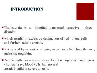
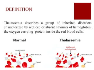
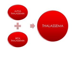
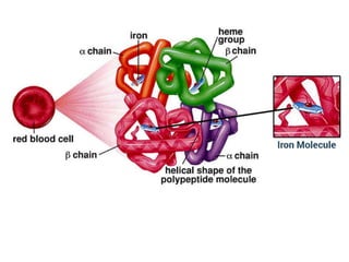
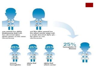
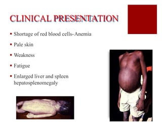
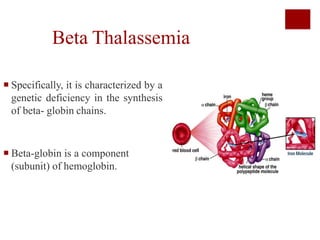
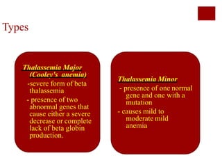
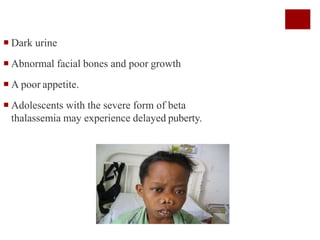
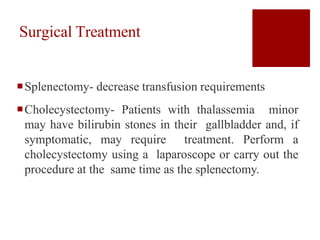
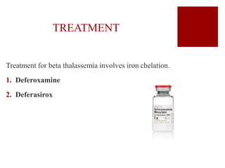
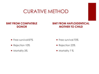
Mr. Abhay Rajpoot
INTRODUCTION
Thalassemia is an inherited autosomal recessive blood
disorder.
which results in excessive destruction of red blood cells
and further leads to anemia.
It is caused by variant or missing genes that affect how the body
make haemoglobin.
People with thalassemia make less haemoglobin and fewer
circulating red blood cells than normal
,result in mild or severe anemia.
DEFINITION
Thalassemia describes a group of inherited disorders
characterized by reduced or absent amounts of hemoglobin ,
the oxygen carrying protein inside the red blood cells.
INCIDENCE
There are about 240 million carriers of β
thalassemia worldwide, i.e. 1.5% of world population,
and in India alone, the number is approximately 30
million with 505 in S.E.Asia. The burden of
hemoglobinopathies in India is high with nearly 12,000
infants being born every year with a severe disorder.
ALPHA
THALASSEMIA
BETA
THALASSEMIA
THALASSEMA
Alpha Thalassemia
Alpha thalassemia is the result of changes in the
genes for the alpha globin component in hemoglobin.
The worldwide distribution of inherited Alpha-
Thalassemia is corresponding to areas of Malaria
exposure.
ETIOLOGY
 Mutation in the DNA of cells that produce
hemoglobin
 It is a form of thalassemia involving the
genes HB1 and HB2
 It is most commonly inherited in a
Mendelian recessive fashion
PATHPHYSIOLOGY
 Alpha thalassemia results when there is
disturbance in production of α-globin from any
or all four of the α-globin genes.
 Genes are responsible for regulating the
synthesis and structure of different globins which
are divided into 2 clusters.
 The α-globin genes are encoded on chromosome
16 and the γ, δ, and β-globin genes are encoded
on chromosome 11
 A normal person carries a linked pair of alpha
globin genes, 2 each from maternal and paternal
chromosome.
 Therefore, alpha thalassemia occurs when there
is a disturbance in production of α-globin from
any or all four of the α-globin genes.
 When functional point mutations, frame shift
mutations, nonsense mutations, and chain
termination mutations occur within or around the
coding sequences of the alpha-globin gene
cluster hemoglobin is impaired.
 When that occurs, protein synthesis may be
inhibited.
CLINICAL PRESENTATION
 Shortage of red blood cells-Anemia
 Pale skin
 Weakness
 Fatigue
 Enlarged liver and spleen
hepatosplenomegaly
 Heart defects
 Abnormalities of the urinary system or genitalia
 Hb Bart syndrome can cause complications in pregnancy such
as
o High blood pressure
o Premature delivery
o Abnormal bleeding
o Jaundice
Treatment of AlphaThalassemia
 Treatment for thalassemia often involves regular
blood transfusions and folate supplements.
 If you receive blood transfusions, you should not take iron
supplements. Doing so can cause a high amount of iron to build up in
the body, which can be harmful.
 Persons who receive significant numbers of blood transfusions need a
treatment called chelation therapy to remove excess iron from the
body.
 Bone marrow transplant may help treat the disease in some patients,
especially children.
Surgical Treatment
Perform splenectomy if transfusion
requirements are increasing.
Surgical or orthodontic correction may be
necessary to correct skeletal deformities of the
skull and maxilla caused by erythroid hyperplasia.
DEFEROXAMINE - INJECTION
Medications
FOLIC ACID- ORAL
FOLIC ACID - INJECTION
FOLIC ACID - ORAL
 BRAND NAME(S): FA-8
 USES
 Folic acid is the man-made form of folate which
is a B6-vitamin naturally found in some foods.
 It is needed to form healthy cells, especially
red blood cells.
 Active forms of folic acid are: L-methylfolate
and levomefolate
 Folic acid supplements are used to treat or
prevent low folate levels.
SIDE EFFECTS
Folic acid usually has very few side effects
Possible side effects include:
Serious allergic reaction, including: rash,
itching/swelling (especially of the
face/tongue/throat), dizziness, trouble
breathing
Folic acid is safe to take during pregnancy when used
as directed. It is included in prenatal vitamin products.
Certain spinal cord birth defects may be prevented by
taking adequate amounts of folic acid during
pregnancy.
Beta Thalassemia
 Specifically, it is characterized by a
genetic deficiency in the synthesis
of beta- globin chains.
 Beta-globin is a component
(subunit) of hemoglobin.
Types
Thalassemia Major
(Cooley's anemia)
-severe form of beta
thalassemia
- presence of two
abnormal genes that
cause either a severe
decrease or complete
lack of beta globin
production.
Thalassemia Minor
- presence of one normal
gene and one with a
mutation
- causes mild to
moderate mild
anemia.
ETIOLOGY
Beta thalassemia is caused by a deficiency of Beta
globin inherited in an autosomal recessive pattern,
which means both copies of the
HBB(Hemoglobin beta) gene in each cell have
mutations.
The parents of an individual with an autosomal recessive
condition each carry one copy of the mutated gene, but
they typically do not show signs and symptoms of the
condition.
 A lack of beta-globin leads to a reduced amount of functional
hemoglobin. Without sufficient hemoglobin, red blood cells do
not develop normally, causing a shortage of mature red blood
cells.
 The low number of mature red blood cells leads to anemia and
other associated health problems in people with beta
thalassemia.
Clinical Presentations
Thalassemia minor- characterized by mild anemia
Symptoms of beta thalassemia major appear in the first two
years of life.
 Fatigue and weakness
 Pale skin or jaundice (yellowing of the skin)
 Protruding abdomen with enlarged spleen and liver
 Dark urine
 Abnormal facial bones and poor growth
 A poor appetite.
 Adolescents with the severe form of beta
thalassemia may experience delayed puberty.
PATHOPHYSIOLOGY
 In Beta thalassemia major, patients have severe anemia, ineffective
erythropoiesis, extramedullary hematopoiesis, and iron overload
resulting from transfusion and increased iron absorption.
 The skin may show pallor from anemia and jaundice from
hyperbilirubinemia.
 The skull and other bones may be deformed secondary to erythroid
hyperplasia with intramedullary expansion and cortical bone
thinning.
 Heart examination may reveal findings of cardiac failure and
arrhythmia, related to either severe anemia or iron overload.
 Abdominal examination may reveal changes in the liver,
gallbladder, and spleen.
 Patients who have received blood transfusions may have
hepatomegaly or chronic hepatitis due to iron overload.
 The gallbladder may contain bilirubin stones formed as a
result of the patient's lifelong hemolytic state.
 Splenomegaly typically is observed as part of the extramedullary
hematopoiesis or as a hypertrophic response related to the
extravascular hemolysis.
 In addition to cardiac dysfunction, hepatomegaly, and hepatitis, iron
overload can also cause endocrine dysfunction, especially affecting
the pancreas, testes, and thyroid.
 Transfusion-associated viral hepatits resulting in cirrhosis or
portal hypertension also may be seen.
Surgical Treatment
Splenectomy- decrease transfusion requirements
Cholecystectomy- Patients with thalassemia minor
may have bilirubin stones in their gallbladder and, if
symptomatic, may require treatment. Perform a
cholecystectomy using a laparoscope or carry out the
procedure at the same time as the splenectomy.
TREATMENT
Treatment for beta thalassemia involves iron chelation.
1. Deferoxamine
2. Deferasirox
CURATIVE METHOD
BMT FROM COMPATIBLE
DONOR
 Free survival 87%
 Rejection 10%
 Mortality 3%
BMT FROM HAPLOIDENTICAL
MOTHER TO CHILD
 Free survival 70%
 Rejection 23%
 Mortality 7 %
Thalassemia

Thalassemia

  • 1.
  • 2.
    INTRODUCTION Thalassemia is aninherited autosomal recessive blood disorder. which results in excessive destruction of red blood cells and further leads to anemia. It is caused by variant or missing genes that affect how the body make haemoglobin. People with thalassemia make less haemoglobin and fewer circulating red blood cells than normal ,result in mild or severe anemia.
  • 3.
    DEFINITION Thalassemia describes agroup of inherited disorders characterized by reduced or absent amounts of hemoglobin , the oxygen carrying protein inside the red blood cells.
  • 4.
    INCIDENCE There are about240 million carriers of β thalassemia worldwide, i.e. 1.5% of world population, and in India alone, the number is approximately 30 million with 505 in S.E.Asia. The burden of hemoglobinopathies in India is high with nearly 12,000 infants being born every year with a severe disorder.
  • 5.
  • 8.
    Alpha Thalassemia Alpha thalassemiais the result of changes in the genes for the alpha globin component in hemoglobin. The worldwide distribution of inherited Alpha- Thalassemia is corresponding to areas of Malaria exposure.
  • 9.
    ETIOLOGY  Mutation inthe DNA of cells that produce hemoglobin  It is a form of thalassemia involving the genes HB1 and HB2  It is most commonly inherited in a Mendelian recessive fashion
  • 10.
    PATHPHYSIOLOGY  Alpha thalassemiaresults when there is disturbance in production of α-globin from any or all four of the α-globin genes.  Genes are responsible for regulating the synthesis and structure of different globins which are divided into 2 clusters.  The α-globin genes are encoded on chromosome 16 and the γ, δ, and β-globin genes are encoded on chromosome 11  A normal person carries a linked pair of alpha globin genes, 2 each from maternal and paternal chromosome.
  • 11.
     Therefore, alphathalassemia occurs when there is a disturbance in production of α-globin from any or all four of the α-globin genes.  When functional point mutations, frame shift mutations, nonsense mutations, and chain termination mutations occur within or around the coding sequences of the alpha-globin gene cluster hemoglobin is impaired.  When that occurs, protein synthesis may be inhibited.
  • 13.
    CLINICAL PRESENTATION  Shortageof red blood cells-Anemia  Pale skin  Weakness  Fatigue  Enlarged liver and spleen hepatosplenomegaly
  • 14.
     Heart defects Abnormalities of the urinary system or genitalia  Hb Bart syndrome can cause complications in pregnancy such as o High blood pressure o Premature delivery o Abnormal bleeding o Jaundice
  • 15.
    Treatment of AlphaThalassemia Treatment for thalassemia often involves regular blood transfusions and folate supplements.  If you receive blood transfusions, you should not take iron supplements. Doing so can cause a high amount of iron to build up in the body, which can be harmful.  Persons who receive significant numbers of blood transfusions need a treatment called chelation therapy to remove excess iron from the body.  Bone marrow transplant may help treat the disease in some patients, especially children.
  • 16.
    Surgical Treatment Perform splenectomyif transfusion requirements are increasing. Surgical or orthodontic correction may be necessary to correct skeletal deformities of the skull and maxilla caused by erythroid hyperplasia. DEFEROXAMINE - INJECTION
  • 17.
  • 18.
    FOLIC ACID -ORAL  BRAND NAME(S): FA-8  USES  Folic acid is the man-made form of folate which is a B6-vitamin naturally found in some foods.  It is needed to form healthy cells, especially red blood cells.  Active forms of folic acid are: L-methylfolate and levomefolate  Folic acid supplements are used to treat or prevent low folate levels.
  • 19.
    SIDE EFFECTS Folic acidusually has very few side effects Possible side effects include: Serious allergic reaction, including: rash, itching/swelling (especially of the face/tongue/throat), dizziness, trouble breathing
  • 20.
    Folic acid issafe to take during pregnancy when used as directed. It is included in prenatal vitamin products. Certain spinal cord birth defects may be prevented by taking adequate amounts of folic acid during pregnancy.
  • 22.
    Beta Thalassemia  Specifically,it is characterized by a genetic deficiency in the synthesis of beta- globin chains.  Beta-globin is a component (subunit) of hemoglobin.
  • 23.
    Types Thalassemia Major (Cooley's anemia) -severeform of beta thalassemia - presence of two abnormal genes that cause either a severe decrease or complete lack of beta globin production. Thalassemia Minor - presence of one normal gene and one with a mutation - causes mild to moderate mild anemia.
  • 24.
    ETIOLOGY Beta thalassemia iscaused by a deficiency of Beta globin inherited in an autosomal recessive pattern, which means both copies of the HBB(Hemoglobin beta) gene in each cell have mutations. The parents of an individual with an autosomal recessive condition each carry one copy of the mutated gene, but they typically do not show signs and symptoms of the condition.
  • 26.
     A lackof beta-globin leads to a reduced amount of functional hemoglobin. Without sufficient hemoglobin, red blood cells do not develop normally, causing a shortage of mature red blood cells.  The low number of mature red blood cells leads to anemia and other associated health problems in people with beta thalassemia.
  • 27.
    Clinical Presentations Thalassemia minor-characterized by mild anemia Symptoms of beta thalassemia major appear in the first two years of life.  Fatigue and weakness  Pale skin or jaundice (yellowing of the skin)  Protruding abdomen with enlarged spleen and liver
  • 28.
     Dark urine Abnormal facial bones and poor growth  A poor appetite.  Adolescents with the severe form of beta thalassemia may experience delayed puberty.
  • 29.
    PATHOPHYSIOLOGY  In Betathalassemia major, patients have severe anemia, ineffective erythropoiesis, extramedullary hematopoiesis, and iron overload resulting from transfusion and increased iron absorption.  The skin may show pallor from anemia and jaundice from hyperbilirubinemia.  The skull and other bones may be deformed secondary to erythroid hyperplasia with intramedullary expansion and cortical bone thinning.  Heart examination may reveal findings of cardiac failure and arrhythmia, related to either severe anemia or iron overload.
  • 30.
     Abdominal examinationmay reveal changes in the liver, gallbladder, and spleen.  Patients who have received blood transfusions may have hepatomegaly or chronic hepatitis due to iron overload.  The gallbladder may contain bilirubin stones formed as a result of the patient's lifelong hemolytic state.
  • 31.
     Splenomegaly typicallyis observed as part of the extramedullary hematopoiesis or as a hypertrophic response related to the extravascular hemolysis.  In addition to cardiac dysfunction, hepatomegaly, and hepatitis, iron overload can also cause endocrine dysfunction, especially affecting the pancreas, testes, and thyroid.  Transfusion-associated viral hepatits resulting in cirrhosis or portal hypertension also may be seen.
  • 32.
    Surgical Treatment Splenectomy- decreasetransfusion requirements Cholecystectomy- Patients with thalassemia minor may have bilirubin stones in their gallbladder and, if symptomatic, may require treatment. Perform a cholecystectomy using a laparoscope or carry out the procedure at the same time as the splenectomy.
  • 33.
    TREATMENT Treatment for betathalassemia involves iron chelation. 1. Deferoxamine 2. Deferasirox
  • 34.
    CURATIVE METHOD BMT FROMCOMPATIBLE DONOR  Free survival 87%  Rejection 10%  Mortality 3% BMT FROM HAPLOIDENTICAL MOTHER TO CHILD  Free survival 70%  Rejection 23%  Mortality 7 %