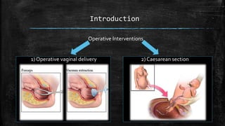
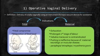
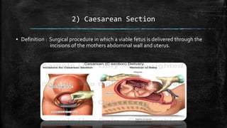
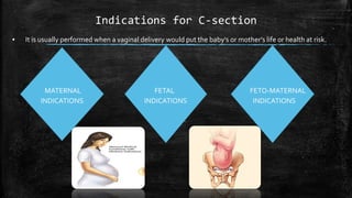
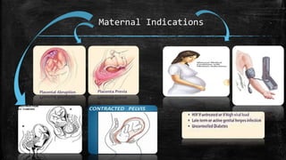
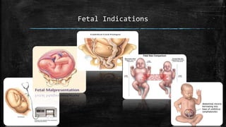
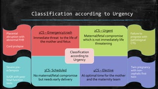
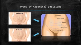
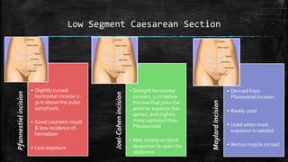
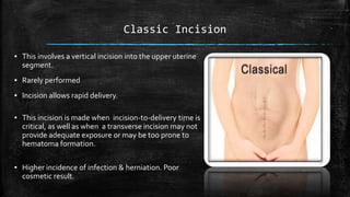
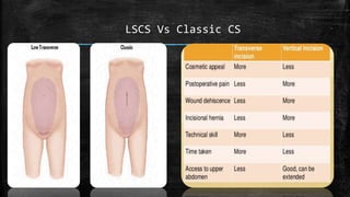
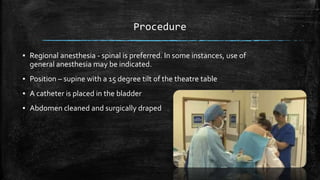
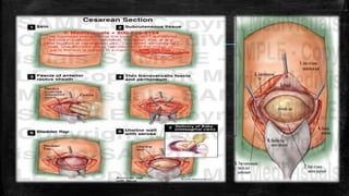
Operative Interventions
In Obstetrics
- Dr.Mani Smk
Introduction
Operative Interventions
1) Operative vaginal delivery 2) Caesarean section
OPERATIVE VAGINAL
DELIVERY
1) Operative Vaginal Delivery
▪ Definition : Delivery of a baby vaginally using an instrument (forceps/ vacuum device) for assistance.
Indications
Fetal Maternal
*Fetal compromise * Exhaustion
*Malposition of fetal head *Prolonged 2nd stage of labour
*Valsalva maneuver is contraindicated
* Pushing is ineffective because of maternal
neurologic or muscular disease.
- paraplegia/ tetraplegia / myasthenia gravis
CONTRAINDICATIONS
▪ ●Fetal demineralizing disease (eg, osteogenesis imperfecta)
▪ ●Unknown fetal position.
▪ ●Brow or face presentation.
▪ ● Suspected fetal-pelvic disproportion.
PREREQUISITES
▪ Fetus alive
▪ Cervix is fully dilated.
▪ Membranes are ruptured.
▪ Fetal head descended and is engaged.
▪ Fetal presentation, position, station known.
▪ Gestational age >33wks to use vacuum devices.
i) FORCEPS DELIVERY
i)Forceps Delivery
▪ Parts of the forceps :
Types of Forceps
CLASSIFICATION OF FORCEPS DELIVERY
▪ It is based on station and amount of
rotation which correlate with the degree of
difficulty and risk of the procedure.
▪ Station is measured in centimeters, −5 to 0
to +5. Rotation </> 45 degrees.
▪ Deliveries are categorized as outlet, low,
and midpelvic procedures.
Outlet forceps Low forceps Mid-forceps
•The leading point of the fetal skull
has reached the pelvic floor and at
or on the perineum
•The scalp is visible at the introitus
without separating the labia.
•Rotation does not exceed 45
degrees
•The leading point of the fetal skull
is ≥2 cm beyond the ischial spines,
but not on the pelvic floor (ie,
station is at least +2/5 cm ).
•Low forceps have two
subdivisions:
-Rotation ≤45 degrees
-Rotation >45 degrees
•The head is engaged(ie, at least 0
station), but the leading point of the
skull is <2 cm beyond the ischial spines
(ie, station is 0/5 cm or +1/5 cm).
Application of the Forceps
The left branch is held Place the right hand on the The left blade is introduced Repeat the same manoeuvre
in the left hand vertically left side, behind fetal head into the left side of the pelvis on the right side, using the
right hand & right branch
Lock the forceps Traction - horizontal Vertical traction As the fetal head is
reached, the forceps are
removed
FORCEPS DELIVERY : COMPLICATIONS
▪ FETAL COMPLICATIONS
– Injury to facial nerves
– Lacerations & bruising of the face and scalp
– Fractures of the face and skull
▪ MATERNAL COMPLICATIONS
– Tears of the genital tract may occur
– Trauma to soft tissue
ALPHABETS
MNEMONICS
FOR FORCEPS
APPLICATION
ii) VACUUM DELIVERY
ii)Vacuum Extraction - Dr.Mani Smk
▪ The vacuum device consists of the
– suction cup and the hand-pump.
▪ Check all connections before application.
▪ Assess the position of the fetal head.
▪ Identify the posterior fontanelle
Suction cup
Hand-pump
Application of the device
▪ Insert the cup into the vagina
in an oblique angle
▪ Apply the cup, with the center of the
cup over the flexion point.
▪ After applying the cup move a finger
around the cup to ensure there is no
maternal soft tissue (cervix or vagina)
within the rim.
▪ With the pump, create a vacuum of 0.2 kg/cm2
negative pressure and check the application.
▪ Increase the vacuum to 0.8 kg/cm2 and check
the application.
▪ After maximum negative pressure, start
traction in the line of the pelvic axis and
perpendicular to the cup.
▪ Remove the cup when the fetal jaw is
reachable.
VACUUM EXTRACTION : COMPLICATIONS
▪ FETAL COMPLICATIONS
– Localized scalp oedema
– Cephalohematoma
– Retinal haemorrhage
– Scalp abrasions and lacerations
– Intracranial bleeding
▪ MATERNAL COMPLICATIONS
– Tears of the genital tract may occur due to entrapment of vaginal mucosa between suction
cup & fetal head
ALPHABETS
MNEMONICS
FOR
VACUUM
APPLICATION
Forceps Vs Vacuum
▪ More likely to cause
maternal genital tract
trauma.
▪ Doesn’t need the help of the
mother, so done under
anaesthetics
▪ Can be used on premature
fetuses & to actively rotate
the fetal head
▪ More likely to cause fetal
trauma like:
cephalohaematoma & retinal
haemorrhage
▪ Need the mothers assistance
during delivery. So
anaesthetics are not given
▪ Fetal age:>33weeks of ges.
New Devices
▪ Odon device :
▪ Was introduced by theWHO for use in areas that
have limited or no access to cesarean birth.
▪ Made of film-like polyethylene material that creates a
sac filled with air that surrounds the entire fetal head
and enables extraction when traction is applied.
CAESAREAN
SECTION
Dr.Mani Smk
2) Caesarean Section
▪ Definition : Surgical procedure in which a viable fetus is delivered through the
incisions of the mothers abdominal wall and uterus.
Indications for C-section
▪ It is usually performed when a vaginal delivery would put the baby's or mother's life or health at risk.
MATERNAL FETAL FETO-MATERNAL
INDICATIONS INDICATIONS INDICATIONS
Maternal Indications
Fetal Indications
Contraindications
▪ Intrauterine fetal death
▪ Coagulation defects
Classification according to Urgency
1CS – Emergency/crash
Immediate threat to the life of
the mother and fetus
2CS – Urgent
Maternal/fetal compromise
which is not immediately life
threatening
3CS- Scheduled
No maternal/fetal compromise
but needs early delivery
4CS – Elective
At optimal time for the mother
and the maternity team
Classification
according to
Urgency
Placental
abruption with
abnormal FHR
Cord prolapse
Failure to
progress with
pathological
CTG
Severe pre-
eclampsia
IUGR with poor
fetal function
tests
Twin pregnancy
with non-
cephalic first
twin
Types of Abdominal Incisions
Low Segment Caesarean SectionPfannestielincision
• Slightly curved
horizontal incision 2-
3cm above the pubic
symphysis.
• Good cosmetic result
& less incidence of
herniation
• Less exposure
Joel-Cohenincision
• Straight horizontal
incision, 3 cm below
the line that joins the
anterior superior iliac
spines, and slightly
more cephalad than
Pfannenstiel
• Rely mostly on blunt
dissection to open the
abdomen
MaylardIncision
• Derived from
Pfannestiel incision.
• Rarely used
• Used when more
exposure is needed.
• Rectus muscle incised
Classic Incision
▪ This involves a vertical incision into the upper uterine
segment.
▪ Rarely performed
▪ Incision allows rapid delivery.
▪ This incision is made when incision-to-delivery time is
critical, as well as when a transverse incision may not
provide adequate exposure or may be too prone to
hematoma formation.
▪ Higher incidence of infection & herniation. Poor
cosmetic result.
LSCS Vs Classic CS
Procedure
▪ Regional anesthesia - spinal is preferred. In some instances, use of
general anesthesia may be indicated.
▪ Position – supine with a 15 degree tilt of the theatre table
▪ A catheter is placed in the bladder
▪ Abdomen cleaned and surgically draped
▪ The uterine incision can be low transverse, classic vertical, low vertical,
J shaped or T shaped.
1 2 3
4 5 6
DELIVERYOF FETUS CUTTINGTHE UMBILICAL CORD
DELIVERYOF PLACENTA
▪ Some obstetricians repair the
uterus by uterine
exteriorization, and some
repair it while it is still in the
abdomen.
▪ The uterus is closed with one
or two layers of suture
▪ The layers of the abdominal
wall are sutured and then the
skin closed
Complications of Caesarean Section
 Uterine lacerations
 Heavy blood loss.(>1L)
 Bladder injury.
 Post anesthetic complications – respiratory difficulty
 Wound Infection.
 Deep venous thrombosis
 Risk of incisional hernia
 Rupture of uterus at the site of the scar in future pregnancies
Thank You …
Mom !!!

Operative Interventions In Obstetrics

  • 1.
  • 2.
    Introduction Operative Interventions 1) Operativevaginal delivery 2) Caesarean section
  • 3.
  • 4.
    1) Operative VaginalDelivery ▪ Definition : Delivery of a baby vaginally using an instrument (forceps/ vacuum device) for assistance. Indications Fetal Maternal *Fetal compromise * Exhaustion *Malposition of fetal head *Prolonged 2nd stage of labour *Valsalva maneuver is contraindicated * Pushing is ineffective because of maternal neurologic or muscular disease. - paraplegia/ tetraplegia / myasthenia gravis
  • 5.
    CONTRAINDICATIONS ▪ ●Fetal demineralizingdisease (eg, osteogenesis imperfecta) ▪ ●Unknown fetal position. ▪ ●Brow or face presentation. ▪ ● Suspected fetal-pelvic disproportion.
  • 6.
    PREREQUISITES ▪ Fetus alive ▪Cervix is fully dilated. ▪ Membranes are ruptured. ▪ Fetal head descended and is engaged. ▪ Fetal presentation, position, station known. ▪ Gestational age >33wks to use vacuum devices.
  • 7.
  • 8.
  • 9.
  • 10.
    CLASSIFICATION OF FORCEPSDELIVERY ▪ It is based on station and amount of rotation which correlate with the degree of difficulty and risk of the procedure. ▪ Station is measured in centimeters, −5 to 0 to +5. Rotation </> 45 degrees. ▪ Deliveries are categorized as outlet, low, and midpelvic procedures.
  • 11.
    Outlet forceps Lowforceps Mid-forceps •The leading point of the fetal skull has reached the pelvic floor and at or on the perineum •The scalp is visible at the introitus without separating the labia. •Rotation does not exceed 45 degrees •The leading point of the fetal skull is ≥2 cm beyond the ischial spines, but not on the pelvic floor (ie, station is at least +2/5 cm ). •Low forceps have two subdivisions: -Rotation ≤45 degrees -Rotation >45 degrees •The head is engaged(ie, at least 0 station), but the leading point of the skull is <2 cm beyond the ischial spines (ie, station is 0/5 cm or +1/5 cm).
  • 12.
  • 13.
    Application of theForceps The left branch is held Place the right hand on the The left blade is introduced Repeat the same manoeuvre in the left hand vertically left side, behind fetal head into the left side of the pelvis on the right side, using the right hand & right branch
  • 14.
    Lock the forcepsTraction - horizontal Vertical traction As the fetal head is reached, the forceps are removed
  • 15.
    FORCEPS DELIVERY :COMPLICATIONS ▪ FETAL COMPLICATIONS – Injury to facial nerves – Lacerations & bruising of the face and scalp – Fractures of the face and skull ▪ MATERNAL COMPLICATIONS – Tears of the genital tract may occur – Trauma to soft tissue
  • 16.
  • 17.
  • 18.
    ii)Vacuum Extraction -Dr.Mani Smk ▪ The vacuum device consists of the – suction cup and the hand-pump. ▪ Check all connections before application. ▪ Assess the position of the fetal head. ▪ Identify the posterior fontanelle Suction cup Hand-pump
  • 19.
    Application of thedevice ▪ Insert the cup into the vagina in an oblique angle ▪ Apply the cup, with the center of the cup over the flexion point. ▪ After applying the cup move a finger around the cup to ensure there is no maternal soft tissue (cervix or vagina) within the rim.
  • 20.
    ▪ With thepump, create a vacuum of 0.2 kg/cm2 negative pressure and check the application. ▪ Increase the vacuum to 0.8 kg/cm2 and check the application. ▪ After maximum negative pressure, start traction in the line of the pelvic axis and perpendicular to the cup. ▪ Remove the cup when the fetal jaw is reachable.
  • 21.
    VACUUM EXTRACTION :COMPLICATIONS ▪ FETAL COMPLICATIONS – Localized scalp oedema – Cephalohematoma – Retinal haemorrhage – Scalp abrasions and lacerations – Intracranial bleeding ▪ MATERNAL COMPLICATIONS – Tears of the genital tract may occur due to entrapment of vaginal mucosa between suction cup & fetal head
  • 22.
  • 23.
    Forceps Vs Vacuum ▪More likely to cause maternal genital tract trauma. ▪ Doesn’t need the help of the mother, so done under anaesthetics ▪ Can be used on premature fetuses & to actively rotate the fetal head ▪ More likely to cause fetal trauma like: cephalohaematoma & retinal haemorrhage ▪ Need the mothers assistance during delivery. So anaesthetics are not given ▪ Fetal age:>33weeks of ges.
  • 24.
    New Devices ▪ Odondevice : ▪ Was introduced by theWHO for use in areas that have limited or no access to cesarean birth. ▪ Made of film-like polyethylene material that creates a sac filled with air that surrounds the entire fetal head and enables extraction when traction is applied.
  • 29.
  • 30.
    2) Caesarean Section ▪Definition : Surgical procedure in which a viable fetus is delivered through the incisions of the mothers abdominal wall and uterus.
  • 31.
    Indications for C-section ▪It is usually performed when a vaginal delivery would put the baby's or mother's life or health at risk. MATERNAL FETAL FETO-MATERNAL INDICATIONS INDICATIONS INDICATIONS
  • 32.
  • 33.
  • 34.
    Contraindications ▪ Intrauterine fetaldeath ▪ Coagulation defects
  • 35.
    Classification according toUrgency 1CS – Emergency/crash Immediate threat to the life of the mother and fetus 2CS – Urgent Maternal/fetal compromise which is not immediately life threatening 3CS- Scheduled No maternal/fetal compromise but needs early delivery 4CS – Elective At optimal time for the mother and the maternity team Classification according to Urgency Placental abruption with abnormal FHR Cord prolapse Failure to progress with pathological CTG Severe pre- eclampsia IUGR with poor fetal function tests Twin pregnancy with non- cephalic first twin
  • 36.
  • 37.
    Low Segment CaesareanSectionPfannestielincision • Slightly curved horizontal incision 2- 3cm above the pubic symphysis. • Good cosmetic result & less incidence of herniation • Less exposure Joel-Cohenincision • Straight horizontal incision, 3 cm below the line that joins the anterior superior iliac spines, and slightly more cephalad than Pfannenstiel • Rely mostly on blunt dissection to open the abdomen MaylardIncision • Derived from Pfannestiel incision. • Rarely used • Used when more exposure is needed. • Rectus muscle incised
  • 38.
    Classic Incision ▪ Thisinvolves a vertical incision into the upper uterine segment. ▪ Rarely performed ▪ Incision allows rapid delivery. ▪ This incision is made when incision-to-delivery time is critical, as well as when a transverse incision may not provide adequate exposure or may be too prone to hematoma formation. ▪ Higher incidence of infection & herniation. Poor cosmetic result.
  • 39.
  • 40.
    Procedure ▪ Regional anesthesia- spinal is preferred. In some instances, use of general anesthesia may be indicated. ▪ Position – supine with a 15 degree tilt of the theatre table ▪ A catheter is placed in the bladder ▪ Abdomen cleaned and surgically draped
  • 42.
    ▪ The uterineincision can be low transverse, classic vertical, low vertical, J shaped or T shaped. 1 2 3 4 5 6
  • 43.
    DELIVERYOF FETUS CUTTINGTHEUMBILICAL CORD DELIVERYOF PLACENTA
  • 44.
    ▪ Some obstetriciansrepair the uterus by uterine exteriorization, and some repair it while it is still in the abdomen. ▪ The uterus is closed with one or two layers of suture ▪ The layers of the abdominal wall are sutured and then the skin closed
  • 45.
    Complications of CaesareanSection  Uterine lacerations  Heavy blood loss.(>1L)  Bladder injury.  Post anesthetic complications – respiratory difficulty  Wound Infection.  Deep venous thrombosis  Risk of incisional hernia  Rupture of uterus at the site of the scar in future pregnancies
  • 46.