Telomeres cap the ends of chromosomes and protect them from degradation during cell division. As cells divide, telomeres shorten due to the inability of DNA replication enzymes to fully copy chromosome ends. This limits a cell to around 50-70 divisions before entering senescence. Cancer cells activate telomerase to maintain telomere length, allowing unlimited division. Telomeres play a key role in both aging and cancer - their shortening limits the lifespan of normal cells but cancer cells overcome this via telomerase to achieve immortality and uncontrolled growth. Measuring and targeting telomerase may provide new strategies for cancer detection and treatment.
This presentation provides an overview of Cell senescence, Aging, Theories of Aging,principle of senescence, Mechanism of action, Factors, Diseases caused due to this action, Senescence and cancer, Insulin signalling cascade, Telomere shortening.
It describes about Structure and function of telomere, Telomerase enzyme, How does telomerase works?, Telomere replication, What happens to telomeres as we age?, Factors contribute to telomere shortening
Telomere structure stability, function in plant breedingSachin Dharwad
TELOMERE, TELOMERE STRUCTURE, ITS FUNCTION AND USE IN PLANT BREEDING. Telomere in plant breeding perspective. Case studies related to telomere in case of plant breeding. telomeres can be made use as markers in plant breeding.
Majority of cancer lead by point mutation in p53 gene. which is also known as "guardian of genome". this mutation leads conversion of normal cell into cancerous cell.
Chromatin is the complex combination of DNA and proteins that makes up chromosomes. It can be made visible by staining with specific techniques and stain (thus the name chromatin which literally means colored material). The major proteins involved in chromatin are histone proteins; although many other chromosomal proteins have prominent roles too. The functions of chromatin is to package DNA into smaller volume to fit in the cell, to strengthen the DNA to allow mitosis and meiosis and to serve as a mechanism to control gene expression and DNA replication.
This presentation describes the structure and function of telomeres ,their role in various disease.The structure and function of telomerase is also described ,together with its possible role in therapy .
The presentation outlines aspects of immunity against cancer, evasion strategies by cells, immunotherapy in cancer, cancer vaccines etc. Download and view the slideshow for better experience.
Prepared in Sept 2014
ONCOGENE AND PROTOONCOGENE
P53 GENE AND ITS APPLICATION IN CANCER ETIOLOGY
TUMOUR SUPPRESSOR GENE AND BCA AND BAC GENE AND ITS APPLICATION ON THE APOPTOSIS AND DEATH RECEPTORS
Comparative genomic hybridization is a molecular cytogenetic method for analysing copy number variations (CNVs) relative to ploidy level in the DNA of a test sample compared to a reference sample, without the need for culturing cells
Telomere is the end part of a chromosome.its length is maintained by na enzyme called telomerase.if telomerase is lacking,many genetic diseases may result( like progeria)
In this way, you can take care of your health and learn more about telomeres. Consider telomere testing and talk to medical experts who focus on telomere research. By working together, we can unlock the mysteries of aging and create a world where healthy longevity is the norm.
TA Sciences presents their latest product TA-65. To learn more about TA-65, check out their website at http://www.tasciences.com TA-65 can help your immune system, vision, male sexual performance, skin appearance and more!
Dr. Al Sears explains the Nobel Prize winning breakthrough telomere technology. This opened the way for Harvard researcher, Dr. Ronal DePinho to find a way to activate telomerase. Telomerase is the enzyme that signals your telomeres to grow longer, unfortunately, it shuts down while you are still in your mother's womb.
Once Nobel Prize winning research identified that telomeres are the protective tips at each end of the strands of your DNA, and as your cells replicate, gradully your telomeres grow shorter. They are the "aging-clocks" inside your DNA.
Once Dr. DePinho found a way to reactivate the telomerase enzyme, he turned old mice into young mice again.
Not long after, scientists discovered ways to do this in humans as well, and today, the discovery of the telomere and telomerase are the most important anti-aging breakthrough of our time.
What is the consequence when a chromosome loses its telomeresSol.pdfarchiesgallery
What is the consequence when a chromosome loses its telomeres?
Solution
Telomeres are DNA-protein complexes that contain short repeat sequences added on to the ends
of chromosomes by the enzyme telomerase. Telomeres serve multiple functions, including
protecting the ends of chromosomes and preventing chromosome fusion. In humans, telomeres
are thought to be maintained in germ line cells, but shorten with age in somatic cells due to the
lack of sufficient telomerase activity to compensate for the loss of small amounts of telomeric
repeat sequences with each cell division. Telomere shortening in somatic cells is a signal for cell
senescence, which involves a permanent cell cycle arrest in G1. Primary human fibroblasts that
have lost the ability to senesce continue to show telomere shortening and eventually enter
“crisis”, which involves increased chromosome fusion, aneuploidy, and cell death.
Usually loss of a telomere is associated with extensive chromosome fusion which can be
associated in human epithelial cells failing to senesce and thus entering “agonescence\". Rare
cells that survive crisis are invariably those that have regained the ability to maintain their
telomeres, either through activation of telomerase or through an alternative mechanism involving
recombination.
Spontaneous telomere loss has been proposed as an important mechanism for initiating the
chromosome instability commonly found in cancer cells. Studies have shown that spontaneous
telomere loss in a human cancer cell line initiates breakage/fusion/bridge (B/F/B) cycles that
continue for many cell generations, resulting in DNA amplification and translocations on the
chromosome that lost its telomere. For a chromosome that lost its telomeres, telomere acquisition
during B/F/B cycles occurs mainly through translocations involving either the nonreciprocal
transfer or duplication of the arms of other chromosomes. Telomere acquisition also occurs
through small duplications involving the sub-telomeric region of the other end of the same
chromosome. Although all of these mechanisms stabilized the chromosome that lost its telomere,
they differed in their consequences for the stability of the genome as a whole.
Loss of a telomere on the donor chromosome due to telomere acquisition also resulted into
consequently additional translocations, isochromosome formation, or complete loss of the donor
chromosome..
Similar to Telomere, Functions & Role in Aging & Cancer (20)
Programmed Assembly of Synthetic Protocells into Thermoresponsive PrototissuesZohaib HUSSAIN
Programmed assembly of synthetic protocells into thermoresponsive prototissues
Introduction
Anatomy and Physiology of bone
Bone Tissue Engineering
Recent studies related to bone tissue engineering
Commercialized products and ongoing clinical trials
Biomedical start-ups
Concluding remarks
Introduction
Anatomy and Physiology of bone
Bone Tissue Engineering
Recent studies related to bone tissue engineering
Commercialized products and ongoing clinical trials
Biomedical start-ups
Concluding remarks
Introduction
Anatomy and Physiology of bone
Bone Tissue Engineering
Recent studies related to bone tissue engineering
Commercialized products and ongoing clinical trials
Biomedical start-ups
Concluding remarks
Large-scale Production of Stem Cells Utilizing MicrocarriersZohaib HUSSAIN
Large-scale Production of Stem Cells Utilizing MicrocarriersLarge-scale Production of Stem Cells Utilizing MicrocarriersLarge-scale Production of Stem Cells Utilizing MicrocarriersLarge-scale Production of Stem Cells Utilizing MicrocarriersLarge-scale Production of Stem Cells Utilizing MicrocarriersLarge-scale Production of Stem Cells Utilizing MicrocarriersLarge-scale Production of Stem Cells Utilizing MicrocarriersLarge-scale Production of Stem Cells Utilizing MicrocarriersLarge-scale Production of Stem Cells Utilizing MicrocarriersLarge-scale Production of Stem Cells Utilizing Microcarriers
PHOTOSYNTHESIS: What we have learned so far? Zohaib HUSSAIN
No matter how complex or advanced a machine, such as the latest cellular phone, the device cannot function without energy. Living things, similar to machines, have many complex components; they too cannot do anything without energy, which is why humans and all other organisms must “eat” in some form or another. That may be common knowledge, but how many people realize that every bite of every meal ingested depends on the process of photosynthesis?
Contents
1. Insulin Molecule
2. Effect of Insulin in Body
3. History of Insulin
4. Recent Trends in Insulin Productions and Types
4.1 Animal Insulins
4.2 Long-Acting Insulins
4.3 Human Insulins
4.4 Insulin Analogues
4.5 Biosimilar Insulins
5. Insulin Production (Chain A and Chain B Method)
5.1 Upstream Processing
5.2 Downstream Processing
6. The Proinsulin Process
7. Insulin Available in Market with Different Brand Names
8. References
Oxidation & Reduction involves electron transfer & How enzymes find their sub...Zohaib HUSSAIN
Oxidation is loss of electrons
Reduction is gain of electrons
Oxidation is always accompanied by reduction
The total number of electrons is kept constant
Oxidizing agents oxidize and are themselves reduced
Reducing agents reduce and are themselves oxidized
Cellulase (Types, Sources, Mode of Action & Applications)Zohaib HUSSAIN
Cellulase is a class of enzyme that catalyzes the cellulolysis i.e., hydrolysis of cellulose. Celulase is a multiple enzyme system consisting of endo – 1, 4 –β–D – glucanases and exo – 1, 4 –β– D – glucanases along with cellobiase (β– D – glucosideglucano hydrolase).
Types of Cellulases
On the basis of fractionation studies on culture filtrate have demonstrated that, there are ‘three’ major types of enzymes involved in the hydrolysis of native cellulose to glucose, namely: Others are produced by the some animals and plants.
Amylases (Types, Sources, Mode of Action & Applications)Zohaib HUSSAIN
Amylases are important hydrolase enzymes which have been widely used since many decades. These enzymes randomly cleave internal glycosidic linkages in starch molecules to hydrolyze them and yield dextrins and oligosaccharides. Among amylases α-Amylase is in maximum demand due to its wide range of applications in the industrial front. α-Amylase can be produced by plant or microbial sources. The ubiquitous nature, ease of production and broad spectrum of applications make α-Amylase an industrially important enzyme.
Life on Earth (By Alonso Ricardo and Jack W. Szostak) Summary (By Zohaib Hus...Zohaib HUSSAIN
Life on Earth (By Alonso Ricardo and Jack W. Szostak)
Summary (By Zohaib Hussain)
Life on Earth (By Alonso Ricardo and Jack W. Szostak)
Summary (By Zohaib Hussain)
Life on Earth (By Alonso Ricardo and Jack W. Szostak)
Summary (By Zohaib Hussain)
Life on Earth (By Alonso Ricardo and Jack W. Szostak)
Summary (By Zohaib Hussain)
Layout of the Cell Culture Room Layout of the Cell Culture Room Layout of the Cell Culture Room Layout of the Cell Culture Room Layout of the Cell Culture Room Layout of the Cell Culture Room Layout of the Cell Culture Room Layout of the Cell Culture Room Layout of the Cell Culture Room Layout of the Cell Culture Room Layout of the Cell Culture Room Layout of the Cell Culture Room Layout of the Cell Culture Room Layout of the Cell Culture Room Layout of the Cell Culture Room Layout of the Cell Culture Room Layout of the Cell Culture Room Layout of the Cell Culture Room Layout of the Cell Culture Room Layout of the Cell Culture Room
1. Levels of gene regulation
The observation that differences in the RNA and protein content of different tissues are not paralleled by significant differences in their DNA content indicates that the process whereby DNA produces mRNA must be the level at which gene expression is regulated in eukaryotes. In bacteria this process involves only a single stage, that of transcription, in which RNA copy of the DNA is produced by the enzyme RNA polymerase. Even while this process is still occurring, ribosomes attach to the nascent RNA chain and begin to translate it into protein. Hence cases
of gene regulation in bacteria, such as the switching on of the synthesis of the enzyme β-galactosidase in response to the presence of lactose (its substrate), are mediated by increased transcription of the appropriate gene. Clearly, a similar regulation of gene transcription in different tissues, or in response to substances such as steroid hormones which induce the synthesis of new proteins, represents an attractive method of gene regulation in eukaryotes.
In contrast to the situation in bacteria, however, a number of stages intervene between the initial synthesis of the primary RNA transcript and the eventual production of mRNA (Fig. 1).
The initial transcript is modified at its 5′ end by the addition of a cap structure containing a modified guanosine residue and is subsequently cleaved near its 3′ end, followed by the addition of up to 200 adenosine residues in a process known as polyadenylation. Subsequently, intervening sequences or introns, which interrupt the protein-coding sequence in both the DNA and the primary transcript of many genes. Although this produces a functional mRNA, the spliced molecule must then be transported from the nucleus, where these processes occur, to the cytoplasm where it can be translated into protein.
Eukaryotic and Prokaryotic Chromosomes Eukaryotic and Prokaryotic Chromosomes Eukaryotic and Prokaryotic Chromosomes Eukaryotic and Prokaryotic Chromosomes Eukaryotic and Prokaryotic Chromosomes Eukaryotic and Prokaryotic Chromosomes Eukaryotic and Prokaryotic Chromosomes
Chromosomes are bundles of tightly coiled DNA located within the nucleus of almost every cell in our body. A chromosome is a DNA molecule with part or all of the genetic material (genome) of an organism. Chromosomes are normally visible under a light microscope only when the cell is undergoing the metaphase of cell division. Before this happens, every chromosome is copied once (S phase), and the copy is joined to the original by a centromere, resulting in an X-shaped structure. The original chromosome and the copy are now called sister chromatids. During metaphase, when a chromosome is in its most condensed state, the X-shape structure is called a metaphase chromosome.
Richard's entangled aventures in wonderlandRichard Gill
Since the loophole-free Bell experiments of 2020 and the Nobel prizes in physics of 2022, critics of Bell's work have retreated to the fortress of super-determinism. Now, super-determinism is a derogatory word - it just means "determinism". Palmer, Hance and Hossenfelder argue that quantum mechanics and determinism are not incompatible, using a sophisticated mathematical construction based on a subtle thinning of allowed states and measurements in quantum mechanics, such that what is left appears to make Bell's argument fail, without altering the empirical predictions of quantum mechanics. I think however that it is a smoke screen, and the slogan "lost in math" comes to my mind. I will discuss some other recent disproofs of Bell's theorem using the language of causality based on causal graphs. Causal thinking is also central to law and justice. I will mention surprising connections to my work on serial killer nurse cases, in particular the Dutch case of Lucia de Berk and the current UK case of Lucy Letby.
Seminar of U.V. Spectroscopy by SAMIR PANDASAMIR PANDA
Spectroscopy is a branch of science dealing the study of interaction of electromagnetic radiation with matter.
Ultraviolet-visible spectroscopy refers to absorption spectroscopy or reflect spectroscopy in the UV-VIS spectral region.
Ultraviolet-visible spectroscopy is an analytical method that can measure the amount of light received by the analyte.
A brief information about the SCOP protein database used in bioinformatics.
The Structural Classification of Proteins (SCOP) database is a comprehensive and authoritative resource for the structural and evolutionary relationships of proteins. It provides a detailed and curated classification of protein structures, grouping them into families, superfamilies, and folds based on their structural and sequence similarities.
Cancer cell metabolism: special Reference to Lactate PathwayAADYARAJPANDEY1
Normal Cell Metabolism:
Cellular respiration describes the series of steps that cells use to break down sugar and other chemicals to get the energy we need to function.
Energy is stored in the bonds of glucose and when glucose is broken down, much of that energy is released.
Cell utilize energy in the form of ATP.
The first step of respiration is called glycolysis. In a series of steps, glycolysis breaks glucose into two smaller molecules - a chemical called pyruvate. A small amount of ATP is formed during this process.
Most healthy cells continue the breakdown in a second process, called the Kreb's cycle. The Kreb's cycle allows cells to “burn” the pyruvates made in glycolysis to get more ATP.
The last step in the breakdown of glucose is called oxidative phosphorylation (Ox-Phos).
It takes place in specialized cell structures called mitochondria. This process produces a large amount of ATP. Importantly, cells need oxygen to complete oxidative phosphorylation.
If a cell completes only glycolysis, only 2 molecules of ATP are made per glucose. However, if the cell completes the entire respiration process (glycolysis - Kreb's - oxidative phosphorylation), about 36 molecules of ATP are created, giving it much more energy to use.
IN CANCER CELL:
Unlike healthy cells that "burn" the entire molecule of sugar to capture a large amount of energy as ATP, cancer cells are wasteful.
Cancer cells only partially break down sugar molecules. They overuse the first step of respiration, glycolysis. They frequently do not complete the second step, oxidative phosphorylation.
This results in only 2 molecules of ATP per each glucose molecule instead of the 36 or so ATPs healthy cells gain. As a result, cancer cells need to use a lot more sugar molecules to get enough energy to survive.
introduction to WARBERG PHENOMENA:
WARBURG EFFECT Usually, cancer cells are highly glycolytic (glucose addiction) and take up more glucose than do normal cells from outside.
Otto Heinrich Warburg (; 8 October 1883 – 1 August 1970) In 1931 was awarded the Nobel Prize in Physiology for his "discovery of the nature and mode of action of the respiratory enzyme.
WARNBURG EFFECT : cancer cells under aerobic (well-oxygenated) conditions to metabolize glucose to lactate (aerobic glycolysis) is known as the Warburg effect. Warburg made the observation that tumor slices consume glucose and secrete lactate at a higher rate than normal tissues.
Introduction:
RNA interference (RNAi) or Post-Transcriptional Gene Silencing (PTGS) is an important biological process for modulating eukaryotic gene expression.
It is highly conserved process of posttranscriptional gene silencing by which double stranded RNA (dsRNA) causes sequence-specific degradation of mRNA sequences.
dsRNA-induced gene silencing (RNAi) is reported in a wide range of eukaryotes ranging from worms, insects, mammals and plants.
This process mediates resistance to both endogenous parasitic and exogenous pathogenic nucleic acids, and regulates the expression of protein-coding genes.
What are small ncRNAs?
micro RNA (miRNA)
short interfering RNA (siRNA)
Properties of small non-coding RNA:
Involved in silencing mRNA transcripts.
Called “small” because they are usually only about 21-24 nucleotides long.
Synthesized by first cutting up longer precursor sequences (like the 61nt one that Lee discovered).
Silence an mRNA by base pairing with some sequence on the mRNA.
Discovery of siRNA?
The first small RNA:
In 1993 Rosalind Lee (Victor Ambros lab) was studying a non- coding gene in C. elegans, lin-4, that was involved in silencing of another gene, lin-14, at the appropriate time in the
development of the worm C. elegans.
Two small transcripts of lin-4 (22nt and 61nt) were found to be complementary to a sequence in the 3' UTR of lin-14.
Because lin-4 encoded no protein, she deduced that it must be these transcripts that are causing the silencing by RNA-RNA interactions.
Types of RNAi ( non coding RNA)
MiRNA
Length (23-25 nt)
Trans acting
Binds with target MRNA in mismatch
Translation inhibition
Si RNA
Length 21 nt.
Cis acting
Bind with target Mrna in perfect complementary sequence
Piwi-RNA
Length ; 25 to 36 nt.
Expressed in Germ Cells
Regulates trnasposomes activity
MECHANISM OF RNAI:
First the double-stranded RNA teams up with a protein complex named Dicer, which cuts the long RNA into short pieces.
Then another protein complex called RISC (RNA-induced silencing complex) discards one of the two RNA strands.
The RISC-docked, single-stranded RNA then pairs with the homologous mRNA and destroys it.
THE RISC COMPLEX:
RISC is large(>500kD) RNA multi- protein Binding complex which triggers MRNA degradation in response to MRNA
Unwinding of double stranded Si RNA by ATP independent Helicase
Active component of RISC is Ago proteins( ENDONUCLEASE) which cleave target MRNA.
DICER: endonuclease (RNase Family III)
Argonaute: Central Component of the RNA-Induced Silencing Complex (RISC)
One strand of the dsRNA produced by Dicer is retained in the RISC complex in association with Argonaute
ARGONAUTE PROTEIN :
1.PAZ(PIWI/Argonaute/ Zwille)- Recognition of target MRNA
2.PIWI (p-element induced wimpy Testis)- breaks Phosphodiester bond of mRNA.)RNAse H activity.
MiRNA:
The Double-stranded RNAs are naturally produced in eukaryotic cells during development, and they have a key role in regulating gene expression .
THE IMPORTANCE OF MARTIAN ATMOSPHERE SAMPLE RETURN.Sérgio Sacani
The return of a sample of near-surface atmosphere from Mars would facilitate answers to several first-order science questions surrounding the formation and evolution of the planet. One of the important aspects of terrestrial planet formation in general is the role that primary atmospheres played in influencing the chemistry and structure of the planets and their antecedents. Studies of the martian atmosphere can be used to investigate the role of a primary atmosphere in its history. Atmosphere samples would also inform our understanding of the near-surface chemistry of the planet, and ultimately the prospects for life. High-precision isotopic analyses of constituent gases are needed to address these questions, requiring that the analyses are made on returned samples rather than in situ.
Earliest Galaxies in the JADES Origins Field: Luminosity Function and Cosmic ...Sérgio Sacani
We characterize the earliest galaxy population in the JADES Origins Field (JOF), the deepest
imaging field observed with JWST. We make use of the ancillary Hubble optical images (5 filters
spanning 0.4−0.9µm) and novel JWST images with 14 filters spanning 0.8−5µm, including 7 mediumband filters, and reaching total exposure times of up to 46 hours per filter. We combine all our data
at > 2.3µm to construct an ultradeep image, reaching as deep as ≈ 31.4 AB mag in the stack and
30.3-31.0 AB mag (5σ, r = 0.1” circular aperture) in individual filters. We measure photometric
redshifts and use robust selection criteria to identify a sample of eight galaxy candidates at redshifts
z = 11.5 − 15. These objects show compact half-light radii of R1/2 ∼ 50 − 200pc, stellar masses of
M⋆ ∼ 107−108M⊙, and star-formation rates of SFR ∼ 0.1−1 M⊙ yr−1
. Our search finds no candidates
at 15 < z < 20, placing upper limits at these redshifts. We develop a forward modeling approach to
infer the properties of the evolving luminosity function without binning in redshift or luminosity that
marginalizes over the photometric redshift uncertainty of our candidate galaxies and incorporates the
impact of non-detections. We find a z = 12 luminosity function in good agreement with prior results,
and that the luminosity function normalization and UV luminosity density decline by a factor of ∼ 2.5
from z = 12 to z = 14. We discuss the possible implications of our results in the context of theoretical
models for evolution of the dark matter halo mass function.
(May 29th, 2024) Advancements in Intravital Microscopy- Insights for Preclini...Scintica Instrumentation
Intravital microscopy (IVM) is a powerful tool utilized to study cellular behavior over time and space in vivo. Much of our understanding of cell biology has been accomplished using various in vitro and ex vivo methods; however, these studies do not necessarily reflect the natural dynamics of biological processes. Unlike traditional cell culture or fixed tissue imaging, IVM allows for the ultra-fast high-resolution imaging of cellular processes over time and space and were studied in its natural environment. Real-time visualization of biological processes in the context of an intact organism helps maintain physiological relevance and provide insights into the progression of disease, response to treatments or developmental processes.
In this webinar we give an overview of advanced applications of the IVM system in preclinical research. IVIM technology is a provider of all-in-one intravital microscopy systems and solutions optimized for in vivo imaging of live animal models at sub-micron resolution. The system’s unique features and user-friendly software enables researchers to probe fast dynamic biological processes such as immune cell tracking, cell-cell interaction as well as vascularization and tumor metastasis with exceptional detail. This webinar will also give an overview of IVM being utilized in drug development, offering a view into the intricate interaction between drugs/nanoparticles and tissues in vivo and allows for the evaluation of therapeutic intervention in a variety of tissues and organs. This interdisciplinary collaboration continues to drive the advancements of novel therapeutic strategies.
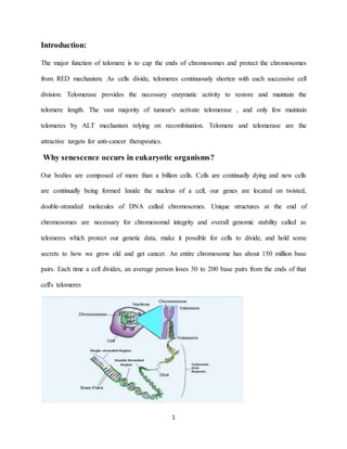
1. 1
Introduction:
The major function of telomere is to cap the ends of chromosomes and protect the chromosomes
from RED mechanism. As cells divide, telomeres continuously shorten with each successive cell
division. Telomerase provides the necessary enzymatic activity to restore and maintain the
telomere length. The vast majority of tumour's activate telomerase , and only few maintain
telomeres by ALT mechanism relying on recombination. Telomere and telomerase are the
attractive targets for anti-cancer therapeutics.
Why senescence occurs in eukaryotic organisms?
Our bodies are composed of more than a billion cells. Cells are continually dying and new cells
are continually being formed Inside the nucleus of a cell, our genes are located on twisted,
double-stranded molecules of DNA called chromosomes. Unique structures at the end of
chromosomes are necessary for chromosomal integrity and overall genomic stability called as
telomeres which protect our genetic data, make it possible for cells to divide, and hold some
secrets to how we grow old and get cancer. An entire chromosome has about 150 million base
pairs. Each time a cell divides, an average person loses 30 to 200 base pairs from the ends of that
cell's telomeres
2. 2
This is because enzymes that duplicate DNA cannot continue their duplication all the way to the
end of chromosomes. If cells divided without telomeres, they would lose their ends of
chromosomes and necessary information they contain.Cells normally can divide only about 50 to
70 times, with telomeres getting progressively shorter until the cells become senescent, die or
sustain genetic damage that can cause cancer.
Example: In human blood cells, the length of telomeres ranges from 8,000 base pairs at birth to
3,000 base pairs as people age and as low as 1,500 in elderly people. Telomeres do not shorten
with age in tissues such as heart muscle in which cells do not continually divide.
Telomere structure:
3. 3
Telomeres are comprised of repeat sequences and bound by multiple telomeric interacting
proteins. In mammalian cells, telomere DNA contains double-stranded tandem repeats of
TTAGGG followed by terminal3¹ G-rich single-stranded over- hangs. Telomere DNA is thought
to adopt the T-loop structure, where the telomere end folds back on itself and the3¹ G strand
overhang invades into the double-stranded DNA(these-called D- loop).
Why do telomeres get shorter each time a cell divides?
Before a cell can divide, the chromosomes within it are duplicated so that each of the two
new cells contains identical genetic material. A chromosome's two strands of DNA must
unwind and separate.
While replicating DNA, the eukaryotic DNA replicating enzymes, cannot replicate the
sequences present at the end of chromosomes. Hence these sequences and the information
they carry may get lost.
They cap the end sequences and themselves get lost in the process of DNA replication.
In 1972, James Watson called this as End-replication problem.
The first step is to unwind their double helices into separate strands. As the double helix of
DNA unwinds into two parent strands, the ends of the different bases are exposed. Due to the
obligatory pairing of A-T and G-C, each parent strand becomes a template for copying a
whole new DNA helix.
Since the DNA structure can be rebuilt on both parent strands, two identical DNA helices
are produced, each containing one original parent strand and one newly synthesized strand,
called a complementary strand.
Due to the nature of the mechanism via which DNA is replicated, one strand of the DNA is
left with an incompletely replicated end. Without specialized means of
4. 4
maintaining chromosomes, this causes chromosome ends to shrink with each successive cell
division.
What role do telomeres play in cancer?
Telomeres were first discovered in cancer cells because, cancer cells are saturated with an
enzyme called telomerase.
Telomerase is the key enzyme for human cells to accquire immortality.
As a cell begins to cancerous, it divides more often and its telomere becomes very short. If its
telomeres get too short, the cell may die, whereas normal cell is devoid of telomerase
activity.
It can escape this fate by becoming cancerous cell by activating telomerase (or) ALT
pathway is activated, resulting in abnormal telomere lengthen & proliferative growth
Telomerase is over expressed in many cancers cells.
When cells lose the function of P53 pathway, they can no longer arrest cells in G1 an
important point in cell cycle for repairing DNA damage response. Cells without P53 are able
to divide with deprotected telomeres, which cause genomic instability a common feature of
malignant cells.
Role of telomeres in aging?
Aging is a degenerative process that is associated with progressive accumulation of deleterious
changes with time, reduction of physiological function and increase in the chance of disease and
death.
Some long lived species like human have telomeres that are much shorter than species like
mice, which live only few years.
5. 5
But its evidence shows that telomeres alone, do not reduce the life span, but there are some
factors which also plays an important role in aging.
Cawthon´s study, found that, when people are divided into 2 groups based on telomere
length, the half with longer telomere lives five years longer than the shorter telomeres. That
suggests lifespan could be increased five years by increasing the length of telomeres in
shorter one.
Short telomeres are linked to higher risk of age related diseases.
Stressful life experiences in childhood and adulthood have been linked to accelerate telomere
shortening.
Long term unemployment may accelerate aging in men.
The major cause of aging is ʻʻOxidative stressʼʼ and ʻʻGlycationʼʼ.
Mitochondrial dysfunction also plays an important role in aging and age related diseases.
Protein misfolding can also cause age related disease as we grow old
The below graphs shows human life span has increased from1700ˊS with an average of
5years
6. 6
Conclusion:
Measuring telomerase may be a new way to detect cancer.
If scientists can learn how to stop telomerase, they might be able to fight with cancer by
making cancer cells age and die.
Some of the drugs are showed positive results by inhibiting telomerase and associated
proteins and finding the way to shortening of telomere which results in cell death/apoptosis.
Most of anti-telomerase drugs are still in Clinical phases I and II.