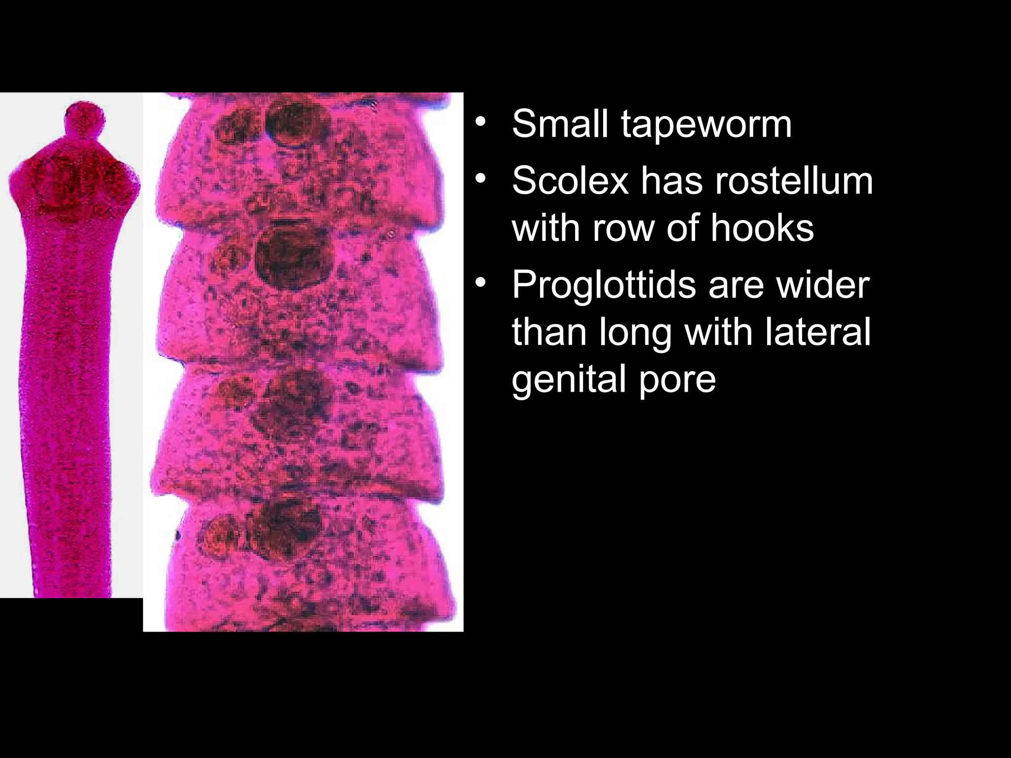
Class Cestoidea
by Asa Masese
The Tapeworms
General characteristics:
Adult
• Segmented long tape-like
worms
• Vary from few mm to several
meters
• No mouth/digestive system.
• Obtains its nutrient by
absorption through body
surface.
Body is divided into three main regions
a. Scolex (head)
b. Neck
c. Strobila made
up of proglottids
Morphology
Morphology
Scolex
– “head” of the organism
– Has holdfast organs to keep the tapeworm in
place
– Three main types of Scolex
• Three main types of Scolex
Cyclophyllidae
A-Globular head with 4
Muscular Sucker
B- 4 Sucker +Rostellum
armed with hooks
Pseudohyllidea
Bothria-Shallow grooves
or pits
• Area where new
segments are
created
• Give rise to
proglottids in
strobila.
Neck
Proglottids/segment
• Proglottid
– Set of reproductive organs
– Includes male and female organs and
genital pore
• Segment
– Segments may have one or more
proglottids
– Body divisisions
– More mature as gets farther from neck
• Size and shape of segment along with
number of proglottids and location of
genital pore key to identification many of the
tapeworms.
Reproduction
• Sex:- Hermaphrodites
• Have well developed reproductive
system.
• Reproduction
–Sexual-Oviporous
–Asexual-Sometimes multiplication
with in larval forms
Strobila • Is the entire chain of
proglottids
Strobila is divided into three regions:
• a/ Immature segment:
– near neck, sex organs are
immature.
• b/ mature segment:
– large segment, sex organs are
fully mature.
• c/ gravid segment:
– found at the tail end, uterus is
filled with eggs
Egg: -
• Two type
– Operculated, immature when voided to the
external environment.
– Non-operculated ,fully embryonated when
voided to the external environment.
Operculated
Operculum
Non-operculated
Classification
1. Order Cyclophyllidea
1. T. saginata,
2. T. solium,
3. Hymenelopis sps
4. Echinococcus
granulosus
2. Order Pseudophyllidea
1. Diphyllobothrium latum
• Less medically important:
– Order Cyclophyllidea
• T. multiceps
• Dipylidium caninum
• Echinococcus
multicuralis.
– Order Pseudophyllidea
• Spirometra species
Life cycle
• Complete in two host (exception H.nana)
• Habitat:- Adult live in small intestine
• Man is:-
• The only/main Definitive Host for T. saginata, T. solium,
H. nana and D. latum
• Intermittent Host for E. granulosus and E. multilcolaris
• DH & IH for H. nana and T. solium
Order- Pseudophillidea Order –Cyclophillidea
1. Scolex Spoonshaped, grooves - globular with 4 suckers
2. Genital pore - venteral - marginal
3. Utrine pore -Present(ventral) - absent
4. Uterus - coiled - sacular tubular or branched
5. Ova - operculated - non-operculated
6. Onchosphere - ciliated - non-ciliated
7. Rostellum - absent - present
8. Progilottids - broader than long - longer than broader
9. Larval forms - solid - cystic
• Geographical Distribution:-
– T. saginata
• World wide distribution where cattle are raised and beef is eaten raw or under
cooked.
• Very common in Ethiopia
– T. solium
• Not widely distributed as T saginata.
• Common in all areas where raw or partially cooked pork is eaten.
• Common throughout Mexico, South America and southern Africa & southern
Europe.
• Not reported from Ethiopia
Taenia species
Morphology:
• Adult:
– Size: 4-10 m long (can
reach up to 20 m)
– Colour: ivory white
– Strobila : 1000-2000
proglottides
– Mature segment: 1-2cm
long
T. saginata
• Adult
– Size: 2-3m
– Colour: pale blue
– Strobila: 800-1000
Proglottides
– Mature segment :0.5-1.5
cm
T. solium
Taenia saginata
Scolex (head):
• Quadrate, with four suckers, no
hooks, no rostellum on scolex
• Size-2mm across
Taenia solium
Scolex has
• Four sucker
• two rows of hooks on a prominent
rostellum
• Size-1mm
Taenia sp.
• Larval stage is cysticercus
– Invaginated scolex in fluid filled body
– Cysticercus bovis-T saginata
– Cysticercus celluless-T solium
• Eggs are very round with very thick walls.
Transmission and life cycle
• Transmission
– Humans become infected by ingesting raw or
undercooked meat infected with cystcerus
larvae:
• Beef- T saginata
• pork meat –T.solium
– T. solium can also be transmitted by :
• Ingesting ova in food or water
• Internal autoinfections
Taenia solium
• Distinct difference with T. saginata is that humans
can be infected with egg stage and onocosphere
migrates to some site in body and develops into
cycticercus
• This can be serious, called Cysticercosis
Clinical manifestation
• T saginata
–Taeniasis.
• Usually asymptomatic but may
cause dizziness, abdominal pain,
diarrhea, headache and nausea.
–Proglottids obvious in feces.
– Proglottides have a strong tendency
to crawl from the anus during the day
when its host is active
• T. solium
–Taeniasis
• Major symptoms of taeniasis are as a
result of the adult worm.
• These include abdominal pain, loss
of appetite, and
–Cysticercosis
• T. solium ( when infected by eggs)
cause cysticercosis (larval cysts in
lung, liver, eye and brain) resulting in
blindness and neurological disorders.
Laboratory Diagnosis
• Detecting eggs in faeces .
• Identifying macroscopically
– gravid segments in faeces
– scolex recovered from clothing or
passed in faeces.
• In addition
• T.saginata-
– ova on perianal skin (cellotape slide)
• T.solium ( cysticercosis)
– Finding calcified larvae in histological
or X-rays examination .
Egg : T.solium &T.saginata
• Size: - 33-40 m
• Shape: -Round
• Colour: - Shell-dark
yellowish-brown,
• Content: light yellowish
gray.
• Shell:-Thick, Smooth,
brown, radially straited
(embryophore)
• Content: - A round granular
mass enclosed by a fine
membrane with six hooklets
• T. saginata ova stains red
(acid fast) in Ziehl-
Neelsen stain
• this character helps to
differentiate it from
T.solium which do not
have red color in such
staining ( not acid fast)
Morphologically eggs of T.saginata and T. solium are
indistinguishable unless stained by AFB
Taenia saginata
• Gravid proglottide
• Detach when fully develop
and pass through the
anus independently.
• Color- white and opaque
• Size- 20mmX6mm
• Uterus- > 13 main (15-
30) lateral uterine
branches.
Taenia solium
• Gravid proglottides-
• Grey-blue and transluscent
• Size-13mmX8m
• 7 to 12, on average 10 lateral
compound uterine branches.
• Small chains of 3-4 rectangular
segments found in the faeces
Control- Taenia saginata
Inspection of beef for cysticerci is the best
preventive measure. Beef must be thoroughly
cooked in endemic areas—to at least 56°C
throughout the meat, which may be difficult to
accomplish with large cuts of fatty meat,
particularly pork. Freezing at 10°C for 10 days
usually is lethal to Taenia cysticerci, but they can
withstand 70 days at 0°C.
Treatment is readily available for the
intestinal adult worms. Niclosamide, is a
nonabsorbed oxidative phosphorylation
inhibitor that kills the scolex and anterior
segments on contact, after which the worm is
expelled. Praziquantel, a synthetic
isoquinoline-pyrazine derivative, is an
equally effective and relatively nontoxic
cesticidal compound..
Since the scolex is usually but not always
destroyed, and a new worm can regenerate
if the scolex and a minute portion of the
neck survive, the patient should be
observed for several months, as egg-
bearing segments can reappear in 10-12
weeks
Control- Taenia solium, The
Pork Tapeworm
The control of infection of humans as definitive hosts is
the same as that for T saginata, except that the control
measures apply to pork not beef. In addition, human
sewage from infected individuals may contaminate the
source of drinking water. The eggs are highly resistant
and can withstand many months of environmental
exposure over a broad temperature range. Treatment
for adult T solium is the same as for T saginata.
Cysticercosis may require surgery for ophthalmic
or brain involvement, but chemotherapy should
precede surgery when possible. Tissue infection
can be treated with albendazol (taken with a fatty
meal to increase absorption or praziquantel
(combined with corticosteroids to reduce the
inflammatory response to the dead cysticerci).
Praziquantel should not be used for ocular or
spinal cord infections.
• Hymenolepis nana-
Dwarf Tape Worm
Hymenolepis nana
• Dwarf Tapeworm
– Vampirolepis nana
• Definitive Host: Humans, rodents
– Most common tapeworm of humans in the
world
– 1% rate of infection in the southern U.S.
– 97.3% rate of infection in Moscow, Russia
• Intermediate Host: Larval and adult
beetles (but optional)
– Larval stage, cysticercoid, can develop in
D.H. if it eats the eggs
• Probably a recent evolutionary event
• Small tapeworm
• Scolex has rostellum
with row of hooks
• Proglottids are wider
than long with lateral
genital pore
• Geographical Distribution:-
– H.nana is widely distributed in countries
with warm climates than in cold climates
and fairly common in Ethiopia.
– Children are more commonly infected than
adults.
• Mode of Transmisssion: -
– Ingestion of egg with contaminated food,
drink or finger.
– Autoinfection.
• Life Cycle: H. nana has a direct life
cycle with a human host serving as both
definitive and intermediate host.
Hymenolepis
dimunata
• Slight larger than H.
nana but still very
small.
• Scolex has very
small rostellum with
no hooks
• Proglottid same as
H. nana but larger.
• Egg: H nana
– Size: 35-50m
– Shape: oval, almost round
– Shell: double; thin external
membrane and internal membrane
often thicker at the poles. Thread like
polar filaments coming from both
poles
– Colour: colour less or very pale gray
– Content: Rounded mass (embryo)
with six refractile hooklets arranged
in fan shaped.
Egg: H dimunata
• Color:-Yellow-brown or
bile pigmented.
• Size:-70 by 60μm
• Shell with double shell
and with out thread like
polar filaments.
• Content: A rounded
embryo containing six
hooklets arranged in fan
shape.
Control
Preventing fecal contamination of food and
water in institutions and crowded areas is of
primary importance. General sanitation and
rodent and insect control (especially control
of fleas and grain insects) are also essential
for prevention of H nana infection. Treatment
with praziquantel or niclosamide is usually
effective, and can be repeated if necessary.
Echinococcus granulosus
• Sheep Tapeworm
• Definitive Host: Carnivores including dogs,
wolves, and coyotes
• Intermediate Host: Herbivores including
sheep and mice.
• Occasionally infect humans.
• Hydatidosis is a zoonotic parasitic disease
caused by larval stages (hydatid cysts) of
cestodes belonging to the genus
Echinococcus and the family Taeniidae.
• Hydatid cyst, which is the larval stage of
Echinococcus, is a bladder-like cyst formed in
various organs and tissues following the growth
of the oncospheres of an Echninoccus tape
worm in that specific organ or tissue.
Geographic Distribution: Most common in
sheep raising countries
– New Zealand and Australia highest incidence
MORPHOLOGY
• Smallest of all tapeworms
– Scolex, neck, 3 segments
– Segments look like Taenia
sp.
• Largest larval stage of all
tapeworms
– Hydatid cyst
Clinical feature and Pathology:
• The symptoms, depend upon the location of the cyst.
• Large abdominal cysts produce increasing
discomfort.
• Liver cysts cause obstructive jaundice.
• Peribronchial cysts may produce pulmonary
abscesses.
• Brain cysts produce intracranial pressure and
Jacksonian epilepsy.
• Kidney cysts cause renal dysfunction.
• The contents of a cyst may produce anaphylactic
responses
E.
granulosus
hydatid
cyst
Laboratory Diagnosis
• Histological examination to find larvae
• X-ray examination to find larvae
• Examination of cystic fluid for brood
capsules and protoscoleces
• Casoni's skin test
Control
Effective control is chiefly epidemiologic: denying
sheep dogs access to carcasses of infected sheep,
obligatory testing and treatment of all sheep dogs,
prevention of contact of children with possibly
infected sheep dogs, and widespread education on
the danger and method of spread of hydatid
disease.
Treatment is chiefly by surgical resection
(with extreme care to avoid or decontaminate
spillage). Recent work suggests that a long
course of albendazole may kill the scoleces
within the cyst and even reduce the size of
the cyst. Long, continued use of mebendazole
has also proved effective, although the
results are variable.
A recent approach involves percutaneous
puncture under sonographic guidance,
aspiration of cyst fluid, instillation of a
protoscolecidal agent such as 95%
methanol or cetrimide, and respiration
(PAIR), along with albendazole treatment
to reduce the danger of subsequently
renewed disease from spillage (see Giorgio
et al., 1992).
Dipylidium caninum
Dog Tapeworm
• World wide
Distribution
• Dogs or cats
(humans rarely) as
the definitive host
• Fleas or lice are
the intermediate
host.
• Habitat:
• Adult: mucus membrane of
small intestine of carnivores
such as dog, cat, Man
• Cysticercoid larvae: In the
body cavity of insects
• Egg: in the faeces of dog,
cat, man
Dipylidium caninum
• Flea or louse
ingests the eggs in
the perianal region
of the dog or cat.
• The dog or cat (or
human) is infected
when they ingest a
flea or louse
infected with the
metacestode state
(cysticercoid) Dog flea
Dipylidium caninum
Dipylidium caninum
Proglottids of Dipylidium
caninum compared to a
match stick.
These are often passed
intact in the feces of an
infected dog.
When the proglottids
dry, their appearance is
similar to grains of rice.
Dipylidium
caninum
• Egg: 5-15 eggs in
capsule
• 40m in size and
yellowish brown in
color
Eggs
Taenia sp.
Diphyllobothrium
latum
H. nana
Dipylidium
caninum
H. dimunata
Contro;
The infection is usually asymptomatic and is
self-limited, although praziquantel would
probably be an effective treatment. Flea
control of pets would largely eliminate the
infection from household pets and children.
Diphyllobothrium latum
Common name: Fish tapeworm
• Geographical Distribution:-
– Widely distributed in the lake areas of
Europe, Asia, Far East, North America,
South America and Central Africa .
Diphyllobothrium latum
Fish tapeworm
• Important parasite of
man.
• Definitive hosts can be
humans, dogs, foxes,
cats, mink, bears, and
seals.
• Site of attachment :
small intestine.
Diphyllobothrium latum
Fish tapeworm
• humans are infected
with the plerocercoid .
• In humans the
tapeworm can reach
a length of 10
meters (>30 feet)
and produce over a
million eggs a day! .
Diphyllobothrium
latum
• Scolex has bothria
– Shallow groove
• Segments have one
proglottid
– Wider than long
egg-
58-76µm by 40-51µm
Broadly ovoid
Light golden yellow,
Operculated
Thick shell
Contains immature
• Clinical feature and Pathology :
– Clinical symptoms may be mild, depending on the
number of worms.
– They include abdominal discomfort, loss of weight,
loss of appetite and some malnutrition.
– Anemia and neurological problems associated
with vitamin B12 deficiency are seen in heavily
infected individuals.
• Laboratory Diagnosis
– Eggs in the faeces
– Scolex in the faeces
– Adult worms in the faeces
Egg:
• 58-76m by 40-51m
• Broadly ovoid
• Light golden yellow,
Operculated
• Thick shell
• Contains immature
embryo
Control
Plerocercoids in fish are quickly killed by
thorough cooking, freezing at -10°C for 15
minutes, or thorough pickling. Treatment of
sewage before it enters lakes greatly reduces
the prevalence of infection, as has been
demonstrated in Finland. Treatment with
praziquantel or niclosamide is effective and
nontoxic.

Cestodes by Dr. KennedyMunisi MD. MMed. Msc.ppt

  • 1.
    Class Cestoidea by AsaMasese The Tapeworms
  • 2.
    General characteristics: Adult • Segmentedlong tape-like worms • Vary from few mm to several meters • No mouth/digestive system. • Obtains its nutrient by absorption through body surface.
  • 3.
    Body is dividedinto three main regions a. Scolex (head) b. Neck c. Strobila made up of proglottids Morphology Morphology
  • 4.
    Scolex – “head” ofthe organism – Has holdfast organs to keep the tapeworm in place – Three main types of Scolex
  • 5.
    • Three maintypes of Scolex Cyclophyllidae A-Globular head with 4 Muscular Sucker B- 4 Sucker +Rostellum armed with hooks Pseudohyllidea Bothria-Shallow grooves or pits
  • 6.
    • Area wherenew segments are created • Give rise to proglottids in strobila. Neck
  • 7.
    Proglottids/segment • Proglottid – Setof reproductive organs – Includes male and female organs and genital pore • Segment – Segments may have one or more proglottids – Body divisisions – More mature as gets farther from neck • Size and shape of segment along with number of proglottids and location of genital pore key to identification many of the tapeworms.
  • 8.
    Reproduction • Sex:- Hermaphrodites •Have well developed reproductive system. • Reproduction –Sexual-Oviporous –Asexual-Sometimes multiplication with in larval forms
  • 9.
    Strobila • Isthe entire chain of proglottids
  • 10.
    Strobila is dividedinto three regions: • a/ Immature segment: – near neck, sex organs are immature. • b/ mature segment: – large segment, sex organs are fully mature. • c/ gravid segment: – found at the tail end, uterus is filled with eggs
  • 11.
    Egg: - • Twotype – Operculated, immature when voided to the external environment. – Non-operculated ,fully embryonated when voided to the external environment.
  • 12.
  • 13.
  • 14.
    Classification 1. Order Cyclophyllidea 1.T. saginata, 2. T. solium, 3. Hymenelopis sps 4. Echinococcus granulosus 2. Order Pseudophyllidea 1. Diphyllobothrium latum • Less medically important: – Order Cyclophyllidea • T. multiceps • Dipylidium caninum • Echinococcus multicuralis. – Order Pseudophyllidea • Spirometra species
  • 15.
    Life cycle • Completein two host (exception H.nana) • Habitat:- Adult live in small intestine • Man is:- • The only/main Definitive Host for T. saginata, T. solium, H. nana and D. latum • Intermittent Host for E. granulosus and E. multilcolaris • DH & IH for H. nana and T. solium
  • 16.
    Order- Pseudophillidea Order–Cyclophillidea 1. Scolex Spoonshaped, grooves - globular with 4 suckers 2. Genital pore - venteral - marginal 3. Utrine pore -Present(ventral) - absent 4. Uterus - coiled - sacular tubular or branched 5. Ova - operculated - non-operculated 6. Onchosphere - ciliated - non-ciliated 7. Rostellum - absent - present 8. Progilottids - broader than long - longer than broader 9. Larval forms - solid - cystic
  • 17.
    • Geographical Distribution:- –T. saginata • World wide distribution where cattle are raised and beef is eaten raw or under cooked. • Very common in Ethiopia – T. solium • Not widely distributed as T saginata. • Common in all areas where raw or partially cooked pork is eaten. • Common throughout Mexico, South America and southern Africa & southern Europe. • Not reported from Ethiopia Taenia species
  • 18.
    Morphology: • Adult: – Size:4-10 m long (can reach up to 20 m) – Colour: ivory white – Strobila : 1000-2000 proglottides – Mature segment: 1-2cm long T. saginata • Adult – Size: 2-3m – Colour: pale blue – Strobila: 800-1000 Proglottides – Mature segment :0.5-1.5 cm T. solium
  • 19.
    Taenia saginata Scolex (head): •Quadrate, with four suckers, no hooks, no rostellum on scolex • Size-2mm across
  • 20.
    Taenia solium Scolex has •Four sucker • two rows of hooks on a prominent rostellum • Size-1mm
  • 21.
    Taenia sp. • Larvalstage is cysticercus – Invaginated scolex in fluid filled body – Cysticercus bovis-T saginata – Cysticercus celluless-T solium • Eggs are very round with very thick walls.
  • 22.
    Transmission and lifecycle • Transmission – Humans become infected by ingesting raw or undercooked meat infected with cystcerus larvae: • Beef- T saginata • pork meat –T.solium – T. solium can also be transmitted by : • Ingesting ova in food or water • Internal autoinfections
  • 24.
    Taenia solium • Distinctdifference with T. saginata is that humans can be infected with egg stage and onocosphere migrates to some site in body and develops into cycticercus • This can be serious, called Cysticercosis
  • 26.
    Clinical manifestation • Tsaginata –Taeniasis. • Usually asymptomatic but may cause dizziness, abdominal pain, diarrhea, headache and nausea. –Proglottids obvious in feces. – Proglottides have a strong tendency to crawl from the anus during the day when its host is active
  • 27.
    • T. solium –Taeniasis •Major symptoms of taeniasis are as a result of the adult worm. • These include abdominal pain, loss of appetite, and –Cysticercosis • T. solium ( when infected by eggs) cause cysticercosis (larval cysts in lung, liver, eye and brain) resulting in blindness and neurological disorders.
  • 28.
    Laboratory Diagnosis • Detectingeggs in faeces . • Identifying macroscopically – gravid segments in faeces – scolex recovered from clothing or passed in faeces. • In addition • T.saginata- – ova on perianal skin (cellotape slide) • T.solium ( cysticercosis) – Finding calcified larvae in histological or X-rays examination .
  • 29.
    Egg : T.solium&T.saginata • Size: - 33-40 m • Shape: -Round • Colour: - Shell-dark yellowish-brown, • Content: light yellowish gray. • Shell:-Thick, Smooth, brown, radially straited (embryophore) • Content: - A round granular mass enclosed by a fine membrane with six hooklets
  • 30.
    • T. saginataova stains red (acid fast) in Ziehl- Neelsen stain • this character helps to differentiate it from T.solium which do not have red color in such staining ( not acid fast) Morphologically eggs of T.saginata and T. solium are indistinguishable unless stained by AFB
  • 31.
    Taenia saginata • Gravidproglottide • Detach when fully develop and pass through the anus independently. • Color- white and opaque • Size- 20mmX6mm • Uterus- > 13 main (15- 30) lateral uterine branches.
  • 32.
    Taenia solium • Gravidproglottides- • Grey-blue and transluscent • Size-13mmX8m • 7 to 12, on average 10 lateral compound uterine branches. • Small chains of 3-4 rectangular segments found in the faeces
  • 33.
    Control- Taenia saginata Inspectionof beef for cysticerci is the best preventive measure. Beef must be thoroughly cooked in endemic areas—to at least 56°C throughout the meat, which may be difficult to accomplish with large cuts of fatty meat, particularly pork. Freezing at 10°C for 10 days usually is lethal to Taenia cysticerci, but they can withstand 70 days at 0°C.
  • 34.
    Treatment is readilyavailable for the intestinal adult worms. Niclosamide, is a nonabsorbed oxidative phosphorylation inhibitor that kills the scolex and anterior segments on contact, after which the worm is expelled. Praziquantel, a synthetic isoquinoline-pyrazine derivative, is an equally effective and relatively nontoxic cesticidal compound..
  • 35.
    Since the scolexis usually but not always destroyed, and a new worm can regenerate if the scolex and a minute portion of the neck survive, the patient should be observed for several months, as egg- bearing segments can reappear in 10-12 weeks
  • 36.
    Control- Taenia solium,The Pork Tapeworm The control of infection of humans as definitive hosts is the same as that for T saginata, except that the control measures apply to pork not beef. In addition, human sewage from infected individuals may contaminate the source of drinking water. The eggs are highly resistant and can withstand many months of environmental exposure over a broad temperature range. Treatment for adult T solium is the same as for T saginata.
  • 37.
    Cysticercosis may requiresurgery for ophthalmic or brain involvement, but chemotherapy should precede surgery when possible. Tissue infection can be treated with albendazol (taken with a fatty meal to increase absorption or praziquantel (combined with corticosteroids to reduce the inflammatory response to the dead cysticerci). Praziquantel should not be used for ocular or spinal cord infections.
  • 38.
  • 39.
    Hymenolepis nana • DwarfTapeworm – Vampirolepis nana • Definitive Host: Humans, rodents – Most common tapeworm of humans in the world – 1% rate of infection in the southern U.S. – 97.3% rate of infection in Moscow, Russia • Intermediate Host: Larval and adult beetles (but optional) – Larval stage, cysticercoid, can develop in D.H. if it eats the eggs • Probably a recent evolutionary event
  • 40.
    • Small tapeworm •Scolex has rostellum with row of hooks • Proglottids are wider than long with lateral genital pore
  • 41.
    • Geographical Distribution:- –H.nana is widely distributed in countries with warm climates than in cold climates and fairly common in Ethiopia. – Children are more commonly infected than adults.
  • 42.
    • Mode ofTransmisssion: - – Ingestion of egg with contaminated food, drink or finger. – Autoinfection. • Life Cycle: H. nana has a direct life cycle with a human host serving as both definitive and intermediate host.
  • 44.
    Hymenolepis dimunata • Slight largerthan H. nana but still very small. • Scolex has very small rostellum with no hooks • Proglottid same as H. nana but larger.
  • 46.
    • Egg: Hnana – Size: 35-50m – Shape: oval, almost round – Shell: double; thin external membrane and internal membrane often thicker at the poles. Thread like polar filaments coming from both poles – Colour: colour less or very pale gray – Content: Rounded mass (embryo) with six refractile hooklets arranged in fan shaped.
  • 47.
    Egg: H dimunata •Color:-Yellow-brown or bile pigmented. • Size:-70 by 60μm • Shell with double shell and with out thread like polar filaments. • Content: A rounded embryo containing six hooklets arranged in fan shape.
  • 48.
    Control Preventing fecal contaminationof food and water in institutions and crowded areas is of primary importance. General sanitation and rodent and insect control (especially control of fleas and grain insects) are also essential for prevention of H nana infection. Treatment with praziquantel or niclosamide is usually effective, and can be repeated if necessary.
  • 49.
    Echinococcus granulosus • SheepTapeworm • Definitive Host: Carnivores including dogs, wolves, and coyotes • Intermediate Host: Herbivores including sheep and mice. • Occasionally infect humans. • Hydatidosis is a zoonotic parasitic disease caused by larval stages (hydatid cysts) of cestodes belonging to the genus Echinococcus and the family Taeniidae.
  • 50.
    • Hydatid cyst,which is the larval stage of Echinococcus, is a bladder-like cyst formed in various organs and tissues following the growth of the oncospheres of an Echninoccus tape worm in that specific organ or tissue. Geographic Distribution: Most common in sheep raising countries – New Zealand and Australia highest incidence
  • 51.
    MORPHOLOGY • Smallest ofall tapeworms – Scolex, neck, 3 segments – Segments look like Taenia sp. • Largest larval stage of all tapeworms – Hydatid cyst
  • 53.
    Clinical feature andPathology: • The symptoms, depend upon the location of the cyst. • Large abdominal cysts produce increasing discomfort. • Liver cysts cause obstructive jaundice. • Peribronchial cysts may produce pulmonary abscesses. • Brain cysts produce intracranial pressure and Jacksonian epilepsy. • Kidney cysts cause renal dysfunction. • The contents of a cyst may produce anaphylactic responses
  • 54.
  • 55.
    Laboratory Diagnosis • Histologicalexamination to find larvae • X-ray examination to find larvae • Examination of cystic fluid for brood capsules and protoscoleces • Casoni's skin test
  • 56.
    Control Effective control ischiefly epidemiologic: denying sheep dogs access to carcasses of infected sheep, obligatory testing and treatment of all sheep dogs, prevention of contact of children with possibly infected sheep dogs, and widespread education on the danger and method of spread of hydatid disease.
  • 57.
    Treatment is chieflyby surgical resection (with extreme care to avoid or decontaminate spillage). Recent work suggests that a long course of albendazole may kill the scoleces within the cyst and even reduce the size of the cyst. Long, continued use of mebendazole has also proved effective, although the results are variable.
  • 58.
    A recent approachinvolves percutaneous puncture under sonographic guidance, aspiration of cyst fluid, instillation of a protoscolecidal agent such as 95% methanol or cetrimide, and respiration (PAIR), along with albendazole treatment to reduce the danger of subsequently renewed disease from spillage (see Giorgio et al., 1992).
  • 59.
    Dipylidium caninum Dog Tapeworm •World wide Distribution • Dogs or cats (humans rarely) as the definitive host • Fleas or lice are the intermediate host. • Habitat: • Adult: mucus membrane of small intestine of carnivores such as dog, cat, Man • Cysticercoid larvae: In the body cavity of insects • Egg: in the faeces of dog, cat, man
  • 61.
    Dipylidium caninum • Fleaor louse ingests the eggs in the perianal region of the dog or cat. • The dog or cat (or human) is infected when they ingest a flea or louse infected with the metacestode state (cysticercoid) Dog flea
  • 62.
    Dipylidium caninum Dipylidium caninum Proglottidsof Dipylidium caninum compared to a match stick. These are often passed intact in the feces of an infected dog. When the proglottids dry, their appearance is similar to grains of rice.
  • 63.
    Dipylidium caninum • Egg: 5-15eggs in capsule • 40m in size and yellowish brown in color
  • 64.
  • 65.
    Contro; The infection isusually asymptomatic and is self-limited, although praziquantel would probably be an effective treatment. Flea control of pets would largely eliminate the infection from household pets and children.
  • 66.
    Diphyllobothrium latum Common name:Fish tapeworm • Geographical Distribution:- – Widely distributed in the lake areas of Europe, Asia, Far East, North America, South America and Central Africa .
  • 67.
    Diphyllobothrium latum Fish tapeworm •Important parasite of man. • Definitive hosts can be humans, dogs, foxes, cats, mink, bears, and seals. • Site of attachment : small intestine.
  • 68.
    Diphyllobothrium latum Fish tapeworm •humans are infected with the plerocercoid . • In humans the tapeworm can reach a length of 10 meters (>30 feet) and produce over a million eggs a day! .
  • 69.
    Diphyllobothrium latum • Scolex hasbothria – Shallow groove • Segments have one proglottid – Wider than long egg- 58-76µm by 40-51µm Broadly ovoid Light golden yellow, Operculated Thick shell Contains immature
  • 71.
    • Clinical featureand Pathology : – Clinical symptoms may be mild, depending on the number of worms. – They include abdominal discomfort, loss of weight, loss of appetite and some malnutrition. – Anemia and neurological problems associated with vitamin B12 deficiency are seen in heavily infected individuals. • Laboratory Diagnosis – Eggs in the faeces – Scolex in the faeces – Adult worms in the faeces
  • 72.
    Egg: • 58-76m by40-51m • Broadly ovoid • Light golden yellow, Operculated • Thick shell • Contains immature embryo
  • 74.
    Control Plerocercoids in fishare quickly killed by thorough cooking, freezing at -10°C for 15 minutes, or thorough pickling. Treatment of sewage before it enters lakes greatly reduces the prevalence of infection, as has been demonstrated in Finland. Treatment with praziquantel or niclosamide is effective and nontoxic.

Editor's Notes

  • #39 Large genera with host in wide variety of birds and mammals. All require arthropod intermediate except H. nana.
  • #49 Smallest adult forms the biggest larvae. There may be different strains involved that use wolf-moose, wolf-reindeer, dingo-wallaby, lion-warthog. For humans, the most important is dog-sheep. The strains differ morphologically, development, metabolism, DNA, and host specificity. Worms of one strain do not develop well in other species. Can be important in human infections. In Europe, the strain common in horse and pigs do not infect humans but the strain in sheep and cattle does.