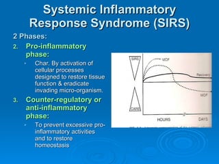
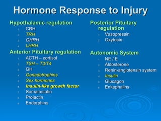
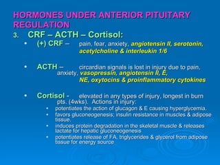
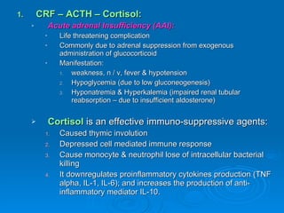
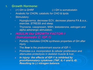
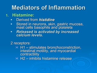
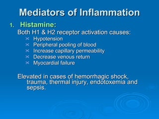
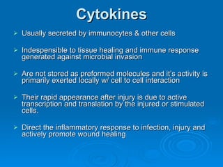
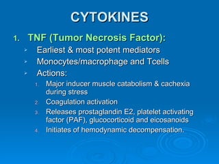
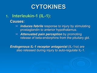
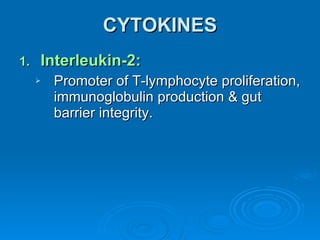
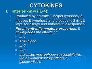
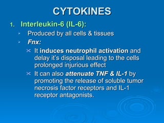
SYSTEMIC RESPONSE TO INJURY James Taclin C. Banez, MD, FPSGS, FPCS
Injury   (surgery, traumatic & infections): Alteration of  neuro-endocrine system, metabolic  and  immunology  ----> causes disequilibrium of internal environment & tries to return to homeostasis.
Minor Injuries : is usually followed by functional restoration w/ minimal intervention. Major injuries : associated with overwhelming inflammatory response ----> failure to give appropriate intervention ----> multiple organ failure ----->  DEATH
Systemic Inflammatory Response Syndrome (SIRS) 2 Phases: Pro-inflammatory phase: Char. By activation of cellular processes designed to restore tissue function & eradicate invading micro-organism. Counter-regulatory or anti-inflammatory phase: To prevent excessive pro-inflammatory activities and to restore homeostasis
Terminologies Term Definition Infection Identifiable source of microbial insult SIRS Two or more following criteria: - Temp >/= 38C or </= 36C - Heart rate >/= 90 beats/min - Respiratory rate >/= 20 breaths/min or PaCO2 </= 32mmHg or mechanical ventilation - WBC >/= 12,000/ul or </= 4000/ul or >/= 10% band forms Sepsis Identifiable source of infection + SIRS Severe sepsis Sepsis + organ dysfunction Septic shock Sepsis + cardiovascular collapse (requiring vasopressor support)
Central Nervous System Regulation of Inflammation Afferent Signals: Circulatory: Areas of CNS devoid of bld-brain barrier admit passage of inflammatory mediators (TNF) Causing fever, anorexia & depression Neural pathways: Afferent stimuli to the  vagus nerve :  cytokines – TNF, IL-1 Baroreceptors Chemoreceptors Thermoreceptors  From the site of injuries
Central Nervous System Regulation of Inflammation Cholinergic Anti-Inflammatory Pathways: Acetycholine of parasympathetic: reduces tissue macrophage activation Reduces tissue macrophage release of inflammatory mediators (TNF alpha, IL1, IL18 & high mobility grp protein (HMG-1), but not the anti-inflammatory cytokine IL10 Vagal stimulation reduces HR, increases gut motility, dilates arterioles, causes pupil constriction &  regulates inflammation
Systemic Neuro-Endocrine Reflexes Stimuli: Effective Circulatory Volume (ECV): Sensed by:  high pressure baroreceptor  (aorta, carotid & renal artery) low pressure stretch receptors  (atrial volume) Decreased ECV ---> release of tonic inhibition ---> (+) receptors ----> (+) ACTH, vasopressin , beta-endorphin, E & NE, renin.  If decrease ECV is < 30% of TBV the neuroendocrine & cardiovascular response can compensate; > 30% ---->  DECOMPENSATE (hypotension)
Systemic Neuro-Endocrine Reflexes Stimuli: Chemoreceptor Relexes: sensed by: a. carotid bodies (inactive) b. aortic bodies (inactive) stimulation of chemoreceptors: decrease oxygen increase CO2 and H Results to: Decrease sympathetic activity (cardiac) & increase in parasympathetic activity. Increase respiratory rate & decrease cardiac rate and contractility.
Systemic Neuro-Endocrine Reflexes Stimuli: Pain & Emotion: Pain ---> (+) thalamus & hypothalamus Emotion ---> (+) limbic ----> (+) hypothalamus Substrate alteration: Alteration plasma glucose concentration activates neuroendocrine reflexes thru it’s receptors: Hypothalamus -----> pituitary Pancreas Temperature: Changes in core temperature, sensed by the pre-optic area of the hypothalamus ---> alters secretion of several hormones
Systemic Neuro-Endocrine Reflexes CNS Centers:  (hypothalamus) Posterior Hypothalamus:  ACTH, sympathetic activity Paraventricular Nucleus: vasopressin, oxytocin & ACTH Ventromedial Nucleus: GH, ACTH Supraoptic Nucleus vasopressin & oxytocin Suprachiasmatic Nucleus: ACTH & gonadotrophin
Systemic Neuro-Endocrine Reflexes Efferent Output: Autonomic response: Hormonal response: Hypothalamic – anterior pituitary control Posterior pituitary control Autonomic control Local tissue response:
Hormone Response to Injury Hypothalamic regulation CRH TRH GHRH LHRH Anterior Pituitary regulation ACTH – cortisol TSH – T3/T4 GH Gonadotrophins Sex hormones Insulin-like growth factor Somatostatin Prolactin Endorphins Posterior Pituitary regulation Vasopressin Oxytocin Autonomic System NE / E Aldosterone Renin-angiotensin system Insulin Glucagon Enkephalins
HORMONES UNDER ANTERIOR PITUITARY REGULATION CRF – ACTH – Cortisol: (+) CRF  –  pain, fear, anxiety,  angiotensin II, serotonin,  acetylcholine & interleukin 1/6 ACTH  –  circardian signals is lost in injury due to pain,  anxiety,  vasopressin, angiotensin II, E,  NE, oxytocins & proinflammatory cytokines Cortisol  -  elevated in any types of injury, longest in burn  pts. (4wks).  Actions in injury: potentiates the action of glucagon & E causing hyperglycemia. favors gluconeogenesis; insulin resistance in muscles & adipose tissue. induces protein degradation in the skeletal muscle & releases lactate for hepatic gluconeogenesis  potentiates release of FA, triglycerides & glycerol from adipose tissue for energy source
CRF – ACTH – Cortisol: Acute adrenal Insufficiency (AAI): Life threatening complication Commonly due to adrenal suppression from exogenous administration of glucocorticoid Manifestation: weakness, n / v, fever & hypotension Hypoglycemia (due to low gluconeogenesis) Hyponatremia & Hyperkalemia (impaired renal tubular reabsorption – due to insufficient aldosterone)  Cortisol  is an effective immuno-suppressive agents: Caused thymic involution Depressed cell mediated immune response Cause monocyte & neutrophil lose of intracellular bacterial killing It downregulates proinflammatory cytokines production (TNF alpha, IL-1, IL-6); and increases the production of anti-inflammatory mediator IL-10.
Growth Hormone: (+) GH is GHRF (-) GH is somatostatin Anabolic for CHON; catabolic for CHO & lipids Stimulatory: Hypoglycemia, decrease ECV, decrease plasma FA & a.a., exercise, STRESS and sleep. Thyroxine, vasopressin, MSH, testosterone, estrogen and alpha adrenergic stimulation. INSULIN like GROWTH FACTOR-1 (Somatomedin C; IGF-1) Partially mediates CHON synthesis properties of GH after injury The  liver  is the predominant source of IGF-1. Promotes a.a. incorporation & cellular proliferation and attenuates proteolysis in skeletal muscle & liver. In injury: the effects of IGF-1 is inhibited by proinflammatory cytokines (TNF, IL-1 and IL-6). Resulting to (-) nitrogen balance.
Macrophage Inhibitory Factor: Produced by: Anterior pituitary gland T lymphocytes at the site of inflammation. Actions: A glucocorticoid antagonist (suppresses the immunosuppresive effects of cortisol). It is a proinflammatory mediator that potentiates gm (-) & (+) septic shock. Endogenous Opiods: Endorphins, enkephalins Elevated after injury & surgery Endorphins ----> attenuate pain perceptions / hypotension Enkephalins ----> HPN, decrease peristalsis and secretion of GIT
Thyroid Hormone (T4 / T3): In injury: Low T3 (-) TSH release Conversion of T4 – T3 in the target organs are impaired due to cortisol. T4 is converted to an inactive T3 called rT3 Gonadotrophins (LHRH/GnRH) & (FSH/LH): Injury, stress or severe illness ----> (-) GRH ----> (-) LH and  (-) FSH ---> decrease estrogen and androgen secretions. Causes menstrual irregularities and decrease libido.  Prolactin: Produced by anterior pituitary gld and T lymphocytes Elevated level after injury in adults not seen in children Causes amenorrhea
HORMONES OF AUTONOMIC SYSTEM: Catecholamines (E / NE): Causes hypermetabolic state following severe injury 3 – 4fold increase of E & NE in the plasma for 24 – 48 hrs. Causes: Promotes glycogenolysis, gluconeogenesis, lipolysis and ketogenesis. Decreased insulin release & increase glucagon secretion. Peripherally, it increases lipolysis in adipose tissue and induces insulin resistance in skeletal muscle It inhibit the release of aldosterone. Immune function: -- enhances neutrophilia and lymphocytosis
Aldosterone (Mineralocorticoid): Released by adrenal zona glomerulosa Release is caused by: Angiotensin II Hyperkalemia Aldosterone stimulating factor (ASF) in pituitary ACTH (is the most potent stimulant). Major function is to maintain intravascular volume by conserving Na & eliminating potassium and H+ in the early distal convoluted tubules of nephron
Renin – Angiotensin: Renin in Juxtaglomerular apparatus is released by: ACTH, Glucagon, porstagladin, K+, Mg+, and Ca+ Baroreceptor – respond to decrease blood pressure Macula densa detects changes in chloride concentration. Action of angiotensin II: Vasoconstrictor (+) aldosterone (+) ADH (+) E Increase heart rate and contractility
Glucagon: alpha islet cell catabolic role elevated release after injury Insulin: Inhibit its release in injury: Catecholamine Glucagon Somatostatin Beta endorphins IL-1
Hormones Under Posterior Pituitary Regulation: Vasopressin / ADH / Arginine Vasopressin (AVP): Causes   readsorption of H2O in DCT Vasoconstriction peripherally Stimulates hepatic glycogenolysis & gluconeogenesis Elevated plasma osmolality is its major stimulus: Location of osmoreceptors:  hypothalamus, portal circulation
Hormones Under Posterior Pituitary Regulation: Vasopressin / ADH / Arginine Vasopressin (AVP): Its release also happens in 10% loss of ECV stimulating the baroreceptor in the left atrium Other stimulus: PAIN Beta adrenergic Angiotensin II Opiods Elevated glucose
Oxytocin: Its release caused by SUCKING the nipple Stimulates contraction of mammary gland and uterus during parturition No known function in males Role in injury is unknown
Mediators of Inflammation Cytokines: Most potent mediator of the inflammatory response Eradicates invading microorganism and promotes wound healing Overwhelming productions of proinflammatory cytokines can cause: Hemodynamic instability (septic shock) Metabolic derangement (muscle wasting) Can exaggerate to multiple organ failure  and death. Inappropriate anti-inflammatory mediator release can lead to immunocompromised and susceptible to overwhelming infection.
Mediators of Inflammation Heat Shock Proteins: Intracellular protein modifiers and transporters that protect cells from the deleterious effects of traumatic stress. The following induced its production: Hypoxia Trauma Heavy metals Local trauma Hemorrhage
Mediators of Inflammation Reactive Oxygen Metabolites: Oxygen radicals  are produced by reduction of oxygen to superoxide anion, and further metabolized to form  H2O2 & hydroxyl radicals Causes injury by oxidation of unsaturated fatty acids w/in cell membranes. Activated leukocytes  are potent generators for reactive oxygen metabolites. Cells are protected from this metabolite by oxygen scavengers:  GLUTATHIONE  &  CATALASES.
Mediators of Inflammation Eicosanoids: Are oxidation derivatives of membrane  phospholipid arachidonic acids Secreted by nucleated cells, except lymphocytes Not stored w/in cells but are  synthesized rapidly upon stimulation by hypoxic injury, tissue injury, endotoxin, NE, vasopressin angiotensin II, bradykinin, serotonin, acetylcholine, cytokines and histamine
Mediators of Inflammation Eicosanoids: Actions: PGE 2   increases fluid leakage from bld vessels, inhibit gluconeogenesis and hormone stimulated lipolysis. Leukotrienes  are  1000x more potent than histamine promoting capillary leakage, vasoconstriction, bronchoconstriction, neutrophil activation. Products of cyclooxygenase  inhibit pancreatic beta cell release of insulin.
Mediators of Inflammation Kallikrein-Kinin System: Bradykinin: potent vasodilators  Release is caused by hypoxic and ischemic injury Kinins:   increase capillary permeability and tissue edema,  evoke pain,  Inhibit gluconeogenesis  increase bronchoconstriction.  increase renal vasodilation and reduces renal perfusion pressure ----> (+) renin-angiotensin system.
Mediators of Inflammation Serotonin: neurotransmitter (5hydrohytryptamine) tryptophan derivative  found in chromaffin cells of the intestine (carcinoid tumors). Vasoconstrictions, bronchoconstriction & platelet aggregations myocardial chronotrope and inotrope
Mediators of Inflammation Histamine: Derived from  histidine Stored in neurons, skin, gastric mucosa, mast cells basophils and platelets Released is activated by increased calcium levels. 2 receptors: H1 – stimulates bronchoconstriction, intestinal motility, and myocardial contractility H2 – inhibits histamine release
Mediators of Inflammation Histamine: Both H1 & H2 receptor activation causes: Hypotension Peripheral pooling of blood Increase capillary permeability Decrease venous return Myocardial failure Elevated in cases of hemorrhagic shock, trauma, thermal injury, endotoxemia and sepsis.
Cytokines Usually secreted by immunocytes & other cells Indespensible to tissue healing and immune response generated against microbial invasion Are not stored as preformed molecules and it’s activity is primarily exerted locally w/ cell to cell interaction Their rapid appearance after injury is due to active transcription and translation by the injured or stimulated cells. Direct the inflammatory response to infection, injury and actively promote wound healing
CYTOKINES TNF (Tumor Necrosis Factor): Earliest & most potent mediators Monocytes/macrophage and Tcells Actions: Major inducer muscle catabolism & cachexia during stress Coagulation activation Releases prostaglandin E2, platelet activating factor (PAF), glucocorticoid and eicosanoids Initiates of hemodynamic decompensation.
CYTOKINES Interleukin-1 (IL-1):  Causes: Induces febrile  response to injury by stimulating prostaglandin to anterior hypothalamus. Attenuated pain perception  by promoting release of beta-endorphins from the pituitary gld. Endogenous IL-1 receptor antagonist  (IL-1ra) are also released during injury to auto-regulate IL-1.
CYTOKINES Interleukin-2: Promoter of T-lymphocyte proliferation, immunoglobulin production & gut barrier integrity.
CYTOKINES Interleukin-4 (IL-4): Produced by activate T-helper lymphocyte. Induces B lymphocyte to produce IgG & IgE impt. for allergy and anthelmintic responses. Potent anti-inflammatory properties , it  downgrades the effects of: IL-1 TNF-alpha IL-6 IL-8 Increases macrophage susceptibility to the anti-inflammatory effects of glucocorticoid
CYTOKINES Interleukin-6 (IL-6): Produced by all cells & tissues Fnx: It  induces neutrophil activation  and delay it’s disposal leading to the cells prolonged injurious effect It can also  attenuate TNF & IL-1  by promoting the release of soluble tumor necrosis factor receptors and IL-1 receptor antagonists.
CYTOKINES Interleukin-8 (IL-8): Activity is similar to IL-6 Biomarker for the risk of multiple organ failure Does not produced hemodynamic instability but is a chemoattractant and activator of neutrophil
CYTOKINES Interleukin-10 (IL-10): It reduces TNF-alpha Attenuate systemic inflammatory response  and reduces mortality during septic peritonitis Associated w/ increased bacterial load & mortality
CYTOKINES Interleukin-12 (IL-12): Primary role in cell-mediated immunity & promotes differentiation of T H 1 cells. Inducing an inflammatory response for 48hrs, independently from TNF & IL-1. Promotes neutrophil & coagulation activation Toxicity is synergistic w/ IL-2 .
CYTOKINES Interleukin-13 (IL-13): Structural & functional similarities w/ IL-4 Modulate macrophage function Inhibit nitric oxide production & the expression of proinflammatory cytokines and enhance production of IL-1ra It attenuates leukocyte interaction w/ activated endothelial surfaces
CYTOKINES Interleukin-15 (IL-15): Potent autocrine regulatory properties. Possess similar bioactivity in promoting lymphocyte activation & proliferation Induces IL-8 production . Interleukin-18 (IL-18): Formerly interferon (IFN)-y-inducing factor Proinflammatory cytokine Structurally similar to IL-1beta & functionally similar to IL-12.
CYTOKINES Interferon-y: Produced by Helper T lymphocytes when activated by bacterial antigens, IL-2, IL-12, IL-18 Can also induce production of IL-2, IL-12,  IL-18 Elevated for as long as 8 days. Can activate circulating and tissue macrophage  Alveolar macrophage activation may induce acute lung inflammation after major surgery or trauma.
CYTOKINES Granuloctye-Macrophage Colony-Stimulating Factor (GMC-SF): Delays apoptosis  of macrophages and neutrophils;  contribute to organ injury (ARDS) Promote maturation and recruitment of functional leukocytes needed for normal inflammatory cytokine response & potentially in wound healing.
Cell-Mediated Inflammatory Response Platelets: Clot formed at the site of injury releases inflammatory mediators w/c serves as the principal chemo-attractant for neutrophils and monocytes. Migration of platelets & neutrophils through the vascular endothelium occurs w/in 3 hrs of injury and mediated by: Serotonin Platelet-activating factor Prostaglandin E2
Cell-Mediated Inflammatory Response Lymphocytes & T-cell Immunity: Injury associated w/ Acute impairment of cell-mediated immunity and macrophage funct ion 2 subgroups of T-helper lymphocytes: T H 1   T H 2
Cell-Mediated Inflammatory Response Eosinophils: Migrate to inflammed endothelium and release cytoplasmic granules that are cytotoxic It preferentially migrate to sites of parasitic infection and allergy Resides in GIT, lung and genitourinary tissues Major activators: IL-3 IL-5 Platelet-activating factor Complement anaphylatoxins C3a and C5a
Cell-Mediated Inflammatory Response Mast Cells: When activated it produce: Histamine Cytokines (IL-3, IL4, IL-5, IL-6, IL-10, IL-13, IL-14 & migration-inhibitory factor (MIF). Eicosanoids Proteases Chemokines
Cell-Mediated Inflammatory Response Mast Cells: Immediate results: Vasodilation Recruitment of other immunocytes Capillary leakage TNF-alpha secreted rapidly by this cell bec. of its abundant source
Cell-Mediated Inflammatory Response Monocytes: There is down regulation in monocyte and neutrophil TNFR expression In none surviving pts w/ severe sepsis and failed to recover, an immediate reduction in monocyte surface TNFR expression was observed, while surviving pts have normal or near normal receptor levels  Neutrophils: Inflammatory mediators from site of injury induces neutrophil adherence to the injured tissue. It’s function is mediated by vast array of intracellular granules that are chemotactic or cytotoxic to local tissue & invading microorganisms.
Endothelium-Mediated Injury Neutrophil-Endothelium Interaction: Inc. vascular permeability during inflammation is intended to facilitate O2 delivery and immunocyte migration to the site of injury. However accumulation & infiltration of leukocytes (neutrophil) contribute to the cytotoxicity of vital tissue ---> MOF. Ischemia/reperfusion injury potentiates this response by: unleashing oxygen metabolites Lysosomal enzymes that degrade tissue basal membranes Cause microvascular thrombosis Activate myeloperoxidases.
Endothelium-Mediated Injury Recruitment of circulating neutrophils to endothelial surfaces is mediated by actions of adhesive molecules called  SELECTINS (L,P,E)
Endothelium-Mediated Injury Nitric Oxide: Formed from oxidation of L-arginine Derived from endothelial surfaces Cells that produces this subs: Neutrophil monocytes Renal cells Kupffer cells Cerebellar neurons Action: Maintain normal smooth muscle relaxation Reduce thrombosis by reducing platelet adhesions and aggregation Mediates protein synthesis in hepatocyte
Endothelium-Mediated Injury Prostacyclin: Induces vaso-relaxation and platelet deactivation by increasing cAMP Endothelins: Formed by vascular endothelial cells in response to: Injury Thrombin Transforming growth factor-B (TGF-B) IL-1 Angiotensin II Vasopressin Catecholamine Anoxia Action: The MOST POTENT vasoconstriction  (10 x more angiotensin II) Vasoconstriction is reversed by acetylcholine
Endothelium-Mediated Injury Platelet-Activating Factor: Released by neutrophils, platelets, mast cells and monocytes It activate neutrophils and platelets and increase vascular permeability. Atrial Natriuretic Peptides: Released by atrial tissue, gut, kidney, brain, adrenal glds and endothelium Actions: Vasodilator and induce fluid and electrolyte excretion Inhibits aldosterone secretion Prevent reabsorption of sodium
END OF FIRST PART
Metabolic Changes and Nutritional Management of Surgical Patients James Taclin C. Banez, MD, FPSGS, FPCS
Majority of surgical patients: well nourished / healthy uncomplicated major surgical procedure has sufficient fuel reserve can withstand brief period of catabolic insult and starvation of 7 days Postoperatively: can resume normal oral intake supplemental diet is not needed
Surgical Patients that Needs Nutritional Support To shorten the postoperative recovery phase and minimize the number of complications: Chronically debilitated from their diseases or  malnutrition. Suffered severe trauma, sepsis or surgical complications
Metabolic Changes in Surgical Patients Metabolic events brought about by  STIMULI : Injury Starvation Metabolic response is directed to restore: Homeostasis Repair
Metabolic Response to  Starvation HYPOGLYCEMIA  – is primary stimulus Hormonal Changes: increase  cortisol, catecholamines, glucagon, growth hormones Primary gluconeogenic precursors by the liver & kidney: a.  lactate   b.  glycerol  c.  amino acid (alanine & glutamine)
Proteolysis increase due to increase CORTISOL ------> inc. urinary nitrogen first 4 days of starvation (8-12g/day = 6.25g of muscle/g of nitrogen).
Protein catabolism for gluconeogenesis primarily comes from  SKELETAL  muscle, but in pure starvation other organs are involved In liver. CHON loss is selective; spare enzymes for gluconeogenesis and lipolysis. In pancreas and GIT, enzymes for digestion and protein for regeneration of epithelium is involved ->  PARADOXICAL FOOD INTOLERANCE
Rapid proteolysis of body CHON cannot proceed at 75 g/day for long, or else patient will die immediately  RANDLE EFFECT. decrease urinary excretion of nitrogen 2 – 4 gm/day due to keto-adaptation of the brain decrease protein degeneration and  major source of energy is FAT (90%)
Metabolism of  Injured Patient PHASES: Catabolic phase   (Ebb, Adrenergic-Corticoid): immediately following surgery or trauma characterized w/ hyperglycemia, increase secretion of urinary nitrogen beyond the level of starvation caused by increase  glucagon, glucocorticoid, catecholamines and decrease insulin tries to restore circulatory volume and tissue perfusion
Metabolism of Injured Patient PHASES: Early anabolic phase  (flow, corticoid-withdrawal): tissue perfusion has been restored, may last for days to months depending on: severity of injury previous health medical intervention sharp decline in nitrogen excretion nitrogen balance is positive  (4g/day) indicating synthesis of CHON and there is a rapid and progressive gain in weight and muscular strength
Metabolism of Injured Patient PHASES: Late anabolic phase:   several months after injury occurs once volume deficit have been restored slower re-accumulation of CHON re-accumulation of body fat
Metabolism of Injured Patient Carbohydrate Metabolism in Injured Patient: Hyperglycemia  = proportional to the severity of injury Importance: Homeostatic significance Ready source of energy to the brain Adequate delivery Caused by: Increased catecholamine (primarily), cortisol, glucagon, GH, vasopressin, angiotensin II, somatostatin and decrease insulin.
Metabolism of Injured Patient Carbohydrate Metabolism: Hyperglycemia: Caused by: Increased catecholamine (primarily), cortisol, glucagon, GH, vasopressin, angiotensin II, somatostatin and decrease insulin. Gluconeogenesis in liver and kidney and impaired peripheral uptake of glucose
Metabolism of Injured Patient Carbohydrate Metabolism: Hyperglycemia: Insulin resistance: During the Ebb phase there is reduction in beta cell sensitivity to glucose due to  Catecholamine, somatostatin and reduced pancreatic blood flow Resistance to exogenous administration  on insulin in both EBB and early FLOW phases In middle and late Flow phase, beta cell sensitivity return to normal and it’s level is higher, but hyperglycemia persist because of continuous gluconeogenesis
Metabolism of Injured Patient Carbohydrate Metabolism: Glucose metabolism in wounded tissue : Increase glucose uptake and  lactate  production because of anaerobic glycolysis due to local tissue hypoxia (+) insulin insensitivity
Metabolism of Injured Patient Lipid metabolism: primary source of energy Best stimulus for hormone-sensitive lipase is  CATECHOLAMINE RANDLE EFFECT  is not present
Metabolism of Injured Patient Protein Metabolism: Nitrogen urine excretion 30-50g/day due to  proteolysis ;  20% utilized for energy (calories)  the rest for  gluconeogenesis  by liver and kidney (cortisol, glucagon, catecholamine). Primary source of protein is the  skeletal muscle  and the visceral organs are spared. Ketoadaptation  is  inhibited  ----> gluconeogenesis persist ---> proteolysis persist  (INTERLEUKIN I). The degree and duration (-) nitrogen balance is related to  severity of injury . The net CHON catabolism depends on the  age, sex and physical condition of the patient  (> in young, healthy and male)  (-) nitrogen balance can be reduced by  high caloric nitrogen supplement
Traumatized Man
Injury of any type is associated with: Immobilization Starvation Repair the first two are associated with reduction in energy requirement. While the third is associated w/ increase energy requirement The amount of energy produced in injured pt. is not optimum, to supply necessary energy for the repair due to: reduced or absent nutritional intake significant reduction of energy charge and ATP content during shock, hypoxia, sepsis, ischemia and wound -    anaerobic metabolism
REE (Resting energy expenditure) by Harris and Benedict: (MEN)   66.47 + 13.75 (W) + 5.0 (H) – 6.76 (A)  =  Kcal/day (Female)   65.51 + 9.56 (W) + 1.85 (H) – 4.68 (A)  =  Kcal/day Fever: increase resting energy expenditure of approximately  7%  for each degree of F of fever.
 

Systemic Response To Injury

  • 1.
    SYSTEMIC RESPONSE TOINJURY James Taclin C. Banez, MD, FPSGS, FPCS
  • 2.
    Injury (surgery, traumatic & infections): Alteration of neuro-endocrine system, metabolic and immunology ----> causes disequilibrium of internal environment & tries to return to homeostasis.
  • 3.
    Minor Injuries :is usually followed by functional restoration w/ minimal intervention. Major injuries : associated with overwhelming inflammatory response ----> failure to give appropriate intervention ----> multiple organ failure -----> DEATH
  • 4.
    Systemic Inflammatory ResponseSyndrome (SIRS) 2 Phases: Pro-inflammatory phase: Char. By activation of cellular processes designed to restore tissue function & eradicate invading micro-organism. Counter-regulatory or anti-inflammatory phase: To prevent excessive pro-inflammatory activities and to restore homeostasis
  • 5.
    Terminologies Term DefinitionInfection Identifiable source of microbial insult SIRS Two or more following criteria: - Temp >/= 38C or </= 36C - Heart rate >/= 90 beats/min - Respiratory rate >/= 20 breaths/min or PaCO2 </= 32mmHg or mechanical ventilation - WBC >/= 12,000/ul or </= 4000/ul or >/= 10% band forms Sepsis Identifiable source of infection + SIRS Severe sepsis Sepsis + organ dysfunction Septic shock Sepsis + cardiovascular collapse (requiring vasopressor support)
  • 6.
    Central Nervous SystemRegulation of Inflammation Afferent Signals: Circulatory: Areas of CNS devoid of bld-brain barrier admit passage of inflammatory mediators (TNF) Causing fever, anorexia & depression Neural pathways: Afferent stimuli to the vagus nerve : cytokines – TNF, IL-1 Baroreceptors Chemoreceptors Thermoreceptors From the site of injuries
  • 7.
    Central Nervous SystemRegulation of Inflammation Cholinergic Anti-Inflammatory Pathways: Acetycholine of parasympathetic: reduces tissue macrophage activation Reduces tissue macrophage release of inflammatory mediators (TNF alpha, IL1, IL18 & high mobility grp protein (HMG-1), but not the anti-inflammatory cytokine IL10 Vagal stimulation reduces HR, increases gut motility, dilates arterioles, causes pupil constriction & regulates inflammation
  • 8.
    Systemic Neuro-Endocrine ReflexesStimuli: Effective Circulatory Volume (ECV): Sensed by: high pressure baroreceptor (aorta, carotid & renal artery) low pressure stretch receptors (atrial volume) Decreased ECV ---> release of tonic inhibition ---> (+) receptors ----> (+) ACTH, vasopressin , beta-endorphin, E & NE, renin. If decrease ECV is < 30% of TBV the neuroendocrine & cardiovascular response can compensate; > 30% ----> DECOMPENSATE (hypotension)
  • 9.
    Systemic Neuro-Endocrine ReflexesStimuli: Chemoreceptor Relexes: sensed by: a. carotid bodies (inactive) b. aortic bodies (inactive) stimulation of chemoreceptors: decrease oxygen increase CO2 and H Results to: Decrease sympathetic activity (cardiac) & increase in parasympathetic activity. Increase respiratory rate & decrease cardiac rate and contractility.
  • 10.
    Systemic Neuro-Endocrine ReflexesStimuli: Pain & Emotion: Pain ---> (+) thalamus & hypothalamus Emotion ---> (+) limbic ----> (+) hypothalamus Substrate alteration: Alteration plasma glucose concentration activates neuroendocrine reflexes thru it’s receptors: Hypothalamus -----> pituitary Pancreas Temperature: Changes in core temperature, sensed by the pre-optic area of the hypothalamus ---> alters secretion of several hormones
  • 11.
    Systemic Neuro-Endocrine ReflexesCNS Centers: (hypothalamus) Posterior Hypothalamus: ACTH, sympathetic activity Paraventricular Nucleus: vasopressin, oxytocin & ACTH Ventromedial Nucleus: GH, ACTH Supraoptic Nucleus vasopressin & oxytocin Suprachiasmatic Nucleus: ACTH & gonadotrophin
  • 12.
    Systemic Neuro-Endocrine ReflexesEfferent Output: Autonomic response: Hormonal response: Hypothalamic – anterior pituitary control Posterior pituitary control Autonomic control Local tissue response:
  • 13.
    Hormone Response toInjury Hypothalamic regulation CRH TRH GHRH LHRH Anterior Pituitary regulation ACTH – cortisol TSH – T3/T4 GH Gonadotrophins Sex hormones Insulin-like growth factor Somatostatin Prolactin Endorphins Posterior Pituitary regulation Vasopressin Oxytocin Autonomic System NE / E Aldosterone Renin-angiotensin system Insulin Glucagon Enkephalins
  • 14.
    HORMONES UNDER ANTERIORPITUITARY REGULATION CRF – ACTH – Cortisol: (+) CRF – pain, fear, anxiety, angiotensin II, serotonin, acetylcholine & interleukin 1/6 ACTH – circardian signals is lost in injury due to pain, anxiety, vasopressin, angiotensin II, E, NE, oxytocins & proinflammatory cytokines Cortisol - elevated in any types of injury, longest in burn pts. (4wks). Actions in injury: potentiates the action of glucagon & E causing hyperglycemia. favors gluconeogenesis; insulin resistance in muscles & adipose tissue. induces protein degradation in the skeletal muscle & releases lactate for hepatic gluconeogenesis potentiates release of FA, triglycerides & glycerol from adipose tissue for energy source
  • 15.
    CRF – ACTH– Cortisol: Acute adrenal Insufficiency (AAI): Life threatening complication Commonly due to adrenal suppression from exogenous administration of glucocorticoid Manifestation: weakness, n / v, fever & hypotension Hypoglycemia (due to low gluconeogenesis) Hyponatremia & Hyperkalemia (impaired renal tubular reabsorption – due to insufficient aldosterone) Cortisol is an effective immuno-suppressive agents: Caused thymic involution Depressed cell mediated immune response Cause monocyte & neutrophil lose of intracellular bacterial killing It downregulates proinflammatory cytokines production (TNF alpha, IL-1, IL-6); and increases the production of anti-inflammatory mediator IL-10.
  • 16.
    Growth Hormone: (+)GH is GHRF (-) GH is somatostatin Anabolic for CHON; catabolic for CHO & lipids Stimulatory: Hypoglycemia, decrease ECV, decrease plasma FA & a.a., exercise, STRESS and sleep. Thyroxine, vasopressin, MSH, testosterone, estrogen and alpha adrenergic stimulation. INSULIN like GROWTH FACTOR-1 (Somatomedin C; IGF-1) Partially mediates CHON synthesis properties of GH after injury The liver is the predominant source of IGF-1. Promotes a.a. incorporation & cellular proliferation and attenuates proteolysis in skeletal muscle & liver. In injury: the effects of IGF-1 is inhibited by proinflammatory cytokines (TNF, IL-1 and IL-6). Resulting to (-) nitrogen balance.
  • 17.
    Macrophage Inhibitory Factor:Produced by: Anterior pituitary gland T lymphocytes at the site of inflammation. Actions: A glucocorticoid antagonist (suppresses the immunosuppresive effects of cortisol). It is a proinflammatory mediator that potentiates gm (-) & (+) septic shock. Endogenous Opiods: Endorphins, enkephalins Elevated after injury & surgery Endorphins ----> attenuate pain perceptions / hypotension Enkephalins ----> HPN, decrease peristalsis and secretion of GIT
  • 18.
    Thyroid Hormone (T4/ T3): In injury: Low T3 (-) TSH release Conversion of T4 – T3 in the target organs are impaired due to cortisol. T4 is converted to an inactive T3 called rT3 Gonadotrophins (LHRH/GnRH) & (FSH/LH): Injury, stress or severe illness ----> (-) GRH ----> (-) LH and (-) FSH ---> decrease estrogen and androgen secretions. Causes menstrual irregularities and decrease libido. Prolactin: Produced by anterior pituitary gld and T lymphocytes Elevated level after injury in adults not seen in children Causes amenorrhea
  • 19.
    HORMONES OF AUTONOMICSYSTEM: Catecholamines (E / NE): Causes hypermetabolic state following severe injury 3 – 4fold increase of E & NE in the plasma for 24 – 48 hrs. Causes: Promotes glycogenolysis, gluconeogenesis, lipolysis and ketogenesis. Decreased insulin release & increase glucagon secretion. Peripherally, it increases lipolysis in adipose tissue and induces insulin resistance in skeletal muscle It inhibit the release of aldosterone. Immune function: -- enhances neutrophilia and lymphocytosis
  • 20.
    Aldosterone (Mineralocorticoid): Releasedby adrenal zona glomerulosa Release is caused by: Angiotensin II Hyperkalemia Aldosterone stimulating factor (ASF) in pituitary ACTH (is the most potent stimulant). Major function is to maintain intravascular volume by conserving Na & eliminating potassium and H+ in the early distal convoluted tubules of nephron
  • 21.
    Renin – Angiotensin:Renin in Juxtaglomerular apparatus is released by: ACTH, Glucagon, porstagladin, K+, Mg+, and Ca+ Baroreceptor – respond to decrease blood pressure Macula densa detects changes in chloride concentration. Action of angiotensin II: Vasoconstrictor (+) aldosterone (+) ADH (+) E Increase heart rate and contractility
  • 22.
    Glucagon: alpha isletcell catabolic role elevated release after injury Insulin: Inhibit its release in injury: Catecholamine Glucagon Somatostatin Beta endorphins IL-1
  • 23.
    Hormones Under PosteriorPituitary Regulation: Vasopressin / ADH / Arginine Vasopressin (AVP): Causes readsorption of H2O in DCT Vasoconstriction peripherally Stimulates hepatic glycogenolysis & gluconeogenesis Elevated plasma osmolality is its major stimulus: Location of osmoreceptors: hypothalamus, portal circulation
  • 24.
    Hormones Under PosteriorPituitary Regulation: Vasopressin / ADH / Arginine Vasopressin (AVP): Its release also happens in 10% loss of ECV stimulating the baroreceptor in the left atrium Other stimulus: PAIN Beta adrenergic Angiotensin II Opiods Elevated glucose
  • 25.
    Oxytocin: Its releasecaused by SUCKING the nipple Stimulates contraction of mammary gland and uterus during parturition No known function in males Role in injury is unknown
  • 26.
    Mediators of InflammationCytokines: Most potent mediator of the inflammatory response Eradicates invading microorganism and promotes wound healing Overwhelming productions of proinflammatory cytokines can cause: Hemodynamic instability (septic shock) Metabolic derangement (muscle wasting) Can exaggerate to multiple organ failure and death. Inappropriate anti-inflammatory mediator release can lead to immunocompromised and susceptible to overwhelming infection.
  • 27.
    Mediators of InflammationHeat Shock Proteins: Intracellular protein modifiers and transporters that protect cells from the deleterious effects of traumatic stress. The following induced its production: Hypoxia Trauma Heavy metals Local trauma Hemorrhage
  • 28.
    Mediators of InflammationReactive Oxygen Metabolites: Oxygen radicals are produced by reduction of oxygen to superoxide anion, and further metabolized to form H2O2 & hydroxyl radicals Causes injury by oxidation of unsaturated fatty acids w/in cell membranes. Activated leukocytes are potent generators for reactive oxygen metabolites. Cells are protected from this metabolite by oxygen scavengers: GLUTATHIONE & CATALASES.
  • 29.
    Mediators of InflammationEicosanoids: Are oxidation derivatives of membrane phospholipid arachidonic acids Secreted by nucleated cells, except lymphocytes Not stored w/in cells but are synthesized rapidly upon stimulation by hypoxic injury, tissue injury, endotoxin, NE, vasopressin angiotensin II, bradykinin, serotonin, acetylcholine, cytokines and histamine
  • 30.
    Mediators of InflammationEicosanoids: Actions: PGE 2 increases fluid leakage from bld vessels, inhibit gluconeogenesis and hormone stimulated lipolysis. Leukotrienes are 1000x more potent than histamine promoting capillary leakage, vasoconstriction, bronchoconstriction, neutrophil activation. Products of cyclooxygenase inhibit pancreatic beta cell release of insulin.
  • 31.
    Mediators of InflammationKallikrein-Kinin System: Bradykinin: potent vasodilators Release is caused by hypoxic and ischemic injury Kinins: increase capillary permeability and tissue edema, evoke pain, Inhibit gluconeogenesis increase bronchoconstriction. increase renal vasodilation and reduces renal perfusion pressure ----> (+) renin-angiotensin system.
  • 32.
    Mediators of InflammationSerotonin: neurotransmitter (5hydrohytryptamine) tryptophan derivative found in chromaffin cells of the intestine (carcinoid tumors). Vasoconstrictions, bronchoconstriction & platelet aggregations myocardial chronotrope and inotrope
  • 33.
    Mediators of InflammationHistamine: Derived from histidine Stored in neurons, skin, gastric mucosa, mast cells basophils and platelets Released is activated by increased calcium levels. 2 receptors: H1 – stimulates bronchoconstriction, intestinal motility, and myocardial contractility H2 – inhibits histamine release
  • 34.
    Mediators of InflammationHistamine: Both H1 & H2 receptor activation causes: Hypotension Peripheral pooling of blood Increase capillary permeability Decrease venous return Myocardial failure Elevated in cases of hemorrhagic shock, trauma, thermal injury, endotoxemia and sepsis.
  • 35.
    Cytokines Usually secretedby immunocytes & other cells Indespensible to tissue healing and immune response generated against microbial invasion Are not stored as preformed molecules and it’s activity is primarily exerted locally w/ cell to cell interaction Their rapid appearance after injury is due to active transcription and translation by the injured or stimulated cells. Direct the inflammatory response to infection, injury and actively promote wound healing
  • 36.
    CYTOKINES TNF (TumorNecrosis Factor): Earliest & most potent mediators Monocytes/macrophage and Tcells Actions: Major inducer muscle catabolism & cachexia during stress Coagulation activation Releases prostaglandin E2, platelet activating factor (PAF), glucocorticoid and eicosanoids Initiates of hemodynamic decompensation.
  • 37.
    CYTOKINES Interleukin-1 (IL-1): Causes: Induces febrile response to injury by stimulating prostaglandin to anterior hypothalamus. Attenuated pain perception by promoting release of beta-endorphins from the pituitary gld. Endogenous IL-1 receptor antagonist (IL-1ra) are also released during injury to auto-regulate IL-1.
  • 38.
    CYTOKINES Interleukin-2: Promoterof T-lymphocyte proliferation, immunoglobulin production & gut barrier integrity.
  • 39.
    CYTOKINES Interleukin-4 (IL-4):Produced by activate T-helper lymphocyte. Induces B lymphocyte to produce IgG & IgE impt. for allergy and anthelmintic responses. Potent anti-inflammatory properties , it downgrades the effects of: IL-1 TNF-alpha IL-6 IL-8 Increases macrophage susceptibility to the anti-inflammatory effects of glucocorticoid
  • 40.
    CYTOKINES Interleukin-6 (IL-6):Produced by all cells & tissues Fnx: It induces neutrophil activation and delay it’s disposal leading to the cells prolonged injurious effect It can also attenuate TNF & IL-1 by promoting the release of soluble tumor necrosis factor receptors and IL-1 receptor antagonists.
  • 41.
    CYTOKINES Interleukin-8 (IL-8):Activity is similar to IL-6 Biomarker for the risk of multiple organ failure Does not produced hemodynamic instability but is a chemoattractant and activator of neutrophil
  • 42.
    CYTOKINES Interleukin-10 (IL-10):It reduces TNF-alpha Attenuate systemic inflammatory response and reduces mortality during septic peritonitis Associated w/ increased bacterial load & mortality
  • 43.
    CYTOKINES Interleukin-12 (IL-12):Primary role in cell-mediated immunity & promotes differentiation of T H 1 cells. Inducing an inflammatory response for 48hrs, independently from TNF & IL-1. Promotes neutrophil & coagulation activation Toxicity is synergistic w/ IL-2 .
  • 44.
    CYTOKINES Interleukin-13 (IL-13):Structural & functional similarities w/ IL-4 Modulate macrophage function Inhibit nitric oxide production & the expression of proinflammatory cytokines and enhance production of IL-1ra It attenuates leukocyte interaction w/ activated endothelial surfaces
  • 45.
    CYTOKINES Interleukin-15 (IL-15):Potent autocrine regulatory properties. Possess similar bioactivity in promoting lymphocyte activation & proliferation Induces IL-8 production . Interleukin-18 (IL-18): Formerly interferon (IFN)-y-inducing factor Proinflammatory cytokine Structurally similar to IL-1beta & functionally similar to IL-12.
  • 46.
    CYTOKINES Interferon-y: Producedby Helper T lymphocytes when activated by bacterial antigens, IL-2, IL-12, IL-18 Can also induce production of IL-2, IL-12, IL-18 Elevated for as long as 8 days. Can activate circulating and tissue macrophage Alveolar macrophage activation may induce acute lung inflammation after major surgery or trauma.
  • 47.
    CYTOKINES Granuloctye-Macrophage Colony-StimulatingFactor (GMC-SF): Delays apoptosis of macrophages and neutrophils; contribute to organ injury (ARDS) Promote maturation and recruitment of functional leukocytes needed for normal inflammatory cytokine response & potentially in wound healing.
  • 48.
    Cell-Mediated Inflammatory ResponsePlatelets: Clot formed at the site of injury releases inflammatory mediators w/c serves as the principal chemo-attractant for neutrophils and monocytes. Migration of platelets & neutrophils through the vascular endothelium occurs w/in 3 hrs of injury and mediated by: Serotonin Platelet-activating factor Prostaglandin E2
  • 49.
    Cell-Mediated Inflammatory ResponseLymphocytes & T-cell Immunity: Injury associated w/ Acute impairment of cell-mediated immunity and macrophage funct ion 2 subgroups of T-helper lymphocytes: T H 1 T H 2
  • 50.
    Cell-Mediated Inflammatory ResponseEosinophils: Migrate to inflammed endothelium and release cytoplasmic granules that are cytotoxic It preferentially migrate to sites of parasitic infection and allergy Resides in GIT, lung and genitourinary tissues Major activators: IL-3 IL-5 Platelet-activating factor Complement anaphylatoxins C3a and C5a
  • 51.
    Cell-Mediated Inflammatory ResponseMast Cells: When activated it produce: Histamine Cytokines (IL-3, IL4, IL-5, IL-6, IL-10, IL-13, IL-14 & migration-inhibitory factor (MIF). Eicosanoids Proteases Chemokines
  • 52.
    Cell-Mediated Inflammatory ResponseMast Cells: Immediate results: Vasodilation Recruitment of other immunocytes Capillary leakage TNF-alpha secreted rapidly by this cell bec. of its abundant source
  • 53.
    Cell-Mediated Inflammatory ResponseMonocytes: There is down regulation in monocyte and neutrophil TNFR expression In none surviving pts w/ severe sepsis and failed to recover, an immediate reduction in monocyte surface TNFR expression was observed, while surviving pts have normal or near normal receptor levels Neutrophils: Inflammatory mediators from site of injury induces neutrophil adherence to the injured tissue. It’s function is mediated by vast array of intracellular granules that are chemotactic or cytotoxic to local tissue & invading microorganisms.
  • 54.
    Endothelium-Mediated Injury Neutrophil-EndotheliumInteraction: Inc. vascular permeability during inflammation is intended to facilitate O2 delivery and immunocyte migration to the site of injury. However accumulation & infiltration of leukocytes (neutrophil) contribute to the cytotoxicity of vital tissue ---> MOF. Ischemia/reperfusion injury potentiates this response by: unleashing oxygen metabolites Lysosomal enzymes that degrade tissue basal membranes Cause microvascular thrombosis Activate myeloperoxidases.
  • 55.
    Endothelium-Mediated Injury Recruitmentof circulating neutrophils to endothelial surfaces is mediated by actions of adhesive molecules called SELECTINS (L,P,E)
  • 56.
    Endothelium-Mediated Injury NitricOxide: Formed from oxidation of L-arginine Derived from endothelial surfaces Cells that produces this subs: Neutrophil monocytes Renal cells Kupffer cells Cerebellar neurons Action: Maintain normal smooth muscle relaxation Reduce thrombosis by reducing platelet adhesions and aggregation Mediates protein synthesis in hepatocyte
  • 57.
    Endothelium-Mediated Injury Prostacyclin:Induces vaso-relaxation and platelet deactivation by increasing cAMP Endothelins: Formed by vascular endothelial cells in response to: Injury Thrombin Transforming growth factor-B (TGF-B) IL-1 Angiotensin II Vasopressin Catecholamine Anoxia Action: The MOST POTENT vasoconstriction (10 x more angiotensin II) Vasoconstriction is reversed by acetylcholine
  • 58.
    Endothelium-Mediated Injury Platelet-ActivatingFactor: Released by neutrophils, platelets, mast cells and monocytes It activate neutrophils and platelets and increase vascular permeability. Atrial Natriuretic Peptides: Released by atrial tissue, gut, kidney, brain, adrenal glds and endothelium Actions: Vasodilator and induce fluid and electrolyte excretion Inhibits aldosterone secretion Prevent reabsorption of sodium
  • 59.
  • 60.
    Metabolic Changes andNutritional Management of Surgical Patients James Taclin C. Banez, MD, FPSGS, FPCS
  • 61.
    Majority of surgicalpatients: well nourished / healthy uncomplicated major surgical procedure has sufficient fuel reserve can withstand brief period of catabolic insult and starvation of 7 days Postoperatively: can resume normal oral intake supplemental diet is not needed
  • 62.
    Surgical Patients thatNeeds Nutritional Support To shorten the postoperative recovery phase and minimize the number of complications: Chronically debilitated from their diseases or malnutrition. Suffered severe trauma, sepsis or surgical complications
  • 63.
    Metabolic Changes inSurgical Patients Metabolic events brought about by STIMULI : Injury Starvation Metabolic response is directed to restore: Homeostasis Repair
  • 64.
    Metabolic Response to Starvation HYPOGLYCEMIA – is primary stimulus Hormonal Changes: increase cortisol, catecholamines, glucagon, growth hormones Primary gluconeogenic precursors by the liver & kidney: a. lactate b. glycerol c. amino acid (alanine & glutamine)
  • 65.
    Proteolysis increase dueto increase CORTISOL ------> inc. urinary nitrogen first 4 days of starvation (8-12g/day = 6.25g of muscle/g of nitrogen).
  • 66.
    Protein catabolism forgluconeogenesis primarily comes from SKELETAL muscle, but in pure starvation other organs are involved In liver. CHON loss is selective; spare enzymes for gluconeogenesis and lipolysis. In pancreas and GIT, enzymes for digestion and protein for regeneration of epithelium is involved -> PARADOXICAL FOOD INTOLERANCE
  • 67.
    Rapid proteolysis ofbody CHON cannot proceed at 75 g/day for long, or else patient will die immediately RANDLE EFFECT. decrease urinary excretion of nitrogen 2 – 4 gm/day due to keto-adaptation of the brain decrease protein degeneration and major source of energy is FAT (90%)
  • 68.
    Metabolism of Injured Patient PHASES: Catabolic phase (Ebb, Adrenergic-Corticoid): immediately following surgery or trauma characterized w/ hyperglycemia, increase secretion of urinary nitrogen beyond the level of starvation caused by increase glucagon, glucocorticoid, catecholamines and decrease insulin tries to restore circulatory volume and tissue perfusion
  • 69.
    Metabolism of InjuredPatient PHASES: Early anabolic phase (flow, corticoid-withdrawal): tissue perfusion has been restored, may last for days to months depending on: severity of injury previous health medical intervention sharp decline in nitrogen excretion nitrogen balance is positive (4g/day) indicating synthesis of CHON and there is a rapid and progressive gain in weight and muscular strength
  • 70.
    Metabolism of InjuredPatient PHASES: Late anabolic phase: several months after injury occurs once volume deficit have been restored slower re-accumulation of CHON re-accumulation of body fat
  • 71.
    Metabolism of InjuredPatient Carbohydrate Metabolism in Injured Patient: Hyperglycemia = proportional to the severity of injury Importance: Homeostatic significance Ready source of energy to the brain Adequate delivery Caused by: Increased catecholamine (primarily), cortisol, glucagon, GH, vasopressin, angiotensin II, somatostatin and decrease insulin.
  • 72.
    Metabolism of InjuredPatient Carbohydrate Metabolism: Hyperglycemia: Caused by: Increased catecholamine (primarily), cortisol, glucagon, GH, vasopressin, angiotensin II, somatostatin and decrease insulin. Gluconeogenesis in liver and kidney and impaired peripheral uptake of glucose
  • 73.
    Metabolism of InjuredPatient Carbohydrate Metabolism: Hyperglycemia: Insulin resistance: During the Ebb phase there is reduction in beta cell sensitivity to glucose due to Catecholamine, somatostatin and reduced pancreatic blood flow Resistance to exogenous administration on insulin in both EBB and early FLOW phases In middle and late Flow phase, beta cell sensitivity return to normal and it’s level is higher, but hyperglycemia persist because of continuous gluconeogenesis
  • 74.
    Metabolism of InjuredPatient Carbohydrate Metabolism: Glucose metabolism in wounded tissue : Increase glucose uptake and lactate production because of anaerobic glycolysis due to local tissue hypoxia (+) insulin insensitivity
  • 75.
    Metabolism of InjuredPatient Lipid metabolism: primary source of energy Best stimulus for hormone-sensitive lipase is CATECHOLAMINE RANDLE EFFECT is not present
  • 76.
    Metabolism of InjuredPatient Protein Metabolism: Nitrogen urine excretion 30-50g/day due to proteolysis ; 20% utilized for energy (calories) the rest for gluconeogenesis by liver and kidney (cortisol, glucagon, catecholamine). Primary source of protein is the skeletal muscle and the visceral organs are spared. Ketoadaptation is inhibited ----> gluconeogenesis persist ---> proteolysis persist (INTERLEUKIN I). The degree and duration (-) nitrogen balance is related to severity of injury . The net CHON catabolism depends on the age, sex and physical condition of the patient (> in young, healthy and male) (-) nitrogen balance can be reduced by high caloric nitrogen supplement
  • 77.
  • 78.
    Injury of anytype is associated with: Immobilization Starvation Repair the first two are associated with reduction in energy requirement. While the third is associated w/ increase energy requirement The amount of energy produced in injured pt. is not optimum, to supply necessary energy for the repair due to: reduced or absent nutritional intake significant reduction of energy charge and ATP content during shock, hypoxia, sepsis, ischemia and wound -  anaerobic metabolism
  • 79.
    REE (Resting energyexpenditure) by Harris and Benedict: (MEN) 66.47 + 13.75 (W) + 5.0 (H) – 6.76 (A) = Kcal/day (Female) 65.51 + 9.56 (W) + 1.85 (H) – 4.68 (A) = Kcal/day Fever: increase resting energy expenditure of approximately 7% for each degree of F of fever.
  • 80.

Editor's Notes

  • #77 Ree and duration of (-)