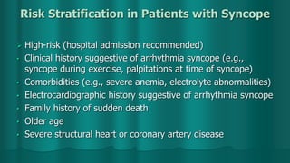
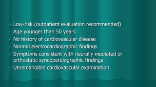
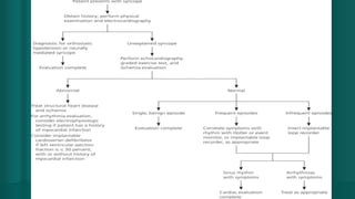
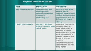
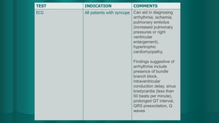
APPROACH TO SYNCOPE
SUPERVISOR :DR HO YI BIN
PRESENTER : ALI HUSAIN
FIRDAUS
SYNCOPE
 Motionless and limp and usually has cool extremities, a
weak pulse, and shallow breathing. Sometimes brief
involuntary muscle jerks occur, resembling a seizure.
NEAR SYNCOPE
 Near-syncope is light-headedness and a sense of an
impending faint without LOC. It is usually classified and
discussed with syncope because the causes are the same
PATOPHYSIOLOGY
 Insufficient cerebral blood flow. Some cases involve adequate flow but with insufficient
cerebral substrate (oxygen, glucose, or both).
 Most deficiencies in cerebral blood flow result from decreased cardiac output (CO).
Decreased CO can be caused by
1. 
Cardiac disorders that obstruct outflow
2. 
Cardiac disorders of systolic dysfunction
3. 
Cardiac disorders of diastolic dysfunction
4. 
Arrhythmias (too fast or too slow)
5. 
Conditions that decrease venous return
Insufficient cerebral substrate
 The CNS requires oxygen and glucose to function.
 Hypoglycemia is the primary cause because hypoxia
rarely develops in a manner causing abrupt LOC (other
than in flying or diving incidents). LOC due to
hypoglycemia is seldom as abrupt as in syncope or
seizures because warning symptoms occur
ETIOLOGY
 The most common causes are

Vasovagal (neurocardiogenic)

Idiopathic
EVALUATION
 History
 Physical examinations
 Interpretation of findings
 Testing
History of present illness
 Patient’s activity (eg, exercising, arguing, in a potentially
emotional situation), position (eg, lying or standing), and,
if standing, for how long.
 Sense of impending LOC, nausea, sweating, blurred or
tunnel vision, tingling of lips or fingertips, chest pain, or
palpitations.
 Length of time recovering should also be ascertained.
Witnesses, if any, should be sought and asked to describe
events, particularly the presence and duration of any
seizure activity.
 Asked about symptoms suggesting possible causes,
including bloody or tarry stools, heavy menses (anemia);
vomiting, diarrhea, or excess urination (dehydration or
electrolyte abnormalities)
Past medical history

 Ask about previous syncopal events, known
cardiovascular disease, and known seizure disorders.
 Drugs used should be identified (particularly
antihypertensives, diuretics, vasodilators, and
antiarrhythmics
 Family history
PHYSICAL EXAMINATIONS
 Vital signs are essential. Heart rate and BP are measured
with the patient supine and after 3min of standing. Pulse
is palpated for irregularity.
 General examination notes patient’s mental status,
including any confusion or hesitancy suggesting a
postictal state and any signs of injury (eg, bruising,
swelling, tenderness, tongue bite).
 The heart is auscultated for murmurs; if present, any
change in the murmur with a Valsalva maneuver,
standing, or squatting is noted.
 Abdomen is palpated for tenderness, and a rectal
examination is done to check for gross or occult blood.
 A full neurologic examination is done to identify any focal
abnormalities, which suggest a CNS cause (eg, seizure
disorder)
BENIGN CAUSES
 Syncope precipitated by unpleasant physical or emotional
stimuli (eg, pain, fright), usually occurring in the upright
position and often preceded by vagally mediated warning
symptoms (eg, nausea, weakness, yawning,
apprehension, blurred vision, diaphoresis), suggests
vasovagal syncope.
 Syncope that occurs most often when assuming an
upright position (particularly in elderly patients after
prolonged bed rest or in patients taking drugs in certain
classes) suggests orthostatic syncope.
 Syncope that occurs after standing for long periods
without moving is usually due to venous pooling.
 LOC that is abrupt in onset; is associated with muscular
jerking or convulsions that last more than a few seconds,
incontinence, drooling, or tongue biting; and is followed
by postictal confusion or somnolence suggests a seizure.
DANGEROUS CAUSES
 Syncope with exertion suggests cardiac outflow
obstruction or exercise-induced arrhythmia. Such patients
sometimes also have chest pain, palpitations, or both.
 
Syncope that begins and ends suddenly and
spontaneously is typical of cardiac causes, most
commonly an arrhythmia.
 Syncope while lying down also suggests an arrhythmia
because vasovagal and orthostatic mechanisms do not
cause syncope in the recumbent position.
 
Syncope accompanied by injury during the episode
increases the likelihood of a cardiac cause or seizure
somewhat, and therefore the event is of greater concern.
RED FLAGS
Certain findings suggest a more serious etiology:
 Syncope during exertion
 
Multiple recurrences within a short time
 
Heart murmur or other findings suggesting structural heart disease (eg, chest pain)
 
Older age
 
Significant injury during syncope
 
Family history of sudden unexpected death, exertional syncope, or unexplained recurrent
syncope or seizures
TESTING
 
ECG
 
Pulse oximetry
 
Sometimes echocardiography
 
Sometimes tilt table testing
 
Blood tests only if clinically indicated
 
CNS imaging rarely indicated
CLASSIFICATION EXAMPLES SCENARIO
Cardiac Arrhythmia (e.g.,
bradyarrhythmias,
ventricular
tachyarrhythmias,
supraventricular
tachyarrhythmias, long
QT syndrome),
pacemaker dysfunction
Generally abrupt and
unprovoked, palpitations
may precede symptoms
Obstructive
cardiomyopathy
Hypertrophic
cardiomyopathy
Structural disease
(cardiac)
Aortic stenosis,
Pulmonary stenosis,
Acute myocardial
infarction/ischemia
Structural disease
(other)
Pulmonary embolus,
Acute aortic dissection,
Pulmonary hypertension
CLASSIFICATION EXAMPLES SCENARIO
Neurally mediated (reflex) Carotid sinus
syndrome/hypersensitivity
Head rotation or pressure on the
carotid sinus (e.g., shaving, tight
collar) can reproduce symptoms;
consider in patients with
unexplained falls
Situational Micturition, post-exercise,
postprandial, gastrointestinal
stimulation, cough, phobia of
needle or blood
Vasovagal Mediated by stress, fear, noxious
stimuli, heat exposure
CLASSIFICATION EXAMPLES SCENARIO
Neurologic/miscellaneou
s
Cerebrovascular Induced by a steal
syndrome
Neurogenic Preceding transient
ischemic
attack/cerebrovascular
injury symptoms; severe
basilar artery disease
Psychogenic Depression, anxiety,
panic disorder,
somatization disorders
CLASSIFICATION EXAMPLES SCENARIO
Orthostatic Drug-induced Alcohol, insulin or
antidiabetic agents,
antihypertensives,
antianginals,
antidepressants,
antiparkinsonian agents
Primary autonomic failure Parkinson
disease/parkinsonism,
multiple system atrophy
(i.e., Shy-Drager
syndrome), multiple
sclerosis, Wernicke
encephalopathy
Secondary autonomic
failure
Diabetes mellitus,
amyloidosis, uremia,
spinal cord injury, chronic
inflammatory
polyneuropathy,
connective tissue diseases
Volume depletion Vomiting, diarrhea, poor
intake, acute blood loss
Risk Stratification in Patients with Syncope
 High-risk (hospital admission recommended)
• Clinical history suggestive of arrhythmia syncope (e.g.,
syncope during exercise, palpitations at time of syncope)
• Comorbidities (e.g., severe anemia, electrolyte abnormalities)
• Electrocardiographic history suggestive of arrhythmia syncope
• Family history of sudden death
• Older age
• Severe structural heart or coronary artery disease
 Low-risk (outpatient evaluation recommended)
• Age younger than 50 years
• No history of cardiovascular disease
• Normal electrocardiographic findings
• Symptoms consistent with neurally mediated or
orthostatic syncoperdiographic findings
• Unremarkable cardiovascular examination
TEST INDICATION COMMENTS
Basic laboratory testing As clinically indicated,
including human
chorionic gonadotropin
in women of
childbearing age
Laboratory evaluation
rarely is helpful;
complete blood count for
anemia; brain natriuretic
peptide testing may be
beneficial for cardiac
etiology
Carotid sinus massage Syncope of unknown
etiology in patients older
than 40 years*
Diagnostic if ventricular
pause is more than
three seconds or if a
decrease in systolic
blood pressure > 50 mm
Hg, Contraindicated in
patients with bruits or a
history of transient
ischemic
attack/cerebrovascular
accident within the past
three months
Diagnostic Evaluation of Syncope
TEST INDICATION COMMENTS
ECG All patients with syncope Can aid in diagnosing
arrhythmia, ischemia,
pulmonary embolus
(increased pulmonary
pressures or right
ventricular
enlargement),
hypertrophic
cardiomyopathy,
Findings suggestive of
arrhythmia include
presence of bundle
branch block,
intraventricular
conduction delay, sinus
bradycardia (less than
50 beats per minute),
prolonged QT interval,
QRS preexcitation, Q
waves
TEST INDICATION COMMENTS
Recurrent syncope with
unremarkable initial
evaluation; clinical or
ECG features suggestive
of arrhythmic syncope;
patients with
unexplained falls*
Holter monitor for 24 to
48 hours, event
recorders for 30 to 60
days, implantable
recorders for up to 14
months,
Consider testing in
patients suspected of
having epilepsy not
responsive to therapy
Echocardiography When history,
examination, and ECG
do not provide a
diagnosis or if structural
cardiac disease is
suspected
Diagnostic in aortic
stenosis, pericardial
tamponade, obstructive
cardiac tumors or
thrombi, aortic
dissection, hypertrophic
cardiomyopathy,
congenital anomalies of
the coronary arteries
TEST INDICATION COMMENTS
Electrophysiology Patients with coronary
artery disease after
ischemic evaluation,
nonischemic dilated
cardiomyopathy, bundle
branch block,* syncope
preceded by
palpitations, Brugada
syndrome,
arrhythmogenic right
ventricular
dysplasia/cardiomyopath
y, or high-risk
occupations
Not recommended in
patients without
underlying heart disease
Consider in high-risk
patients with recurrent
unexplained syncope
Exercise testing Hemodynamic and ECG
abnormalities present
with syncope during
exercise, syncope
reproduced with
exercise, precipitate a
Mobitz type II second-
or third-degree block
Inadequate rise of blood
pressure in younger
patients is suggestive of
hypertrophic
cardiomyopathy or left
main disease; similar
findings in older persons
may suggest autonomic
TEST INDICATION COMMENTS
Neurologic testing Suspicious for seizures,
cerebrovascular event,
neurodegenerative
disorders, increased
intracranial pressure
Seizure can be
confirmed with
electroencephalography
Cranial imaging studies
as clinically indicated
Orthostatic blood
pressure
Evaluate neurally
mediated syncope from
orthostatic hypotension*
Diagnostic if decrease in
systolic blood pressure ≥
20 mm Hg; if systolic
blood pressure < 90 mm
Hg; or if decrease in
diastolic blood pressure
≥ 10 mm Hg with
symptoms
Consider diagnostic
even when patient is
asymptomatic
REFERENCE
1. Parry SW, Tan MP. An approach to the evaluation and
management of syncope in adults. BMJ. 2010;340:c880.
2. John Murtagh’s Patient Education, Fifth edition
3. Andrea D. Thompson
, MD, PhD, Department of Internal Medicine, Division of
Cardiovascular Medicine, University of Michigan;
4.Michael J. Shea , MD, Michigan Medicine at the University
of Michigan

Aproach to syncope

  • 1.
    APPROACH TO SYNCOPE SUPERVISOR:DR HO YI BIN PRESENTER : ALI HUSAIN FIRDAUS
  • 2.
    SYNCOPE  Motionless andlimp and usually has cool extremities, a weak pulse, and shallow breathing. Sometimes brief involuntary muscle jerks occur, resembling a seizure.
  • 3.
    NEAR SYNCOPE  Near-syncopeis light-headedness and a sense of an impending faint without LOC. It is usually classified and discussed with syncope because the causes are the same
  • 4.
    PATOPHYSIOLOGY  Insufficient cerebralblood flow. Some cases involve adequate flow but with insufficient cerebral substrate (oxygen, glucose, or both).  Most deficiencies in cerebral blood flow result from decreased cardiac output (CO). Decreased CO can be caused by 1. 
Cardiac disorders that obstruct outflow 2. 
Cardiac disorders of systolic dysfunction 3. 
Cardiac disorders of diastolic dysfunction 4. 
Arrhythmias (too fast or too slow) 5. 
Conditions that decrease venous return
  • 5.
    Insufficient cerebral substrate The CNS requires oxygen and glucose to function.  Hypoglycemia is the primary cause because hypoxia rarely develops in a manner causing abrupt LOC (other than in flying or diving incidents). LOC due to hypoglycemia is seldom as abrupt as in syncope or seizures because warning symptoms occur
  • 6.
    ETIOLOGY  The mostcommon causes are 
Vasovagal (neurocardiogenic) 
Idiopathic
  • 8.
    EVALUATION  History  Physicalexaminations  Interpretation of findings  Testing
  • 9.
    History of presentillness  Patient’s activity (eg, exercising, arguing, in a potentially emotional situation), position (eg, lying or standing), and, if standing, for how long.  Sense of impending LOC, nausea, sweating, blurred or tunnel vision, tingling of lips or fingertips, chest pain, or palpitations.
  • 10.
     Length oftime recovering should also be ascertained. Witnesses, if any, should be sought and asked to describe events, particularly the presence and duration of any seizure activity.  Asked about symptoms suggesting possible causes, including bloody or tarry stools, heavy menses (anemia); vomiting, diarrhea, or excess urination (dehydration or electrolyte abnormalities)
  • 11.
    Past medical history  Ask about previous syncopal events, known cardiovascular disease, and known seizure disorders.  Drugs used should be identified (particularly antihypertensives, diuretics, vasodilators, and antiarrhythmics  Family history
  • 12.
    PHYSICAL EXAMINATIONS  Vitalsigns are essential. Heart rate and BP are measured with the patient supine and after 3min of standing. Pulse is palpated for irregularity.  General examination notes patient’s mental status, including any confusion or hesitancy suggesting a postictal state and any signs of injury (eg, bruising, swelling, tenderness, tongue bite).
  • 13.
     The heartis auscultated for murmurs; if present, any change in the murmur with a Valsalva maneuver, standing, or squatting is noted.  Abdomen is palpated for tenderness, and a rectal examination is done to check for gross or occult blood.  A full neurologic examination is done to identify any focal abnormalities, which suggest a CNS cause (eg, seizure disorder)
  • 14.
    BENIGN CAUSES  Syncopeprecipitated by unpleasant physical or emotional stimuli (eg, pain, fright), usually occurring in the upright position and often preceded by vagally mediated warning symptoms (eg, nausea, weakness, yawning, apprehension, blurred vision, diaphoresis), suggests vasovagal syncope.
  • 15.
     Syncope thatoccurs most often when assuming an upright position (particularly in elderly patients after prolonged bed rest or in patients taking drugs in certain classes) suggests orthostatic syncope.  Syncope that occurs after standing for long periods without moving is usually due to venous pooling.
  • 16.
     LOC thatis abrupt in onset; is associated with muscular jerking or convulsions that last more than a few seconds, incontinence, drooling, or tongue biting; and is followed by postictal confusion or somnolence suggests a seizure.
  • 17.
    DANGEROUS CAUSES  Syncopewith exertion suggests cardiac outflow obstruction or exercise-induced arrhythmia. Such patients sometimes also have chest pain, palpitations, or both.  
Syncope that begins and ends suddenly and spontaneously is typical of cardiac causes, most commonly an arrhythmia.
  • 18.
     Syncope whilelying down also suggests an arrhythmia because vasovagal and orthostatic mechanisms do not cause syncope in the recumbent position.  
Syncope accompanied by injury during the episode increases the likelihood of a cardiac cause or seizure somewhat, and therefore the event is of greater concern.
  • 19.
    RED FLAGS Certain findingssuggest a more serious etiology:  Syncope during exertion  
Multiple recurrences within a short time  
Heart murmur or other findings suggesting structural heart disease (eg, chest pain)  
Older age  
Significant injury during syncope  
Family history of sudden unexpected death, exertional syncope, or unexplained recurrent syncope or seizures
  • 20.
    TESTING  
ECG  
Pulseoximetry  
Sometimes echocardiography  
Sometimes tilt table testing  
Blood tests only if clinically indicated  
CNS imaging rarely indicated
  • 21.
    CLASSIFICATION EXAMPLES SCENARIO CardiacArrhythmia (e.g., bradyarrhythmias, ventricular tachyarrhythmias, supraventricular tachyarrhythmias, long QT syndrome), pacemaker dysfunction Generally abrupt and unprovoked, palpitations may precede symptoms Obstructive cardiomyopathy Hypertrophic cardiomyopathy Structural disease (cardiac) Aortic stenosis, Pulmonary stenosis, Acute myocardial infarction/ischemia Structural disease (other) Pulmonary embolus, Acute aortic dissection, Pulmonary hypertension
  • 22.
    CLASSIFICATION EXAMPLES SCENARIO Neurallymediated (reflex) Carotid sinus syndrome/hypersensitivity Head rotation or pressure on the carotid sinus (e.g., shaving, tight collar) can reproduce symptoms; consider in patients with unexplained falls Situational Micturition, post-exercise, postprandial, gastrointestinal stimulation, cough, phobia of needle or blood Vasovagal Mediated by stress, fear, noxious stimuli, heat exposure
  • 23.
    CLASSIFICATION EXAMPLES SCENARIO Neurologic/miscellaneou s CerebrovascularInduced by a steal syndrome Neurogenic Preceding transient ischemic attack/cerebrovascular injury symptoms; severe basilar artery disease Psychogenic Depression, anxiety, panic disorder, somatization disorders
  • 24.
    CLASSIFICATION EXAMPLES SCENARIO OrthostaticDrug-induced Alcohol, insulin or antidiabetic agents, antihypertensives, antianginals, antidepressants, antiparkinsonian agents Primary autonomic failure Parkinson disease/parkinsonism, multiple system atrophy (i.e., Shy-Drager syndrome), multiple sclerosis, Wernicke encephalopathy Secondary autonomic failure Diabetes mellitus, amyloidosis, uremia, spinal cord injury, chronic inflammatory polyneuropathy, connective tissue diseases Volume depletion Vomiting, diarrhea, poor intake, acute blood loss
  • 25.
    Risk Stratification inPatients with Syncope  High-risk (hospital admission recommended) • Clinical history suggestive of arrhythmia syncope (e.g., syncope during exercise, palpitations at time of syncope) • Comorbidities (e.g., severe anemia, electrolyte abnormalities) • Electrocardiographic history suggestive of arrhythmia syncope • Family history of sudden death • Older age • Severe structural heart or coronary artery disease
  • 26.
     Low-risk (outpatientevaluation recommended) • Age younger than 50 years • No history of cardiovascular disease • Normal electrocardiographic findings • Symptoms consistent with neurally mediated or orthostatic syncoperdiographic findings • Unremarkable cardiovascular examination
  • 28.
    TEST INDICATION COMMENTS Basiclaboratory testing As clinically indicated, including human chorionic gonadotropin in women of childbearing age Laboratory evaluation rarely is helpful; complete blood count for anemia; brain natriuretic peptide testing may be beneficial for cardiac etiology Carotid sinus massage Syncope of unknown etiology in patients older than 40 years* Diagnostic if ventricular pause is more than three seconds or if a decrease in systolic blood pressure > 50 mm Hg, Contraindicated in patients with bruits or a history of transient ischemic attack/cerebrovascular accident within the past three months Diagnostic Evaluation of Syncope
  • 29.
    TEST INDICATION COMMENTS ECGAll patients with syncope Can aid in diagnosing arrhythmia, ischemia, pulmonary embolus (increased pulmonary pressures or right ventricular enlargement), hypertrophic cardiomyopathy, Findings suggestive of arrhythmia include presence of bundle branch block, intraventricular conduction delay, sinus bradycardia (less than 50 beats per minute), prolonged QT interval, QRS preexcitation, Q waves
  • 30.
    TEST INDICATION COMMENTS Recurrentsyncope with unremarkable initial evaluation; clinical or ECG features suggestive of arrhythmic syncope; patients with unexplained falls* Holter monitor for 24 to 48 hours, event recorders for 30 to 60 days, implantable recorders for up to 14 months, Consider testing in patients suspected of having epilepsy not responsive to therapy Echocardiography When history, examination, and ECG do not provide a diagnosis or if structural cardiac disease is suspected Diagnostic in aortic stenosis, pericardial tamponade, obstructive cardiac tumors or thrombi, aortic dissection, hypertrophic cardiomyopathy, congenital anomalies of the coronary arteries
  • 31.
    TEST INDICATION COMMENTS ElectrophysiologyPatients with coronary artery disease after ischemic evaluation, nonischemic dilated cardiomyopathy, bundle branch block,* syncope preceded by palpitations, Brugada syndrome, arrhythmogenic right ventricular dysplasia/cardiomyopath y, or high-risk occupations Not recommended in patients without underlying heart disease Consider in high-risk patients with recurrent unexplained syncope Exercise testing Hemodynamic and ECG abnormalities present with syncope during exercise, syncope reproduced with exercise, precipitate a Mobitz type II second- or third-degree block Inadequate rise of blood pressure in younger patients is suggestive of hypertrophic cardiomyopathy or left main disease; similar findings in older persons may suggest autonomic
  • 32.
    TEST INDICATION COMMENTS Neurologictesting Suspicious for seizures, cerebrovascular event, neurodegenerative disorders, increased intracranial pressure Seizure can be confirmed with electroencephalography Cranial imaging studies as clinically indicated Orthostatic blood pressure Evaluate neurally mediated syncope from orthostatic hypotension* Diagnostic if decrease in systolic blood pressure ≥ 20 mm Hg; if systolic blood pressure < 90 mm Hg; or if decrease in diastolic blood pressure ≥ 10 mm Hg with symptoms Consider diagnostic even when patient is asymptomatic
  • 33.
    REFERENCE 1. Parry SW,Tan MP. An approach to the evaluation and management of syncope in adults. BMJ. 2010;340:c880. 2. John Murtagh’s Patient Education, Fifth edition 3. Andrea D. Thompson , MD, PhD, Department of Internal Medicine, Division of Cardiovascular Medicine, University of Michigan; 4.Michael J. Shea , MD, Michigan Medicine at the University of Michigan

Editor's Notes

  • #26 ECG of arrhythmia -bifascicular block, sinus bradycardia < 40 beats per minute in absence of sinoatrial block or medications, preexcited QRS complex, abnormal QT interval, ST segment elevation leads V1 through V3 [Brugada syndrome], negative T wave in right precordial leads and epsilon wave [arrhythmogenic right ventricular dysplasia/cardiomyopathy])