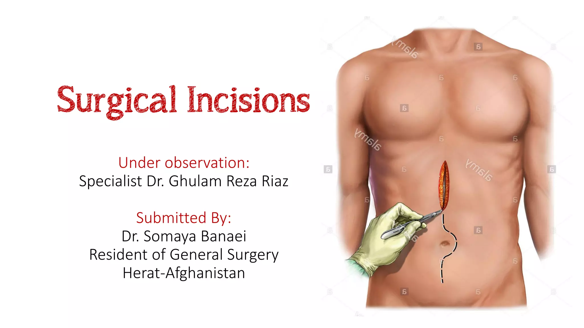
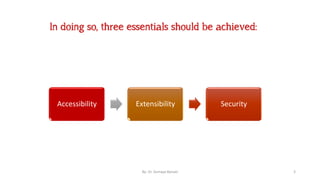
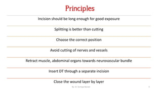
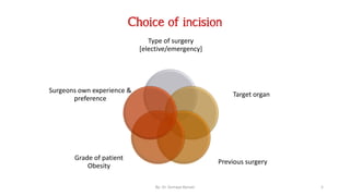
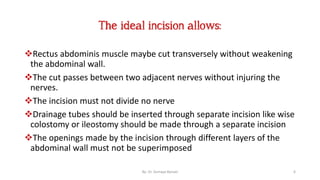
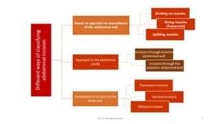
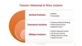
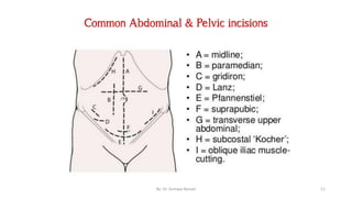
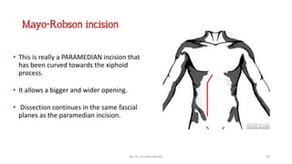
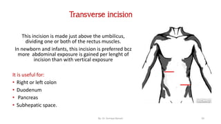
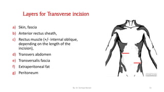
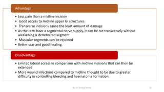
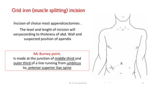
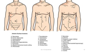
This document discusses surgical incisions, including:
- The importance of choosing an incision that provides accessibility, extensibility, and security for the procedure.
- Common abdominal and pelvic incisions such as vertical, transverse, oblique, midline, paramedian, Pfannenstiel, and flank incisions.
- Factors to consider when choosing an incision, including the type of surgery, target organ, patient characteristics, and surgeon preference.
- Principles for making incisions such as following skin cleavage lines and avoiding cutting nerves and vessels.
The document discusses various types of abdominal incisions used in surgery. It describes incisions like vertical incisions (median, paramedian), transverse incisions (Kocher, McBurney), and pelvic incisions (Pfannenstiel, Maylard). The ideal incision allows easy access to structures, can be extended if needed, and heals with minimal scarring. Factors like the surgery type, target organ, and patient characteristics influence the choice of incision. Complications can include hematoma, wound infections, and hernia.
The document discusses various types of surgical incisions including their purposes, advantages, and disadvantages. It describes abdominal and pelvic incisions such as midline, paramedian, transverse, oblique, Kochler subcostal, McBurney, Pfannenstiel, and Maylard incisions. Langer's lines, which correspond to the natural orientation of collagen fibers, are also mentioned as incisions made parallel to these lines may result in better healing and less scarring. Key layers of the abdominal wall including skin, fascia, muscles and peritoneum are also defined.
LAPAROSCOPIC CHOLECYSTECTOMY- OPERATIVE SURGERY
#surgicaleducator #operativesurgery #laparoscopiccholecystectomy #usmle #babysurgeon #surgicaltutor
Dear viewers,
• Greetings from “Surgical Educator”
• Because of the popular demand by viewers of the YouTube channel “Surgical Educator”, I have decided to create and upload videos on common surgeries.
• I have already uploaded videos on open and Laparoscopic Appendicectomy, Thyroidectomy, Modified Radical Mastectomy and Open Cholecystectomy.
• In this video today, I have discussed Laparoscopic Cholecystectomy- the flagship procedure for laparoscopic surgeries.
• However, these videos are not real surgeries but the theoretical aspect of operative surgery like going through an atlas of operative surgery.
• Along with these videos, I recommend you to watch real operative surgery videos as well and the links are:
• https://www.youtube.com/watch?v=VStEzI1jL8Y
• https://www.youtube.com/watch?v=O8j4kwpzd24
• This will give a very good opportunity for the surgical trainees to mentally rehearse various surgical steps in a sequential manner prior to actual surgery. You can watch the video in the following links:
• surgicaleducator.blogspot.com
• youtube.com/c/surgicaleducator
This document discusses splenectomy, the surgical removal of the spleen. It defines splenectomy and outlines the relevant anatomy of the spleen. The document then discusses the indications for splenectomy, including trauma, hematological disorders, tumors, and vascular abnormalities. It covers the preoperative preparation, anesthesia, positioning, exposure, closure, and postoperative management of splenectomy. Finally, it lists some potential complications of splenectomy.
An exploratory laparotomy is a surgical procedure where the abdomen is opened to examine the internal organs. It is performed under general anesthesia through an upper midline incision. Indications include trauma, infections, malignancy, complications of other procedures, and removal of foreign bodies. During the procedure, the surgeon examines the abdominal organs and treats any issues found. Potential complications include ileus, infection, hernia, and adhesive obstruction.
A modified radical mastectomy removes the breast tissue, nipple, skin, and lymph nodes in levels I and II of the axilla, while the Patey modification also removes the pectoralis minor muscle to access level III lymph nodes; complications can include lymphedema if radiation is also used post-surgery or seromas which are usually drained with needles though drains placed during surgery help prevent them. The procedure involves dissecting the breast from the chest wall and axilla while preserving key nerves and blood vessels.
Burst abdomen, or postoperative separation of an abdominal wound, occurs most commonly between the 5th and 8th postoperative days when wound strength is weakest. It can be partial or complete, and risk factors include preexisting malnutrition or disease, operative issues like poor technique or closure, and post-operative infections. Treatment depends on severity but may involve reapproximating the wound with sutures or a temporary dressing, with prevention prioritizing proper technique, antibiotics, and minimizing intra-abdominal pressure increases.
A nephrectomy is a surgical procedure to remove a kidney. There are several types including simple, partial, and radical nephrectomies. A surgeon must have knowledge of renal anatomy and vasculature. Approaches can be open, laparoscopic, or robotic. Key steps include mobilizing the kidney, isolating and ligating the renal vessels, and closing fascial layers. Complications include bleeding, fistula, and loss of renal function.
An incision in the abdomen is an opening or a cut made by the surgeon. An incision in the abdomen is an opening or a cut made by the surgeon. It is done to permit access to abdominal organs for surgery. The selection of an incision depends on. Underlying condition prompting the surgery.
The document discusses various types of abdominal incisions used in surgery. It describes midline, vertical, transverse, and oblique incisions. Midline incisions provide good access but have a higher risk of hernia. Transverse incisions have better cosmetic outcomes and less risk of complications like hernia compared to vertical incisions. Specific incisions discussed include Kocher for gallbladder surgery, McBurney for appendicectomy, and Pfannenstiel for pelvic operations. Factors affecting incision healing and potential complications are also outlined.
This document discusses prostatectomy procedures including simple and radical prostatectomy. Simple prostatectomy involves removing part of the prostate for benign conditions, while radical prostatectomy removes the entire prostate and surrounding tissues for prostate cancer. The document describes different approaches for radical prostatectomy including radical perineal, supra pubic, and retro pubic. Key instruments used in prostatectomy are also listed such as retractors, forceps, scissors, and hemoclip appliers.
This document provides information on different types of gastric resection surgeries including wedge resection, distal gastrectomy, total gastrectomy, and subtotal gastrectomy. It describes the anatomy of the stomach and surrounding structures. It details the surgical techniques for each type of resection including mobilization, resection, and reconstruction. Common indications for gastric resections are described as peptic ulcer disease and gastric tumors. The history of developments in gastric surgery techniques from the late 19th century onward is also summarized.
1. The document discusses various types of abdominal incisions used in surgery, including midline, para-median, transverse, McBurney, Pfannenstiel, and thoracoabdominal incisions.
2. It provides details on the advantages and disadvantages of each incision type, describing their typical uses in different surgical procedures.
3. The locations and techniques for making each incision are outlined. Midline incisions are described as being along the linea alba, while para-median incisions are made 2-5cm lateral to the umbilicus.
This document provides information on hernias, including their meaning, causes, parts, classifications, and inguinal hernia anatomy and types. Some key points:
1. A hernia is an abnormal protrusion of an organ or tissue through an opening. It is usually defined as a protrusion through the abdominal wall.
2. Hernias can be caused by straining, heavy lifting, coughing, obesity, pregnancy, smoking, and other factors that increase intra-abdominal pressure.
3. Inguinal hernias are the most common type and are classified as direct or indirect based on their anatomy through the inguinal canal.
4. Treatment of hernias
Debridement is an important component of the wound bed preparation (WBP) management Model.
Cause of the wound and patient-centered concerns, debridement is a necessary step in local wound care.
Debridement is the removal of necrotic tissue, exudate, bacteria, and metabolic waste from a wound in order to improve or facilitate the healing process
This document provides information about breast surgery and breast cancer. It discusses the anatomy of the breast including its structure, blood supply, and lymphatic drainage. It also covers common benign breast diseases such as fibroadenoma, duct papilloma, and breast abscess. The document discusses clinical assessment of breast cancer including history, examination, and investigations. It provides details on TNM staging and pathological classification of breast cancer. Finally, it describes different surgical procedures for breast cancer including simple mastectomy, modified radical mastectomy, and breast conserving surgery.
The document provides an outline for performing an open appendectomy surgery. It discusses the relevant anatomy of the appendix, causes of appendicitis, pre-operative care including investigations and antibiotic treatment. It describes the surgical technique including common incisions used, identifying and ligating the appendix and closing the wound. Post-operative care involves monitoring for complications and managing patients depending on whether the case was complicated or uncomplicated.
- Intestinal stomas are surgically created openings of the small or large intestine onto the abdominal wall. There are three main types: colostomy, ileostomy, and loop stoma.
- Complications include prolapse, herniation, stenosis, dermatitis from effluent, and obstruction. Dietary advice focuses on reducing gas, bulk and odorous foods. Management involves properly attaching collection bags and monitoring for complications.
1. Acute appendicitis is caused by obstruction of the appendix lumen, leading to increased intraluminal pressure, edema, and bacterial invasion.
2. The classic presentation includes initially vague periumbilical pain that later localizes to the right lower quadrant, accompanied by anorexia, nausea, and low-grade fever.
3. On examination, tenderness is elicited over McBurney's point with guarding and rebound tenderness. Diagnosis is suggested by clinical scoring systems and confirmed by ultrasound or CT scan showing a thick-walled, inflamed appendix.
Whipple's procedure - Indications, Steps, ComplicationsVikas V
The document describes the Whipple procedure, which was first performed by Dr. Allen Whipple in 1935. It involves removing the head of the pancreas, part of the small intestine, the gallbladder, and bile duct. The original procedure was done in two stages but is now typically done in one stage. The document outlines the key steps of the modern Whipple procedure, including mobilizing tissues, dividing vessels, transecting organs, and reconstructing the digestive and biliary systems with anastomoses. Vascular resection of veins like the splenic vein may sometimes be required as well.
This document describes the procedure for a right hemicolectomy. It involves making a midline abdominal incision, mobilizing the terminal ileum and cecum, ligating and dividing the ileocolic, right colic, and middle colic vessels, removing the ascending colon and portions of the ileum and transverse colon. An end-to-side anastomosis is then performed between the ileum and transverse colon using interrupted sutures in two layers to reconnect the bowel.
This document describes the open cholecystectomy procedure. It indicates that open cholecystectomy is performed to treat conditions like cholecystitis, cholelithiasis, and choledocholithiasis. It outlines the patient preparation, incision type (typically a right subcostal incision), and technique which involves dissecting and ligating/clipping the cystic duct and artery before removing the gallbladder. Potential complications of the open procedure include bleeding, infection, and bile leaks or common bile duct injuries.
This document provides information on the management of inguinal hernias. It discusses the historical development of hernia repair techniques from the 15th century to modern methods. Investigation methods such as ultrasound, CT, MRI, and herniography are outlined. Surgical techniques for hernia repair including herniotomy, herniorrhaphy, hernioplasty, and laparoscopic repair are described in detail. Post-operative complications of open and laparoscopic hernia repair are also reviewed. The conclusion states that laparoscopic and Lichtenstein open mesh repairs have good long-term results and low recurrence rates compared to other open hernia repair techniques.
This document provides an overview of inguinal hernias. It defines a hernia as the protrusion of an organ through a weakness in the muscle wall. It then discusses the epidemiology, types, anatomy, etiology, pathogenesis, signs and symptoms, diagnosis, treatment and complications of inguinal hernias. The treatment options covered are herniotomy, herniorrhaphy, and laparoscopic repair techniques like the Lichtenstein method. The prognosis is typically good, though there is a small risk of recurrence.
Circumcision is the removal of the foreskin from the penis. It is one of the oldest planned operations, dating back 15,000 years. It is commonly performed for religious reasons in Judaism and Islam or for various medical reasons like phimosis, paraphimosis, and infection. The operation involves cutting off the foreskin and sometimes using plastic clamps. Patients are usually able to leave hospital shortly after and should avoid sex and strenuous activity for around 4 weeks to allow healing. Circumcision is contraindicated for preterm infants or those with bleeding disorders or genital abnormalities.
This document provides information about amputation procedures. It describes the indications for amputation, including peripheral vascular disease, diabetic limb disease, trauma, infection, malignancy, and deformity. The goals of amputation are to return the patient to maximum function, ablate diseased tissue, reduce morbidity and mortality, and produce a physiological end organ. Different types of amputations are described for the toes, feet, legs, arms, and limbs. Principles for determining the amputation level and performing the procedure are outlined. Postoperative management focuses on wound healing, edema control, pain management, and rehabilitation to prevent contractures. Potential complications are also reviewed.
1) The document discusses different types of surgical incisions including vertical, transverse, oblique, and flank incisions. It notes key details about each incision like preferred procedures, layers cut, advantages, and disadvantages.
2) Complications from surgical incisions are categorized as immediate, early, or late. Immediate issues include bleeding, organ injury, and anesthesia problems. Early complications involve infections, fluid buildup, and wound separation. Late complications comprise hernia, bowel obstructions, pain, and abnormal healing.
3) Selection of an incision depends on factors like the surgery, patient characteristics, and surgeon preference. Considerations include visibility, extension ability, and minimizing trauma to structures like nerves and blood
Surgical incisions are cuts made through the skin to access organs for operations or procedures. The ideal incision allows easy access to structures, can be extended if needed, and heals quickly with minimal scarring. There are various types of abdominal and pelvic incisions that are chosen based on the surgery, target organ, surgeon's preference, and previous operations. Examples include vertical, transverse, oblique, and muscle-splitting incisions. Factors like wound closure technique and patient health can impact the strength of the resulting scar.
'Surgical Incisions on Abdominal Wall', a Surgical Anatomy Seminar by 1st yr MBBS students of Venkateswara Institute of Medical Science, Galraula, UP. India
An incision is a cut made by a surgeon during surgery. This document describes and compares various types of abdominal incisions including their advantages and disadvantages. Midline incisions provide good access but are cosmetically disapproved. Para-median incisions have weaker muscle repair but access lateral structures. Transverse incisions have the best cosmetic results but take more time. McBurney incisions are best for appendicectomies while Pfannenstiel incisions are commonly used in gynecological and obstetric surgeries.
This document describes various abdominal incisions used in surgery. It discusses the layers of the abdominal wall and names several types of incisions including vertical, transverse, Pfannenstiel, Cherney, Maylard, McBurney and Kochler incisions. For each incision, it provides details on how it is made and indications for its use. It also discusses advantages and disadvantages of the incisions as well as complications that can occur with abdominal incisions. Lastly, it provides tips for proper closure of abdominal incisions.
1. The document discusses various incisions used for abdominal access during surgery.
2. It describes midline, paramedian, transverse, subcostal, McBurney's and Lanz incisions, noting the layers of tissue each passes through and advantages and disadvantages.
3. The ideal incision allows ease of access while minimizing damage to muscles, nerves and risk of infection post-surgery. Location and orientation of the incision depends on the target structures.
Tipo de incisiones abdominales. Cirugía.DeivisGarcia8
This document discusses different types of abdominal incisions used in surgery. It describes midline, paramedian, transverse, Pfannenstiel's, Cherney's, Maylard's, and modified Gibson incisions. The key factors considered in selecting an incision are accessibility to the organ, extensibility of the incision, preservation of abdominal wall function, and secure closure. Complications can include hematoma, infection, wound dehiscence, hernia, and scarring.
This document discusses episiotomy, which is a surgically planned incision made in the perineum and posterior vaginal wall during the second stage of labor. It aims to enlarge the vaginal opening to facilitate delivery and minimize tearing. Common indications include a rigid perineum, operative deliveries, or previous perineal surgery. The main types are mediolateral, median, and lateral incisions. A mediolateral episiotomy has advantages like less blood loss and easier repair compared to other types. The procedure involves preliminaries like anesthesia, followed by the incision and then repair of the vaginal mucosa, muscles, and skin in layers. Post-operative care and potential complications are also
Surgical Options In The Management Of Hernia Repairsafarmas
This document outlines surgical options for inguinal hernias. It discusses the definition and types of hernias, including inguinal and femoral hernias. For surgical management, it describes open hernia repair techniques like Bassini, Shouldice, and tension-free repairs using mesh, as well as laparoscopic approaches like TAPP and TEP. Complications are also outlined. The goal of hernia surgery is to reduce hernia contents and repair the defect using herniorrhaphy or hernioplasty techniques to minimize recurrence.
Recent advances in minimal access surgery.pptxManoj H.V
This document summarizes recent advances in minimally invasive surgery techniques. It discusses laparoscopic inguinal hernia repair procedures like transabdominal preperitoneal repair and total extraperitoneal repair. It also describes newer natural orifice transluminal endoscopic surgery techniques, bikini line laparoscopic cholecystectomy, and transanal total mesorectal excision for rectal cancer surgery. The document provides details of techniques, advantages, and limitations of various minimally invasive procedures.
Ventral hernias occur when abdominal contents protrude through weaknesses in the abdominal wall. There are several types of ventral hernias classified by location and complexity. Examination involves evaluating for reducibility, tenderness, and signs of incarceration or strangulation. Treatment often involves surgical repair using sutures or mesh placement to reinforce the defect. Laparoscopic and open approaches are options depending on hernia characteristics.
1. The anatomy of the abdominal wall includes muscles, fascia, and nerves. Incisions can be vertical, transverse, or oblique and are chosen based on access and cosmesis.
2. Common incisions include midline, paramedian, transverse, and Pfannenstiel incisions. Transverse incisions often allow better cosmetic closure along skin creases.
3. Fascial closure techniques aim to prevent hernia formation and wound complications. Continuous slowly absorbable monofilament sutures with small bites are preferred. Retention sutures may be used in high risk patients.
abdominal incisions wall anatomy and otherfathyabomuch
The document discusses abdominal wall anatomy and various incisions used for abdominal surgeries. It describes the advantages and disadvantages of different incision types including midline, paramedian, transverse, oblique and muscle splitting incisions. It also discusses closure techniques for abdominal fascia including suture materials, continuous versus interrupted closure and the use of retention sutures. Proper closure aims to prevent complications like wound infections and incisional hernias.
Caesarean section is a surgical procedure to deliver a baby through abdominal and uterine incisions after 28 weeks of pregnancy. The incidence of C-sections has increased due to abandoning difficult procedures and increased use for breech births. Indications include maternal conditions like pelvic abnormalities and previous C-sections, as well as fetal indications like distress. A lower segment transverse incision is most common. Complications can include hemorrhage, injury, infection and rupture of the uterine scar in subsequent pregnancies.
The skin is composed of two main layers, the epidermis and dermis. Various types of incisions can be made through the skin for surgical procedures, with considerations including accessibility, extensibility, security, and following natural lines of tissue tension. Different suturing methods can be used to close wounds, including everting, inverting, interrupted, running, and locking stitches.
An abdominal surgery is a surgical repair, resection, or reconstruction of organs inside the abdominal cavity. These surgical wounds made over the abdomen are known as abdominal incisions.
These power-point presentation is precisely made to cover all the aspects of surgical incision required in physiotherapy.
This document discusses various types of ventral hernias. It defines ventral hernias as hernias that occur in the abdominal wall. It then provides details on the anatomy of the abdominal wall and the pathophysiology of hernia formation. Several specific types of ventral hernias are described including epigastric, umbilical, incisional, lumbar, and parastomal hernias. For each type, the document outlines characteristics, clinical presentation, complications, and treatment approaches. Surgical repair is generally recommended, and mesh placement is often used to reinforce repairs.
Surgical management of pheochromocytomakrisshk1989
This document discusses the surgical management of pheochromocytoma. It covers various surgical approaches including open adrenalectomy, flank retroperitoneal approach, transabdominal chevron approach, thoracoabdominal approach, and laparoscopic adrenalectomy. For each approach, it describes the patient positioning, incision details, dissection techniques, and closure. It also lists some operative complications and notes hypotension can occur after tumor removal in pheochromocytoma cases due to alpha blockade.
The document summarizes the anatomy of the liver including its position, lobes, vasculature, ligaments, and biliary system. It describes how the liver receives dual blood supply from the hepatic portal vein and hepatic arteries. It also discusses the liver's unique ability to regenerate lost tissue. Key anatomical structures mentioned include Couinaud's segmentation of the liver into 8 functionally independent segments and structures within the porta hepatis such as the common hepatic duct, hepatic artery, and portal vein.
The document discusses the history and causes of shock, including hypovolemia, vasogenic, cardiogenic, and neurogenic shock. It also mentions the lethal triad of coagulopathy, acidosis, and hypothermia that can occur in shock patients. The document lists references for Harrison's Principles of Internal Medicine and Schwartz's Principles of Surgery.
The document summarizes the surgical anatomy of the anterior abdominal wall according to Dr. Somaya Banaei. It describes the layers that make up the anterior abdominal wall from the skin down to the parietal peritoneum. It discusses the muscles of the anterior abdominal wall including the external oblique, internal oblique, transversus abdominis, and rectus abdominis muscles. It also notes key anatomical structures like the linea alba, inguinal ligament, and Hesselbach's triangle.
The document summarizes various mechanisms of cell injury and adaptation. It discusses how cells respond to physiological stresses through reversible changes like hypertrophy, hyperplasia, atrophy, and metaplasia to maintain homeostasis. Irreversible injury can result from oxidative stress, calcium influx, ATP depletion, and mitochondrial or membrane damage, leading to necrosis or apoptosis. Reversible injury involves structural and functional changes while irreversible injury culminates in cell death.
This document provides information on wound classification, the phases of wound healing, and factors that affect wound healing. It discusses various types of wounds including incised wounds, lacerations, bruises, and puncture wounds. It describes the four main phases of wound healing: hemostasis, inflammation, proliferation, and maturation. Each phase is involved in restoring structure and function to the injured tissue. Chronic wounds may fail to progress through the normal phases due to issues like infection, poor blood supply, or metabolic problems. Careful wound care can help promote proper healing.
This document provides a classification and overview of various surgical instruments. It divides instruments into six categories: cutting and dissecting, grasping and holding, clamping and hemostasis, exposing, retracting and viewing, suturing and stapling, and accessories. Under each category it then lists specific instrument names and types. The document aims to comprehensively cover the range of tools used in surgical procedures.
The document describes the early history of surgery from ancient civilizations like Egypt, Greece, Rome, Persia, and the Middle East. It discusses early surgical techniques and instruments found in texts like the Edwin Smith Papyrus from Ancient Egypt. It then covers the evolution of surgery through the Middle Ages in Europe and early modern period with advances in understanding anatomy, anesthesia, and antisepsis leading to surgery becoming a more scientific discipline.
This document discusses bacteremia, sepsis, and septic shock. It defines bacteremia as bacteria present in the bloodstream, which can occur spontaneously or due to infections, catheters, or medical procedures. Left untreated, bacteremia can cause metastatic infections or sepsis. Sepsis is a life-threatening organ dysfunction caused by an infected response. The document outlines risk factors, signs, diagnosis, and treatment for sepsis and septic shock which involves restoring perfusion with fluids and antibiotics, providing oxygen support, and identifying and controlling infection sources.
Oncology is the branch of medicine that deals with the prevention, diagnosis, and treatment of cancer. A medical professional who practices oncology is called an oncologist. The document discusses cancer epidemiology, etiology, staging, types of tumors, metastasis, tumor markers, roles of surgery in cancer treatment including biopsy techniques, and cancer staging systems.
Intussusception is the telescoping of the proximal bowel inside the distal bowel. It is a common cause of bowel obstruction in infants and toddlers. The classic presenting symptoms are known as the "triad" - crying, currant jelly stools, and a palpable abdominal mass. Ultrasound is the primary diagnostic tool, showing target or doughnut signs. Treatment involves hydrostatic or pneumatic reduction of the intussusception non-operatively. If this fails or signs of perforation are present, surgical reduction or resection is required.
This document discusses perioperative care and preoperative evaluation. It is divided into three phases: preoperative, intraoperative, and postoperative. The preoperative phase begins when the decision for surgery is made and ends when the patient is transferred to the operating room table. This phase involves diagnosis, evaluation of surgical risk factors, preoperative testing, and management of medications. The goal of preoperative evaluation is to optimize the patient's medical condition and minimize risks associated with surgery.
Bowel preparation involves removing feces from the colon prior to a medical procedure. It aims to clean the colon and improve visualization. There are various types of bowel preparations that use osmotic agents like polyethylene glycol (PEG) or stimulant laxatives, sometimes in combination. While generally safe, rare risks include dehydration and kidney damage. Studies have found lower infection rates with the addition of oral antibiotics to mechanical bowel preparation before colorectal surgery.
Shock is classified as either warm or cold shock based on peripheral perfusion and skin characteristics. Warm shock patients are hypotensive and tachycardic with warm, well-perfused extremities and flushed, moist skin. Cold shock patients also exhibit hypotension and tachycardia but have cold, poorly perfused extremities and pale, dry skin.
- Video recording of this lecture in English language: https://youtu.be/kqbnxVAZs-0
- Video recording of this lecture in Arabic language: https://youtu.be/SINlygW1Mpc
- Link to download the book free: https://nephrotube.blogspot.com/p/nephrotube-nephrology-books.html
- Link to NephroTube website: www.NephroTube.com
- Link to NephroTube social media accounts: https://nephrotube.blogspot.com/p/join-nephrotube-on-social-media.html
Promoting Wellbeing - Applied Social Psychology - Psychology SuperNotesPsychoTech Services
A proprietary approach developed by bringing together the best of learning theories from Psychology, design principles from the world of visualization, and pedagogical methods from over a decade of training experience, that enables you to: Learn better, faster!
8 Surprising Reasons To Meditate 40 Minutes A Day That Can Change Your Life.pptxHolistified Wellness
We’re talking about Vedic Meditation, a form of meditation that has been around for at least 5,000 years. Back then, the people who lived in the Indus Valley, now known as India and Pakistan, practised meditation as a fundamental part of daily life. This knowledge that has given us yoga and Ayurveda, was known as Veda, hence the name Vedic. And though there are some written records, the practice has been passed down verbally from generation to generation.
These lecture slides, by Dr Sidra Arshad, offer a quick overview of the physiological basis of a normal electrocardiogram.
Learning objectives:
1. Define an electrocardiogram (ECG) and electrocardiography
2. Describe how dipoles generated by the heart produce the waveforms of the ECG
3. Describe the components of a normal electrocardiogram of a typical bipolar lead (limb II)
4. Differentiate between intervals and segments
5. Enlist some common indications for obtaining an ECG
6. Describe the flow of current around the heart during the cardiac cycle
7. Discuss the placement and polarity of the leads of electrocardiograph
8. Describe the normal electrocardiograms recorded from the limb leads and explain the physiological basis of the different records that are obtained
9. Define mean electrical vector (axis) of the heart and give the normal range
10. Define the mean QRS vector
11. Describe the axes of leads (hexagonal reference system)
12. Comprehend the vectorial analysis of the normal ECG
13. Determine the mean electrical axis of the ventricular QRS and appreciate the mean axis deviation
14. Explain the concepts of current of injury, J point, and their significance
Study Resources:
1. Chapter 11, Guyton and Hall Textbook of Medical Physiology, 14th edition
2. Chapter 9, Human Physiology - From Cells to Systems, Lauralee Sherwood, 9th edition
3. Chapter 29, Ganong’s Review of Medical Physiology, 26th edition
4. Electrocardiogram, StatPearls - https://www.ncbi.nlm.nih.gov/books/NBK549803/
5. ECG in Medical Practice by ABM Abdullah, 4th edition
6. Chapter 3, Cardiology Explained, https://www.ncbi.nlm.nih.gov/books/NBK2214/
7. ECG Basics, http://www.nataliescasebook.com/tag/e-c-g-basics
Does Over-Masturbation Contribute to Chronic Prostatitis.pptxwalterHu5
In some case, your chronic prostatitis may be related to over-masturbation. Generally, natural medicine Diuretic and Anti-inflammatory Pill can help mee get a cure.
Here is the updated list of Top Best Ayurvedic medicine for Gas and Indigestion and those are Gas-O-Go Syp for Dyspepsia | Lavizyme Syrup for Acidity | Yumzyme Hepatoprotective Capsules etc
Cell Therapy Expansion and Challenges in Autoimmune DiseaseHealth Advances
There is increasing confidence that cell therapies will soon play a role in the treatment of autoimmune disorders, but the extent of this impact remains to be seen. Early readouts on autologous CAR-Ts in lupus are encouraging, but manufacturing and cost limitations are likely to restrict access to highly refractory patients. Allogeneic CAR-Ts have the potential to broaden access to earlier lines of treatment due to their inherent cost benefits, however they will need to demonstrate comparable or improved efficacy to established modalities.
In addition to infrastructure and capacity constraints, CAR-Ts face a very different risk-benefit dynamic in autoimmune compared to oncology, highlighting the need for tolerable therapies with low adverse event risk. CAR-NK and Treg-based therapies are also being developed in certain autoimmune disorders and may demonstrate favorable safety profiles. Several novel non-cell therapies such as bispecific antibodies, nanobodies, and RNAi drugs, may also offer future alternative competitive solutions with variable value propositions.
Widespread adoption of cell therapies will not only require strong efficacy and safety data, but also adapted pricing and access strategies. At oncology-based price points, CAR-Ts are unlikely to achieve broad market access in autoimmune disorders, with eligible patient populations that are potentially orders of magnitude greater than the number of currently addressable cancer patients. Developers have made strides towards reducing cell therapy COGS while improving manufacturing efficiency, but payors will inevitably restrict access until more sustainable pricing is achieved.
Despite these headwinds, industry leaders and investors remain confident that cell therapies are poised to address significant unmet need in patients suffering from autoimmune disorders. However, the extent of this impact on the treatment landscape remains to be seen, as the industry rapidly approaches an inflection point.
2. Introduction
Surgical Incision is a cut made through the skin to facilitate an
operation or procedure.
It should be the aim of the surgeon to employ the type of
incision considered to be the most suitable for that particular
operation to be performed.
By: Dr. Somaya Banaei 2
3. In doing so, three essentials should be achieved:
Accessibility Extensibility Security
By: Dr. Somaya Banaei 3
4. Principles
Incision should be long enough for good exposure
Splitting is better than cutting
Choose the correct position
Avoid cutting of nerves and vessels
Retract muscle, abdominal organs towards neurovascular bundle
Insert DT through a separate incision
Close the wound layer by layer
By: Dr. Somaya Banaei 4
5. Choice of incision
Type of surgery
[elective/emergency]
Target organ
Previous surgery
Grade of patient
Obesity
Surgeons own experience &
preference
By: Dr. Somaya Banaei 5
6. Rectus abdominis muscle maybe cut transversely without weakening
the abdominal wall.
The cut passes between two adjacent nerves without injuring the
nerves.
The incision must not divide no nerve
Drainage tubes should be inserted through separate incision like wise
colostomy or ileostomy should be made through a separate incision
The openings made by the incision through different layers of the
abdominal wall must not be superimposed
By: Dr. Somaya Banaei 6
The ideal incision allows:
8. Lines Of Cleavage
Or
Langer’s Line
The natural lines of cleavage in the skin are constant and run
Downward and forward almost horizontally around the trunk.
If possible, all surgical incisions should be made in the lines of
cleavage, Where the bundles of collagen fibers in the dermis
run in parallel rows.
An incision along a cleavage line will heal as a narrow scar.
By: Dr. Somaya Banaei 8
9. Langer’s Line correspond to the natural orientation of collagen fibers
in the dermis, and are generally parallel to the orientation of the
underlying muscle fibers
Incisions made parallel to Langer's lines may heal better and
produce less scarring than those that cut across.
By: Dr. Somaya Banaei 9
Lines Of Cleavage
Or
Langer’s Line
11. By: Dr. Somaya Banaei 11
Common Abdominal & Pelvic incisions
12. Vertical Incisions
Medline Incision
vertical incision which follows the linea alba.
It may be:
Upper Midline Incision
Lower Midline Incision
Single Incision
By: Dr. Somaya Banaei 12
13. Layers of the abdominal wall
• skin, fascia (camper's and scarpa's)
• linea alba
• transversalis fascia
• extraperitoneal fat
• peritoneum
By: Dr. Somaya Banaei 13
It is favored In diagnostic laparotomy, as it
allows wide access to abdominal Cavity.
Vertical Incisions
Medline Incision
14. By: Dr. Somaya Banaei 14
Upper medline
• From xiphoid to above umbilicus.
• Division of the peritoneum is best performed at the
lower end of the incision
• just above the umbilicus ,so that the falciform ligament
can be seen and avoided
• Suitable for Upper GI tract operations
Lower medline
• From the umbilicus superiorly to the pubis symphysis
inferiorly
• Allow access to pelvic organs
• the peritoneum should be opened
• in the uppermost area to avoid injury to the bladder
15. By: Dr. Somaya Banaei 15
• Almost bloodless
• No muscle fibers are divided
• No nerves are injured
• Good access to upper abdominal viscera
• Very quick to make as well as to close
• Can be extended full length of abdomen.
• Supine position
Advantage
• Care needs to be taken just above the umbilicus where the
falciform ligament is.
• Midline scar
• Bladder injury
• Incisional hernia
• Adhesions in lower incisions
Disadvantage
16. Modified Makuuchi Incision
A. The modified incision is used for liver and
right-sided abdominal surgery. This incision
begins cephalad to the xiphoid, extends to 1
cm above the umbilicus, and then extends
laterally to the right.
B. The L incision is used for gastric, pancreatic,
and left-sided abdominal surgery. This
incision is a mirror image of the modified
Makuuchi incision.
By: Dr. Somaya Banaei 16
17. placed 2 to 5 cm lateral to midline
It can be extended from costal margin to pubis
Layers of the abdominal wall:
Skin, fascia (camper's and scarpa's) and the anterior rectus
sheath are incised.
The anterior rectus muscle is freed from the anterior sheath and
retracted laterally.
The posterior rectus sheath (if above the arcuate line) or
transversalis fascia (if below the arcuate line)
Extra peritoneal fat and peritoneum are then excised allowing
entry to the abdominal cavity.
By: Dr. Somaya Banaei 17
Vertical Incisions
Para median Incision
18. By: Dr. Somaya Banaei 18
• Provides access to lateral structures
• Avoids injury to nerves, limits trauma to rectus muscle.
• Permits good restoration of abdominal wall function
• Can be extended by slanting the upper end of the incision
medially towards the xiphoid process if required
Advantage
• Time consuming.
• Bleeding & hematoma
• Incision needs to be closed in layers
• Difficult extension superiorly as limited by the costal margin
• Tends to strip the muscles of their lateral blood and nerve
supply resulting in atrophy of the muscle medial to the
incision
Disadvantage
19. Mayo-Robson incision
• This is really a PARAMEDIAN incision that
has been curved towards the xiphoid
process.
• It allows a bigger and wider opening.
• Dissection continues in the same fascial
planes as the paramedian incision.
By: Dr. Somaya Banaei 19
20. This incision is made just above the umbilicus,
dividing one or both of the rectus muscles.
In newborn and infants, this incision is preferred bcz
more abdominal exposure is gained per lenght of
incision than with vertical exposure
It is useful for:
• Right or left colon
• Duodenum
• Pancreas
• Subhepatic space.
By: Dr. Somaya Banaei 20
Transverse incision
21. Layers for Transverse incision
a) Skin, fascia
b) Anterior rectus sheath,
c) Rectus muscle (+/- internal oblique,
depending on the length of the
incision),
d) Transvers abdomen
e) Transversalis fascia
f) Extraperitoneal fat
g) Peritoneum
By: Dr. Somaya Banaei 21
22. By: Dr. Somaya Banaei 22
• Less pain than a midline incision
• Good access to midline upper GI structures
• Transverse incisions cause the least amount of damage
• As the recti have a segmental nerve supply, it can be cut transversely without
weakening a denervated segment
• Muscular segments can be rejoined
• Better scar and good healing.
Advantage
• Limited lateral access in comparison with midline incisions that can then be
extended
• More wound infections compared to midline thought to be due to greater
difficulty in controlling bleeding and haematoma formation
Disadvantage
23. Pfannenstiel Incision
• Used frequently by gynecologist and
urologist for access to pelvic organ,
bladder, prostate and for c.section.
• A convex 5cm to 12cm incision, located a
the suprapubic skin crease about 2cm to
5cm above the pubic symphysis.
By: Dr. Somaya Banaei 23
25. Maylard Transverse (Muscle Cutting Incision)
• Gives Excellent Exposure To
Pelvic Organ
• Skin Incision Is Placed Above But
Parallel To Traditional Placement
Of Pfannenstiel Incision
By: Dr. Somaya Banaei 25
26. By: Dr. Somaya Banaei 26
• 3cm below the line that joins spina iliaca ant. Sup.
• Slightly higher Pfannenstiel.
• Subsequent layers open bluntly .
• It necessary extended with scissor and not a knife
This incision associated with
Less:
Pain/fever/Analgesic equipment /blood loss
Shorter:
Duration of surgery/Hospital stay
Joel-Cohen incision
27. The Küstner incision
Sometimes incorrectly referred to as
modified pfannenstiel incision
involves a slightly curved skin incision
beginning below the level of the anterior
superior iliac spine and extending just below
the pubic hairline.
This incision is more time‐consuming and
extensibility is limited.
By: Dr. Somaya Banaei 27
28. By: Dr. Somaya Banaei 28
The Cherney incision involves transection of
the rectus muscles at their insertion on the
pubic symphysis and retraction cephalad to
improve exposure.
This can be used for urinary incontinence
procedures to access the space of Retzius and
to gain exposure to the pelvic side‐wall for
hypogastric artery ligation.
The Cherney incision
29. Kocher (Subcostal) Incision
• It affords excellent exposure to gall bladder
and biliary tract and can be made on left side
to afford access to spleen.
• İs started at midline ,2 to 5 cm below the
xiphoid, and extends downwarda, outwards
and paralel to and about 2.5 cm below costal
margin
• Especially used in cholecystectomy
There are two modifications
Chevron (rooftop)modification
Mercedes Benz modification
By: Dr. Somaya Banaei 29
30. Chevron (rooftop) modification
The incision may be continued across the
midline into double Kocher's incision or rooftop
appearance which provide excellent access to
upper abdomen particularly in those with broad
costal margin
Uses:
Total Gastrectomy
Total Oesophagectomy
Extensive Hepatic Resection
Bilateral Adrenectomy
By: Dr. Somaya Banaei 30
31. Mercedes Benz modification
• Consists of bilateral low kocher’s
incision with upper midline incision up
to the xiphi sternum.
• Gives excellent access to the upper
abdominal viscera mainly the
diaphragmatic hiatuses
By: Dr. Somaya Banaei 31
33. Thoraco-abdominal Incisions (left / Right)
They convert the pleural and
peritoneal cavities into one.
They allow good access to the lungs,
liver and spleen.
By: Dr. Somaya Banaei 33
Incision is extended along line of
8th intercostal space
the space immediately distal to inferior
pole of scapula.
34. By: Dr. Somaya Banaei 34
• Right incision may be
particularly useful in elective
and emergency hepatic
resections
• Left incision may be used in
resection of lower end of
esophagus and proximal
portion of stomach.
Thoraco-abdominal Incisions (left / Right)
35. Flank incision (Retroperitoneal approach)
It commences 1.25cm below and lateral to renal
angle and passes towards the anterior superior
iliac spine.
This extends from kidney angle in oblique
direction down wards and outwards toward the
anterior superior spine.
The kidney angle is formed by the outer border
of sacro spinalis muscles at the junction with the
12th rib.
By: Dr. Somaya Banaei 35
36. Flank incision (Retroperitoneal approach)
By: Dr. Somaya Banaei 36
The incision runs in the direction of the
fibres of external oblique muscle.
This incision for open nephrectomy.
Right lateral region of abdomen has been
exposed.
The outline of three lowest ribs made
visible.
37. Grid iron (muscle splitting) incision
İncision of choice most appendicectomies .
The level and lenght of incision will
varyaccording to thickness of abd. Wall and
suspected position of apendix
By: Dr. Somaya Banaei 37
Mc Burney point.
Is made at the junction of middle third and
outer third of a line running from umblicus
to, anterior superior iliac spine
38. By: Dr. Somaya Banaei 38
Originally placed the incision obliquely
from above laterally to below medially.
Also used in left lower quadrant to deal
with certain lesion of sigmoid colon such
as drainage of diverticular abscess.
The level and length of the incision vary
according to thickness of abdominal wall
Suspected position of the appendix
Grid iron (muscle splitting) incision
40. Lanz incision
• It is a variation of McBurneys
incision that is made the same point
but in transverse plane.
• It gives cosmetically good scar
By: Dr. Somaya Banaei 40
41. Rutherford-Morrison Incision
Oblique muscle cutting incision, Extension
of mcburney incision by division of oblique
fossa.
Can be used for:
• Right and left sided colonic resection
• Caecostomy
• sigmoid colostomy.
By: Dr. Somaya Banaei 41
42. Inguinal incision
Done for
Inguinal hernia
Testicular cancer
Cryptorchidism
Hydrocele
Varicocele
UDT
Orchiectomy
By: Dr. Somaya Banaei 42
43. By: Dr. Somaya Banaei 43
In urology, a Gibson incision is used for renal
transplantation or as an extra peritoneal approach
to the distal ureter with low morbidity.
It is started 3 cm above and parallel to the inguinal
ligament and extended vertically 3 cm medial to the
anterior superior iliac spine up to the umbilicus
The Gibson incision cannot be extended easily in
case of unexpected intraoperative situations.
Gibson Incision
46. Complications of
abdominal incision
Hematoma, Stitch abscess, Wound infection
Wound dehiscence
Burst abdomen
Fistula formation
Wound pain
Incisional hernia
Adhesion and its complications
By: Dr. Somaya Banaei 46
Thanks for your
attention
47. William Golden
British Novelist –Playwright and Poet
1911 - 1993
I think women are foolish to pretend they are equal to men.
They are far superior and always have been.
Whatever you give a woman, she will make greater.
If you give her sperm, she will give you a baby.
If you give her a house, she will give you a home.
If you give her groceries, she will give you a meal.
If you give her a smile she will give you her heart.
She multiplies and enlarges what is given to her.
So, if you give her any crap, be ready to receive a ton of shit!”
By: Dr. Somaya Banaei 47